Paper Electrophoresis in Biochemistry Research
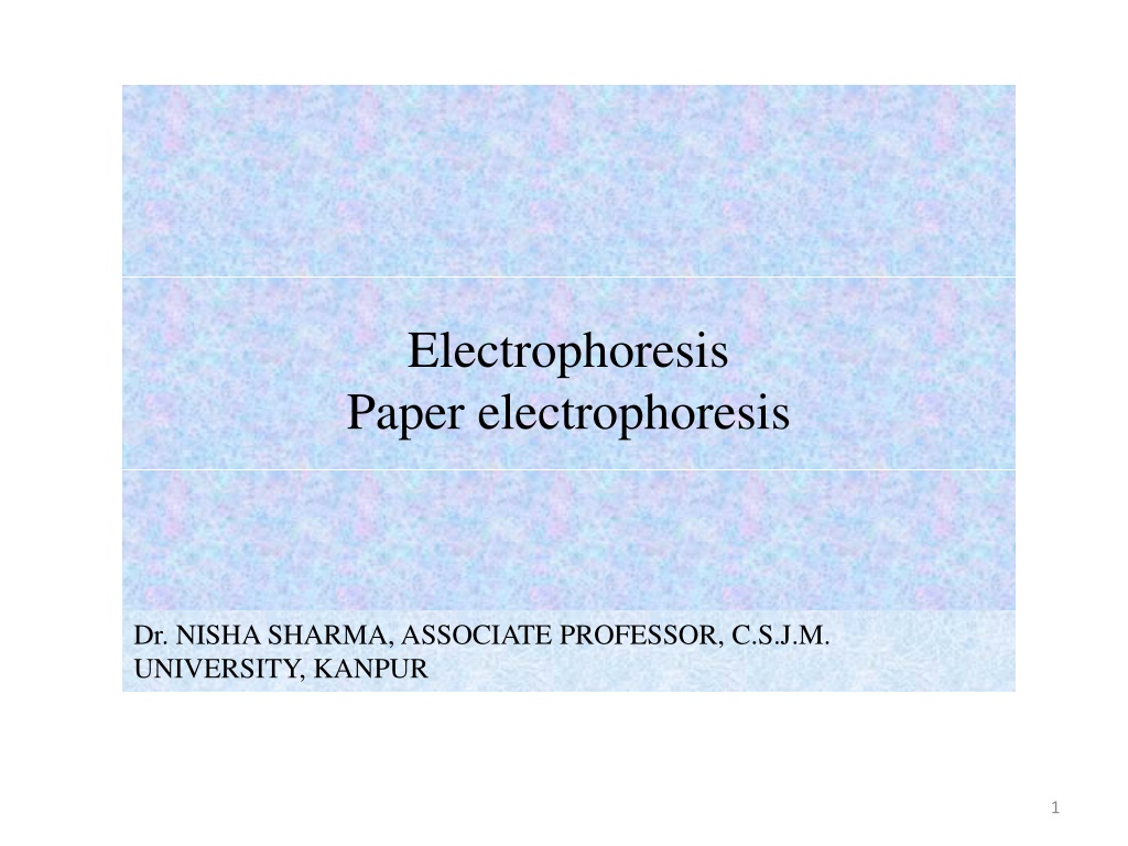


Electrophoresis
Paper electrophoresis

Dr. NISHA SHARMA, ASSOCIATE PROFESSOR, C.S.J.M.
UNIVERSITY, KANPUR
1

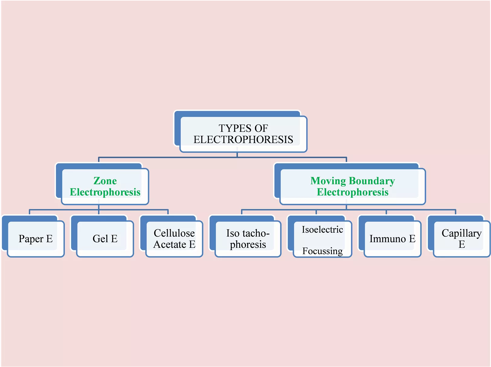

Migration of charged
particles on
supporting media

Migration of charged
particles in solution.
No supporting media
2
PAPER ELECTROPHORESIS
•
Migration or separation of particles takes place along a filter
paper
•
Sample is applied in the paper strip as circular spot / point
•
Whatman filter paper no. 1 and 3mm, 3 or 5 cm wide or
cellulose acetate paper moistened with buffer
•
The end of paper is submerged in separate reservoir containing
buffer & in this electrodes are fitted , current is passed, results
in migration of ions towards oppositely charged electrodes
•
Separation- 12-14 hrs
•
Mainly used for separation of proteins
•
Serum proteins, Hb, Lipoproteins, Isozymes
•
Proteins are charged with NH2 & COOH groups
•
At pH > pI proteins → -ve charge → anode
•
Two types: Horizontal & vertical
3
4
5
PAPER ELECTROPHORESIS
6
7
Paper Electrophoresis
•
The charge carried by a molecule depends on the
pH of the medium.
•
Electrophoresis at low voltage is not usually to
separate low molecular weight compounds
because of diffusion, but it is easier to demonstrate
the relationship b/w charge and pH with amino
acids than with proteins or other macromolecules”
8
Apparatus
•
Power pack DC – 0-500 V or 0-150mA
•
Electrophoretic cell- electrodes, buffer
reservoirs, transparent insulating cover etc.
•
Sample applications: spot/streak
•
Electrophoretic run: sample equillibrated with
buffer
•
Over heating avoided by cold room
•
Paper dried @ 110°C
•
Detection : by comparing standards
•
Fluorescence , UV-absorption, staining
9
Paper Electrophoresis
•
Apparatus:
Consists of Power Pack and an Electrophoretic Cell.
•
Power pack provides stabilized DC & controls V
& I out put, which have an out put of 0-500V and 0-
150mA
•
The Electrophoretic cell has electrodes, buffer
reservoirs, support for paper, supporting transparent
insulating cover. The electrodes- made of platinum.
•
Filter paper:
Paper of good quality, contain at least 95% α-
cellulose, with very slight adsorption capacity.
10
Paper Electrophoresis
•
Application of sample- as spot – 0.5cm dia
•
As a narrow uniform streak.
•
Sample- applied
before
the paper
equilibration with buffer or
after.
•
After sample application, equillibration of
paper with buffer.
•
Current switched on.
11
Buffers used
12
•
Detection:
•
Unknown electrogram compared with std.
•
Individual components identified by
physical properties
•
Fluorescence
•
Staining with Ethinium bromide,
visualization under U.V. light – DNA,
RNA
•
Flurescamine or Dansyl chloride staining-
for amino-acids, peptides, proteins
13
•
Detection:
•
U.V. Absorption: proteins, peptides, nucleic acids-
absorbs- 260-280nm,
•
Quantitative determination
•
Applications:
•
Serum analysis for diagnostic purpose is routinely
carried about by paper electrophoresis.
•
Muscle proteins, egg white proteins, milk proteins &
snake, insect venom analysis done by this technique.
14
•
Advantages
: Economical, easy
•
Disadvantages
: Time taking 14-16 hrs
•
Some compounds- proteins, hydrophillic
molecules, can’t be resolved- adsorptive,
ionogenic prop. of paper- tailing, distortion
of component bands
15
16
Cellulose acetate electrophoresis
•
Modified version of paper electrophoresis
•
Bacteriological acetate membrane filters are taken in
place of regular chromatography paper
•
Advantages of cellulose acetate strips over paper
•
Chemically pure & free of lignin & hemicelluloses
•
low content of glucose, suitable for polysaccaharide
electrophoresis
•
Not hydrophilic, so holds very little buffer so better
resolution in short time
•
Applications: clinical investigation such as separation
of glycoproteins, lipoproteins & haemoglobin from
blood
17
Paper electrophoresis is a powerful technique used in biochemistry to separate charged particles like proteins based on their migration along a filter paper strip. This method, conducted by Dr. Nisha Sharma, an Associate Professor at C.S.J.M. University in Kanpur, involves applying samples to the paper strip and allowing ions to migrate towards oppositely charged electrodes. The technique is mainly utilized for separating proteins, serum proteins, hemoglobin, lipoproteins, and isozymes by taking advantage of their NH2 and COOH groups. The process occurs over 12-14 hours and is classified into horizontal and vertical types. Apparatus like a power pack and electrophoretic cell are essential for conducting paper electrophoresis effectively.

Download Presentation

Please find below an Image/Link to download the presentation.
The content on the website is provided AS IS for your information and personal use only. It may not be sold, licensed, or shared on other websites without obtaining consent from the author.If you encounter any issues during the download, it is possible that the publisher has removed the file from their server.
You are allowed to download the files provided on this website for personal or commercial use, subject to the condition that they are used lawfully. All files are the property of their respective owners.
The content on the website is provided AS IS for your information and personal use only. It may not be sold, licensed, or shared on other websites without obtaining consent from the author.
E N D
Presentation Transcript
Electrophoresis Paper electrophoresis Dr. NISHA SHARMA, ASSOCIATE PROFESSOR, C.S.J.M. UNIVERSITY, KANPUR 1
Migration of charged particles on supporting media Migration of charged particles in solution. No supporting media TYPES OF ELECTROPHORESIS Zone Moving Boundary Electrophoresis Electrophoresis Isoelectric Cellulose Acetate E Iso tacho- phoresis Capillary E Paper E Gel E Immuno E Focussing 2
PAPER ELECTROPHORESIS Migration or separation of particles takes place along a filter paper Sample is applied in the paper strip as circular spot / point Whatman filter paper no. 1 and 3mm, 3 or 5 cm wide or cellulose acetate paper moistened with buffer The end of paper is submerged in separate reservoir containing buffer & in this electrodes are fitted , current is passed, results in migration of ions towards oppositely charged electrodes Separation- 12-14 hrs Mainly used for separation of proteins Serum proteins, Hb, Lipoproteins, Isozymes Proteins are charged with NH2 & COOH groups At pH > pI proteins -ve charge anode Two types: Horizontal & vertical 3
Paper Electrophoresis The charge carried by a molecule depends on the pH of the medium. Electrophoresis at low voltage is not usually to separate low molecular weight compounds because of diffusion, but it is easier to demonstrate the relationship b/w charge and pH with amino acids than with proteins or other macromolecules 8
Apparatus Power pack DC 0-500 V or 0-150mA Electrophoretic cell- electrodes, buffer reservoirs, transparent insulating cover etc. Sample applications: spot/streak Electrophoretic run: sample equillibrated with buffer Over heating avoided by cold room Paper dried @ 110 C Detection : by comparing standards Fluorescence , UV-absorption, staining 9
Paper Electrophoresis Apparatus: Consists of Power Pack and an Electrophoretic Cell. Power pack provides stabilized DC & controls V & I out put, which have an out put of 0-500V and 0- 150mA The Electrophoretic cell has electrodes, buffer reservoirs, support for paper, supporting transparent insulating cover. The electrodes- made of platinum. Filter paper: Paper of good quality, contain at least 95% - cellulose, with very slight adsorption capacity. 10
Paper Electrophoresis Application of sample- as spot 0.5cm dia As a narrow uniform streak. Sample- applied before the paper equilibration with buffer or after. After sample application, equillibration of paper with buffer. Current switched on. 11
Buffers used Separation Buffer pH Ionic str. 0.05 Composition Proteins Barbital 8.6 10.3 g Na-barbiturate 1.84g barbital 0.6g NaH2PO4H2O 2.2G Na2HPO4 3.51g NaCl 3.28g Na-acetate pH-4.5 with HCl 28.46g Na-citrate 20.6g citric acid 20.4gKH2PO4 9.8g sodium barbiturate Phosphate 7.4 -- Nucleoprotei ns Acetate 4.5 0.1 Citrate 4.5 0.13 Amino Acids Phosphate 4.6 8.6 0.15 0.1 Michaelis 12
Detection: Unknown electrogram compared with std. Individual components identified by physical properties Fluorescence Staining with Ethinium bromide, visualization under U.V. light DNA, RNA Flurescamine or Dansyl chloride staining- for amino-acids, peptides, proteins 13
Detection: U.V. Absorption: proteins, peptides, nucleic acids- absorbs- 260-280nm, Quantitative determination Applications: Serum analysis for diagnostic purpose is routinely carried about by paper electrophoresis. Muscle proteins, egg white proteins, milk proteins & snake, insect venom analysis done by this technique. 14
Advantages: Economical, easy Disadvantages: Time taking 14-16 hrs Some compounds- proteins, hydrophillic molecules, can t be resolved- adsorptive, ionogenic prop. of paper- tailing, distortion of component bands 15
Cellulose acetate electrophoresis Modified version of paper electrophoresis Bacteriological acetate membrane filters are taken in place of regular chromatography paper Advantages of cellulose acetate strips over paper Chemically pure & free of lignin & hemicelluloses low content of glucose, suitable for polysaccaharide electrophoresis Not hydrophilic, so holds very little buffer so better resolution in short time Applications: clinical investigation such as separation of glycoproteins, lipoproteins & haemoglobin from blood 17