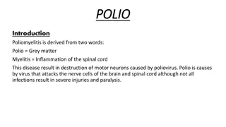
Poliomyelitis: A Comprehensive Overview of the Disease
Poliomyelitis, commonly known as polio, is caused by a non-enveloped, positive, single-stranded RNA virus belonging to the Picornaviridae family. Paralysis is the most severe consequence of polio infection, with transmission occurring through the fecal-oral route. The virus primarily infects the GI tract before spreading to the central nervous system, leading to devastating effects. Vaccination and sanitation play a crucial role in eradicating the disease, which has been successful in most countries but remains endemic in Nigeria, Afghanistan, and Pakistan.
Download Presentation

Please find below an Image/Link to download the presentation.
The content on the website is provided AS IS for your information and personal use only. It may not be sold, licensed, or shared on other websites without obtaining consent from the author.If you encounter any issues during the download, it is possible that the publisher has removed the file from their server.
You are allowed to download the files provided on this website for personal or commercial use, subject to the condition that they are used lawfully. All files are the property of their respective owners.
The content on the website is provided AS IS for your information and personal use only. It may not be sold, licensed, or shared on other websites without obtaining consent from the author.
E N D
Presentation Transcript
Poliomylitis Dr. Abbas
Etiology Non- enveloped,positiv e ,single sranded R.N.A virus Picornaviridae, genus - enterovirus 3- serotypes- 1,2,3 Sensitive heat and light
Epidemiology Paralysis is the most devastating effect of polio virus infection Although 90-95 % infection-subclinical Universal vaccination strategy and improved sanitation: key factor in eradication Eradicated in most of countries except Nigeria, Afghanistan and Pakistan
Transmission virus excreted in feces 2weeks before the onset of paralysis to several weeks after the onset of symptoms Human are only reservoir Spread by feco- oral route
Pathogenesis Infection via GI tract Primary site of infection M cells of mucosa Wild type polio viruses reaches CNS along peripheral nerves Regional lymph node Virus seeds multiple sites :RE systems, brown fat,skeletal muscle Primary viremia after 2-3 days
Pathogenesis(contd..) Vaccine strain of polio do not replicate in CNS. Occasional revertants(by nucleoside subtitution) of these vaccine strain developed a neurovirulent phenotype and cause Vaccine acquired paralytic poliomylitis Reversion occur in small intestine and reaches to CNS via. Peripheral nerves
Pathogenesis(contd..) Infection Traverse neural pathways and multiple site within CNS Perineural inflammation and destruction Petachial hemorrhages and inflammatory edema Primarily infect motor neuron in anterior horn cells and medulla oblongata(cranial nerve nuclei) Involvement of reticular formation that controlled vitals may have catastrophic out come
Involvement of dorsal horn and dorsal root ganglias of spinal cord results in hyperesthesia and myalgias:typical of acute polimyelitis Other neuron affected are the nuclie in the vermis of cerebellum, substantia nigra,thatmus,hypothalmus
Clinical features I.P- 8-12 days ,ranges from 5- 35 days Infection with wild polio virus may follow several courses Inapparant Abortive Non-paralytic Paralytic 90-95% 5% 1% 0.1%
Abortive poliomyelitis Non specific flu like illness. phgysical examination: non specific pharyngitis, abdominal or muscular tenderness and weakness Recovery : complete without sequelae
Non- Paralytic poliomyelitis Sign of abortive poliomyelitis but more intense Headache, nausea, vomiting , sore throat, neck & spinal rigidity, fleeting paralysis of bladder and constipation. Changes in Reflexes may precede before onset of paralysis. Superficial reflexes, cremastric, abdominal and reflexes of ,spinal, gluteal muscle. Spinal and gluteal reflexes disappear before other reflexes. Changes in DTR occur after 8-24 hrs after superficial reflexes diminished. DTR are absent with paralysis Sensory defect don t occur in poliomyelitis Recovery : complete
Paralytic poliomyelitis Spinal Bulbar Encephalitis Paralysis Appears 3-8days after the initial symptoms Clinical features of paralytic polio caused by wild or vaccine strain are comparable
Spinal paralytic polio Biphasic disease ist phase Symptoms similar to abortive polio Patient appear to feel better for 2-5 days Severe headache and fever and exacerbation of previous symptoms. Severe muscle pain sensory and motor phenomenon. Physical examination: distribution of paralysis characteristically spotty After 1-2 days asymmetric flaccid paralysis occur Involvement of 1 leg is most common , followed by involvement of 1 leg and 1 arm Proximal areas of the extremities tend to be involved to a greater extent.
Polio Paralysis Some times biphasic phase absent. 50-60% cases h/o IM injection before paralysis(provocation paralysis) Paralysis start . Little recovery from paralysis noticed during ist few days but not beyond 6 months. Return of strength and reflexes is slow and & may continue to improve as long as 18 months after the acute ds. Atrophy of limb ,growth failure and deformity finally evident
Bulbar Polio Dysfunction of cranial nerve and medullary centers without involving spinal cord Respiratory difficulty, paralysis of extraoccular, facial and masticatory muscles. 1-nasal twang to the voice or cry. 2-inability swallow smoothly , 3-absence of effective coughing 4-nasal regurgitation of saliva 5-deviation of palate, uvula, tongue 6-involvement of vitals centre in the medulla 7-paralysis of 1 or both vocal cords 8- Rope sign: acute angulations b/w chin and larynx caused by weakness of hyoid muscle
Polioencephalitis Rare form of disease Higher centre of brain severely involved Seizure, coma ,spastic paralysis, irritability, disorientation, drowsinesss,cranial nerve paralysis, deaths
Diagnosis Paralysis in any unimmunized or partially immunized children VAPP should be considered in any child with paralysis developed 7-14 days after receiving OPV. Combination of fever ,headache, neck and back pain,assymmetric flaccid paralysis without sensory loss. WHO recommends lab diagnosis of polio must be done by isolation and identification of polio virus in the stool ,with specific identification of wild type and vaccine strains.
In suspected of poliomyelitis two stool sample collected 24-48 hrs apart 80-90% isolation of virus in acute phase. < 20% isolation b/w 3-4 weeks Ideally 8-10gm stool sample Proper cold chain
Differential diagnosis All causes of acute flaccid paralysis
AFP Paralysis of acute onset i.e less than 4weeks and affected limbs are floppy or flaccid or limp. Tone diminished, DTR diminished. Sensation not affected Case definition: any child aged less than 15yrs who has acute onset flaccid paralysis for which no obvious cause(severe trauma, electrolyte imbalance) is found or paralytic illness in a person of any age in which polio is suspected
AFP causes Anterior horn cells disorder Disoreder of neuromuscu lar junction Metabolic disorder Acute traumatic neuritis e.g. GB Neuropathi es poliomyelitis,n onpolioenterov irus,west nile virus syndrome e.g. e.G myasthenia gravis hypokalemia Disorder of muscle: e.g. polymyositis, viral myositis
Treatment only supportive
Prevention WHO recommends 4 strategy for global eradication of polio- 1-routine vaccination 2-NIDs 3-AFP surveillance 3- Mop-up immunization
