Paediatric Fracture Management Guidelines and BOAST Recommendations
This guidance provides tools and sheets for consistent pre-specialist management and referral practices for children's fractures in the Emergency Department (ED) or referring treatment centers. It covers principles for assessing mechanisms, imaging, classification, and definitive management of fractures. The guidelines emphasize appropriate clinical knowledge and discretion. Local modifications and agreement with orthopedic representatives are recommended for the best outcomes.
Download Presentation

Please find below an Image/Link to download the presentation.
The content on the website is provided AS IS for your information and personal use only. It may not be sold, licensed, or shared on other websites without obtaining consent from the author. Download presentation by click this link. If you encounter any issues during the download, it is possible that the publisher has removed the file from their server.
E N D
Presentation Transcript
In line with GIRFT Paediatric Trauma and Orthopaedic recommendations, BSCOS offers this tool set of guidance sheets, designed to help bring consistent pre-specialist management and referral practices to bear on children s fractures in your ED or referring treatment centres. It does not cover major trauma which should have its own specified pathway. ED fracture guidance set Variation in local practice is common and may reflect appropriate and necessary geographical and/or service delivery models. Those centres with virtual fracture clinic review may well have different follow up safety-netting to those without. This guidance is not prescriptive it is a starting point and is best jointly considered, adapted and agreed by representatives from your ED alongside Paediatric Orthopaedics. It is suggested that referral criteria and other local service specific aspects of management are agreed and the sheets modified accordingly, noting the date of agreement and local contacts. Clinical responsibility remains with the treating clinician at all times. BSCOS cannot take any responsibility for the contents of these guidance sheets. BSCOS approved Patient Information Leaflets (PILs) will be hyperlinked to these documents as they become available.
Emergency Dept Fracture Management BOAST guidance The following BOA BOAST documents are relevant to the development of local ED protocols: Management of paediatric acute musculoskeletal infection Management of ACL injury in skeletally immature Early management of paediatric forearm fracture Supracondylar elbow fractures BSCOS has issued guidance on virtual fracture clinics.
Emergency Dept Fracture Management Guidance: Principles Appropriate clinical knowledge and clinical discretion remain essential when using these guidelines. A) Assess NB Mechanism (? Major Trauma), Distracting injuries, Temporary Splint/Analgesia. Consider NAI plausibility of mechanism, age of child, delay to presentation, prior history. High index of suspicion = Major long bone # in <2y &/or multiple fractures of differing healing. B) Best possible imaging - XR - AP & Lateral views shaft fractures must include joint above and below. Ask for repeat views if necessary! C) Classify which bone(s), site (prox/med/distal), displacement (undisplaced, % diameter overlap), communition (fragmentation), angulation (sagittal/coronal/transverse planes), closed/open (contamination?), is a joint &/or physis involved?. D) Definitive management age (of patient), site and type specific. Discuss with Orthopaedics if in doubt Immobilisation 1. Consider - is it necessary? Some injuries will heal with no restriction, or a removable splint 2. Plaster of Paris is best for moulding (ie to hold the position of a fracture that is likely to displace). 3. Softcast is best for removal at home (ie where the fracture is unlikely to displace). 4. Resin is best for lightweight strength (ie when weightbearing) and poor for moulding Ongoing treatment plan - Consider - is review necessary? Some injuries do not require further treatment. Issue patient Info leaflets where relevant. Local modification date/trust/HB: Links to studies (where relevant):
Emergency Dept Fracture Management Guidance: NAI Appropriate clinical knowledge and clinical discretion remain essential when using these guidelines. A) Assess Delayed presentation? Inconsistent mechanism or history changes. Check child at risk register. High index of suspicion = Major long bone # in <2y Multiple fractures of differing healing. Ribs/Skull/Scapula/Sternum fractures Metaphyseal corner fractures / Bucket handle fractures B) Best possible imaging of injured area - XR - AP & Lateral views C) Classify Bone(s) involved, site (prox/mid/distal), comminution, D) Document carefully Discuss with child protection Definitive management refer Ortho Missed NAI carries a high mortality Local modification date/trust/HB: Links to studies (where relevant):
Emergency Dept Fracture Management Guidance: Upper Limb / Clavicle Appropriate clinical knowledge and clinical discretion remain essential when using these guidelines. A) Assess NB Mechanism (? Major Trauma). Caution - Sternoclavicular joint disruption rare - refer urgently if clavicle posteriorly displaced. B) Best possible imaging - XR - AP & 20 cephalad views C) Classify site (med SC Jt/shaft/distal AC Jt), displacement (undisplaced, % diameter overlap), angulation, communition (fragmentation), angulation, closed/open (contamination?). D) Definitive management - Un/Minimal displaced Broad arm sling 2-4 weeks, avoid sports No follow up. - Displaced Figure of 8 brace Instructions to tighten Follow up 1/52 - AC Joint disruption Broad arm sling 2 weeks No follow up under 12y. Links to studies (where relevant): Local modification date/trust/HB:
Emergency Dept Fracture Management Guidance: Upper Limb / Glenohumeral dislocation Appropriate clinical knowledge and clinical discretion remain essential when using these guidelines. A) Assess NB Mechanism Trauma / Previous dislocations. Caution check and document distal NV function before treatment B) Best possible imaging - XR - AP, scapular lateral +/or axillary C) Classify displacement (anterior/posterior- light bulb sign /inferior luxatio erecta ), associated fractures. D) Definitive management Reduction under sedation then polysling - Atraumatic physio OPD referral - Traumatic (eg rugby) consider MRI - Associated fracture refer to Orthopaedics Links to studies (where relevant): Local modification date/trust/HB:
Emergency Dept Fracture Management Guidance: Upper Limb / Humerus Appropriate clinical knowledge and clinical discretion remain essential when using these guidelines. A) Assess NB Mechanism (? Major Trauma). Caution check and document radial N function before treatment B) Best possible imaging - XR - AP, scapular lateral +/or axillary C) Classify site (prox/med/distal), displacement (undisplaced, % diameter overlap), communition (fragmentation), angulation (sagittal/coronal/transverse planes), closed/open (contamination?), is a joint &/or physis involved?. D) Definitive management - Humeral brace or J-slab - Collar & cuff - Fracture clinic 1/52 Links to studies (where relevant): Local modification date/trust/HB:
Emergency Dept Fracture Management Guidance: Upper Limb / Epicondyles of Elbow Appropriate clinical knowledge and clinical discretion remain essential when using these guidelines. A) Assess NB Mechanism typically a fall onto outstretched hand (? Major Trauma). Caution do not mistake condylar for epicondylar fracture B) Best possible imaging - XR - AP & lateral elbow C) Classify note normal ossific nuclei timing Capitellum Radial Head Internal (Medial) Trochlear Olecranon External (lateral) Appears 1 3 5 7 9 11 Fuses 12-14 14-16 16-18 12-14 15-17 12-14 D) Definitive management Medial - Undisplaced (suspected) Broad arm sling 2/52, No FU - Displaced - Backslab, refer Orthopaedics Lateral - <2mm displaced - Backslab, 1/52 FU - >=2mm displaced or suspicion of condylar refer Ortho Links to studies (where relevant): Local modification date/trust/HB:
Emergency Dept Fracture Management Guidance: Upper Limb / Supracondylar elbow Appropriate clinical knowledge and clinical discretion remain essential when using these guidelines. A) Assess NB Mechanism (? Major Trauma). Caution check and document distal NV function before treatment B) Best possible imaging - XR - AP & lateral elbow C) Classify Gartland, closed/open (contamination?) D) Definitive management flexion type (less stable) - apply backslab, refer Ortho - extension type - see below NB see: https://www.boa.ac.uk/static/a240155a-f0dd-4be7- 8c8af7b6cc4da795/BOASTSupracondylarFracturesHumerusChildren2020-v2-FINAL.pdf (or suspected but normal XR) Broad arm sling 1-2/52 No follow up (and/or NV compromise) Backslab Refer Orthopaedics (and/or NV compromise) Backslab Refer Orthopaedics Links to studies (where relevant): Local modification date/trust/HB:
Emergency Dept Fracture Management Guidance: Upper Limb / Lateral condyle of Elbow Appropriate clinical knowledge and clinical discretion remain essential when using these guidelines. A) Assess NB Mechanism typically a fall onto outstretched hand (? Major Trauma). Caution do not mistake for epicondylar fracture B) Best possible imaging - XR - AP & lateral elbow C) Classify Weiss [NB ossific nucleus appears 11yoa, fuses 12-14yoa] D) Definitive management depends on articular disruption, may be obscure if cartilaginous Type 1 - only if certain, backslab, F/U 1/52 Types 2 & 3 (or uncertain) backslab, refer Ortho Links to studies (where relevant): Local modification date/trust/HB:
Emergency Dept Fracture Management Guidance: Upper Limb / Radial head & neck Appropriate clinical knowledge and clinical discretion remain essential when using these guidelines. A) Assess NB Mechanism typically a fall onto outstretched hand (? Major Trauma). Caution watch out for an absent (ie displaced) radial head ossific nucleus after 3yoa B) Best possible imaging - XR - AP & lateral elbow C) Classify Radial head ossific nucleus appears 3yoa, fuses 14-16yoa D) Definitive management Undisplaced/suspected or Angulated <30 deg Broad arm sling (under clothing) for 2-4w, No F/U Displaced or Angulated >30 deg Refer Orthopaedics Links to studies (where relevant): Local modification date/trust/HB:
Emergency Dept Fracture Management Guidance: Upper Limb / Olecranon Appropriate clinical knowledge and clinical discretion remain essential when using these guidelines. A) Assess NB Mechanism (? Major Trauma). Caution check for evidence of subluxation and alignment of radial head with capitellum B) Best possible imaging - XR - AP & lateral elbow C) Classify NB ossific nucleus appears 9yoa, fuses 15-17 D) Definitive management Undisplaced/suspected Backslab/softcast, to remove at home 3/52, No F/U Displaced or Angulated Refer Orthopaedics Any misalignment of radiocapitellar joint? consider Monteggia refer Orthopaedics Links to studies (where relevant): Local modification date/trust/HB:
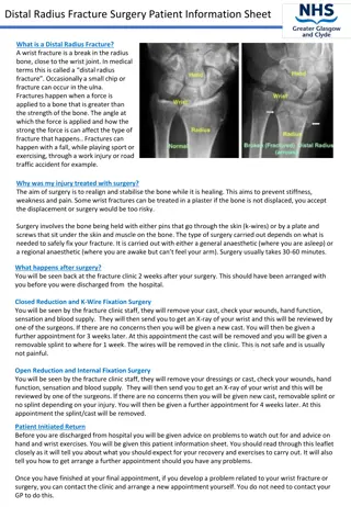
Emergency Dept Fracture Management Guidance: Upper Limb / Forearm Appropriate clinical knowledge and clinical discretion remain essential when using these guidelines. A) Assess NB Mechanism (? Major Trauma). Caution check for evidence of subluxation and alignment at wrist and elbow B) Best possible imaging - XR - AP & lateral forearm (to include elbow and wrist) C) Classify Plastic deformation (younger child, no fracture but NB radiocapitellar and DRUJ) - Ulnar fracture, Radius intact (NB Monteggia radiocapitellar sublux see image below) - Radial fracture, Ulna intact (NB Galeazzi DRUJ sublux) - Both bone forearm displacement, angulation, open/closed D) Definitive management Undisplaced/suspected Backslab/softcast, to remove at home 3/52, No F/U Plastic deformation - Backslab then refer orthopaedics Monteggia/Galeazzi Backslab then refer orthopaedics Displaced or Angulated Backslab then refer orthopaedics Links to studies (where relevant): Local modification date/trust/HB:
Emergency Dept Fracture Management Guidance: Upper Limb / Wrist Appropriate clinical knowledge and clinical discretion remain essential when using these guidelines. A) Assess NB Mechanism fall onto outstretched hand typically. Caution check for evidence of subluxation and alignment at wrist and elbow B) Best possible imaging - XR - AP & lateral centred on wrist [NOT full forearm XR] (consider additional AP & lat elbow if any tenderness/deformity) C) Classify torus (unicortical plastic deformation)/greenstick (torus one cortex, fracture of the other)/bicortical, angulation, displacement, comminution, open/closed D) Definitive management - Torus/Buckle (unicortical) no brace required, futuro splint for comfort - Greenstick/Undisplaced/min angulation <15 deg (bicortical) softcast/backslab, remove at home 3/52 No F/U - Displaced/signif angulation/physis involved backslab, refer to Orthopaedics - consider CRAFFT NB see https://www.boa.ac.uk/static/57ea20ec-8edb-46ce-879222a813ce9af6/BOAST-Paediatric- Forearm.pdf Links to studies (where relevant): Local modification date/trust/HB:
Emergency Dept Fracture Management Guidance: Upper Limb / Carpus Appropriate clinical knowledge and clinical discretion remain essential when using these guidelines. A) Assess NB Mechanism Caution missed scaphoid carries an increased avascular necrosis risk B) Best possible imaging - XR Scaphoid series C) Classify proximal pole / waist / distal pole of scaphoid ? - displacement ? D) Definitive management Scaphoid - <9y - highly unlikely to be fractured (cartilaginous) - seek advice if signif concern. - >9y - clinical suspicion only extended futuro splint 2/52, F/U if ongoing symptoms - undisplaced fracture on XR scaphoid cast 6-8 weeks, 6/52 F/U - displaced fracture on XR refer to ortho - Any other carpal fracture refer to ortho (or hand service) Links to studies (where relevant): Local modification date/trust/HB:
Emergency Dept Fracture Management Guidance: Upper Limb / MCs & phalanges Appropriate clinical knowledge and clinical discretion remain essential when using these guidelines. A) Assess NB nerve and tendon fcn, significant soft tissue injury consider plastic surgery input. Caution check and document rotational deformity of digits B) Best possible imaging - XR PA and true lateral views of any confirmed fractures [obliques are good for diagnostic sensitivity but not for assessment of displacement] C) Classify named digits [use: thumb, index, long, ring, little], prox/mid/distal, open/closed, oblique/tvse/spiral, comminution, displacement. D) Definitive management Scaphoid D) Definitive management - Base of MC ulnar gutter for little/ring finger refer hand service - CMC dislocation - reduce under analgesia refer hand service - Undisplaced MC shaft - buddy strap+futuro No F/U unless stability concern - Multiple MC # - volar slab in position of safe immobilization (CMCjts at 90, IPJs at 180 deg) refer - Undisplaced MC neck - buddy strap+futuro No F/U unless stability concern - Angulated/rotated MC shaft/neck refer hand service - Volar plate injury - buddy strap No F/U - Bony mallet/Nail bed injury/suspected infection refer hand service Links to studies (where relevant): Local modification date/trust/HB:
Emergency Dept Fracture Management Guidance: Spine Appropriate clinical knowledge and clinical discretion remain essential when using these guidelines. A) Assess NB Mech of injury Risk of (missed) major trauma. Document distal NV status on arrival Caution high risk of distracting injury undertake a secondary survey B) Best possible imaging CT vs XR d/w radiology / as per local protocol C) Classify Cervical / Thoracic / Lumbar / Sacral D) Definitive management - Immobilise - Refer to orthopaedics Links to studies (where relevant): Local modification date/trust/HB:
Emergency Dept Fracture Management Guidance: Lower Limb/Pelvis & Hip Appropriate clinical knowledge and clinical discretion remain essential when using these guidelines. A) Assess NB Mech of injury Risk of (missed) major trauma. Caution high risk of distracting injury undertake a secondary survey SUFE is often missed beware groin pain in the adolescent B) Best possible imaging AP Pelvis XR (+ frog lateral in suspected SUFE) C) Classify Minor Avulsions v Injury to Pelvic Ring v SUFE D) Definitive management Avulsion (of apophyses/trochanter) crutches, analgesia, physio referral Major trauma - immobilise,Consider pelvic binder - Refer to orthopaedics SUFE refer to ortho Perthes arrange f/u in elective paeds ortho service Links to studies (where relevant): Local modification date/trust/HB:
Emergency Dept Fracture Management Guidance: Lower Limb / Femur Appropriate clinical knowledge and clinical discretion remain essential when using these guidelines. A) Assess NB record distal neurovasc status. Apply Thomas Splint +/- Fem N Block for pain relief. Caution refer for consideration of NAI in the non-ambulant infant! B) Best possible imaging - XR = PA and true lateral views C) Classify prox/mid/distal, open/closed, oblique/tvse/spiral, comminution, displacement. D) Definitive management - Refer to orthopaedics Links to studies (where relevant): Local modification date/trust/HB:
Emergency Dept Fracture Management Guidance: Lower Limb Patella Appropriate clinical knowledge and clinical discretion remain essential when using these guidelines. A) Assess NB Mech of injury trauma or atraumatic ? Caution always assess integrity of extensor mech with SLR. B) Best possible imaging AP & lat knee (+/- skyline view) C) Classify First time dislocation v recurrent - Displacement of a fracture. D) Definitive management Dislocated patella reduce with sedation - First time dislocation - refer to Orthopaedics - Recurrent dislocation check XR for osteochondral lesions refer to physio Fracture - Undisplaced with intact ext mech resin full leg cast, F/U 1/52 - Displaced refer to orthopaedics. Links to studies (where relevant): Local modification date/trust/HB:
Emergency Dept Fracture Management Guidance: Lower Limb Knee Jt Appropriate clinical knowledge and clinical discretion remain essential when using these guidelines. A) Assess NB Mech of injury trauma or atraumatic ? Caution always assess integrity of extensor mech with SLR. B) Best possible imaging AP & lat knee (+/- skyline view) C) Classify Grade laxity (cf contralat side) if ligamentous injury. - Site of any osteochondral fracture, displacement. Lipohaemarthrosis? - Segond fractures = avulsion of collateral ligament - Tibial spine (ACL) avulsion displacement - Tibial tuberosity fracture displacement D) Definitive management - Soft tissue injury without lipohaemarthrosis - Cricket splint, crutches, urgent physio referral - Lipohaemarthosis w/out obvious # - Cricket splint, crutches - d/w Orthopaedics during working day - Segond or Tib spine or tuberosity fracture - refer Ortho - Osteochondral lesion d/w Orthopaedics during working day NB see: https://www.boa.ac.uk/asset/EA032921%2D2A7F%2D4A15%2D8F7033524E4678D5/ Links to studies (where relevant): Local modification date/trust/HB:
Emergency Dept Fracture Management Guidance: Lower Limb Tibia & Fibula Appropriate clinical knowledge and clinical discretion remain essential when using these guidelines. A) Assess NB Mech of injury trauma or atraumatic ? Caution Beware compartment syndrome. B) Best possible imaging AP & lat (to include knee and ankle joints) C) Classify Bone(s) involved, Site (Prox/Shaft/Distal), Open/closed, Comminution, Displacement, Angulation. D) Definitive management Toddler s fracture - undisplaced <5yo - softcast/backslab x3/52, No F/U - clin suspicion (XR normal) consider analgesia only or as above. Fibular neck # - check comm peroneal nerve, check ankle (Maisonneuve) Tibial prox metaphysis long leg cast, refer Ortho risk of late valgus (Cozen s phenomenon) Tibial shaft undisplaced - long leg cast NWB FU 1/52 - displaced/angulated - AK backslab, refer Ortho Links to studies (where relevant): Local modification date/trust/HB:
Emergency Dept Fracture Management Guidance: Lower Limb Ankle/Hindfoot Appropriate clinical knowledge and clinical discretion remain essential when using these guidelines. A) Assess NB Mech of injury trauma or atraumatic ? Caution Talar neck fractures are easily missed and carry high risk of AVN B) Best possible imaging AP & lat ankle, AP & oblique foot (+ true lat of foot if any tarsal fracture) C) Classify Tillaux = SH 3 anterolat tib epiphyseal avulsion, adolescents only - Triplane = sagittal, coronal and transverse plane fracture, (SH 2 on one view and SH 3 on the other) D) Definitive management - Signif sprain/avulsion/undisplaced # = Splint/Moonboot 2-4/52 No FU - Displaced fracture / Talar shift = Backslab, refer Ortho - Triplane & Tillaux Backslab, refer Ortho, consider CT scan - Talar dome or neck fracture = Backslab, refer Ortho - Tarsal avulsion fractures = WB BK cast, FU 1/52 - Calcaneal fracture elevate, refer Ortho - Ligamentous disruption (Lisfranc injury) true lat XR refer Ortho Links to studies (where relevant): Local modification date/trust/HB:
Emergency Dept Fracture Management Guidance: Lower Limb Foot Appropriate clinical knowledge and clinical discretion remain essential when using these guidelines. A) Assess NB Mech of injury trauma or atraumatic ? Caution B) Best possible imaging AP & oblique [+true lat if fracture is diagnosed] C) Classify Bone(s) involved, site (prox/mid/distal), comminution, open/closed, displacement/angulation D) Definitive management - Significant crush (irrespective of fractures ) consider compartment synd elevate, refer Ortho - MT fractures - Undisplaced/base of 5th avulsion - sturdy (stiff soled) footwear or moonboot x2-3/52, No FU - Displaced refer Ortho - Phalanges - Undisplaced sturdy shoes, No FU - Displaced slipper cast or boot, FU only for Gt toe Links to studies (where relevant): Local modification date/trust/HB:


