Overview of the First Week of Development from Ovulation to Implantation
The first week of development after ovulation involves key processes like fertilization, cleavage, and blastocyst formation. It begins with the maturation of the primary oocyte in the follicle, leading to ovulation and potential fertilization. During this period, hormonal changes in the ovarian cycle play crucial roles in the maturation of the oocyte and ovulation. Understanding these early developmental stages is essential for grasping the complexity of human embryogenesis.

Download Presentation

Please find below an Image/Link to download the presentation.
The content on the website is provided AS IS for your information and personal use only. It may not be sold, licensed, or shared on other websites without obtaining consent from the author.If you encounter any issues during the download, it is possible that the publisher has removed the file from their server.
You are allowed to download the files provided on this website for personal or commercial use, subject to the condition that they are used lawfully. All files are the property of their respective owners.
The content on the website is provided AS IS for your information and personal use only. It may not be sold, licensed, or shared on other websites without obtaining consent from the author.
E N D
Presentation Transcript
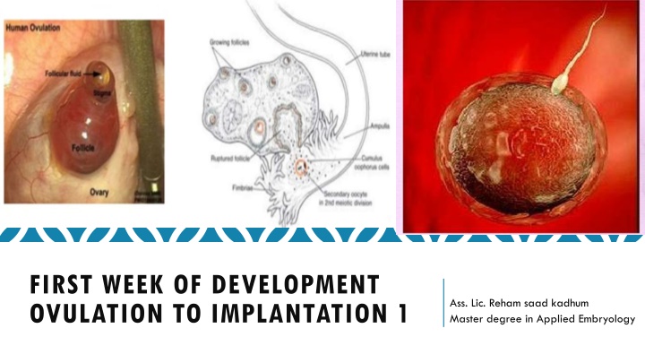
FIRST WEEK OF DEVELOPMENT OVULATION TO IMPLANTATION 1 Ass. Lic. Reham saad kadhum Master degree in Applied Embryology
Initially period Initially period - - the first week Development: the first week Development: Ovulation. Ovulation. Fertilization. Fertilization. Cleavage Cleavage Blastocyst formation. Blastocyst formation.
Introduction 1. The primary oocyte maturates in the mature follicle and stops in the diplotene stage to complete meiosis I resulting in: A. Secondary oocyte. B. First polar body (containing little cytoplasm). 2. The secondary oocyte begins meiosis II, and then the mature follicle raptured and thus ovulation the secondary oocyte outside the ovary as it is in metaphase of meiosis II. 3. If fertilization occurs; the secondary oocyte completed meiosis II resulting in: A. Tertiary mature oocyte. B. Secondary polar body.
OVARIAN CYCLE Its sexual monthly cycle controlled by hypothalamus , begin at puberty Gonadotropin- releasing hormone (GnRH), produced by the hypothalamus, acts on cells of the anterior lobe (adenohypophysis) of the pituitary gland, which in turn secrete gonadotropins. These hormones, follicle-stimulating hormone (FSH) and luteinizing hormone (LH), stimulate and control cyclic changes in the ovary. FSH : Stimulate the development of ovarian follicles and produce estrogen by follicular cells. LH: Stimulate ovulation. Stimulate follicular cells and corpus luteum to produce progesterone.
Estrogen action : The uterine endometrium enters the follicular or proliferative phase. Thinning of the cervical mucus occurs to allow passage of sperm. The anterior lobe of the pituitary gland is stimulated to secrete LH. At mid cycle, there is an LH surge that: Elevates concentrations of maturation- promoting factor, causing oocytes to complete meiosis I and initiate meiosis II Stimulates production of progesterone by follicular stromal cells (luteinization) Causes follicular rupture and ovulation
Ovulation 1. vesicular follicle grows rapidly to a diameter of 25 mm to become a mature vesicular (graafian) follicle. In the days preceding the ovulation under influence of FCH 2. LH surge cause the oocyte complete meiosis I and enter meiosis II but oocyte is arrested in metaphase approximately 3 hours before ovulation the surface of the ovary begins to bulge locally, and at the apex, an avascular spot, the stigma, appears High level of LH : lead to a. increase activity of collagenase enzyme , which lead to digestion of collagen fibers surrounding the follicle. b. Increase level of prostaglandins, which lead to local muscular contraction in the walls of the ovary. Those contractions extrude the oocyte, which together with its surrounding granulosa cells from the regi n of the cumulus oophorus breaks free (ovulation) and floats out of the ovary
Oocyte transport : due to 1. Fimbriae , starts to sweep over the surface of the ovary shortly before ovulation . 2. Rhythmic contraction of uterine tube, this leads to pushing the ova toward the uterus in about 4 days. 3. Movement of the cilia on the uterine tube epithelial lining. Note In humans, the fertilized oocyte reaches the uterine lumen in approximately 3 to 4 days.
Corpus Luteum After ovulation, granulosa cells remaining in the wall of the ruptured follicle, together with cells from the theca interna, are vascularized by surrounding vessels. Under the influence of LH, these cells develop a yellowish pigment and change into lutein cells, which form the corpus luteum and secrete estrogens and progesterone Progesterone, together with some estrogen, causes the uterine mucosa to enter the progestational or secretory stage in preparation for implantation of the embryo.
The corpus luteum contines to grow and forms the corpus luteum of pregnancy (corpus luteum graviditatis). By the end of the third month, this structure may be one third to one half of the total size of the ovary. Yellowish luteal cells continu to secrete progesterone until the end of the fourth month; thereafter, they regress slowly as secretion of progesterone by the trophoblastic component of the placenta becomes adequate for maintenance of pregnancy. Removal of the corpus luteum of pregnancy before the fourth month usually leads to abortion.
Corpus Albicans is the fibrous tissue resulting after generation of the corpus luteum at the 9thday after ovulation if no fertilization occur. It formation accompanied by decreased progesterone secretion resulting in menstrual bleeding. Corpus atreticum: is the connective tissue in the ovary that replaces the degenerating follicles in each ovarian cycle (5-15 follicles grow only once reach maturity).
Fertilization Definition: A process by which the spermatozoon and oocyte unit and give rise to a new organism , the Zygot. Site of fertilization: Spermatozoa ascend from vagina enter the cervix and uterine tube with the help of muscular contraction of the uterus and uterine tube, until they reach the Ampullary region of the uterine tube. Sperm can survive few days inside the female , and the oocyte die 12-24 hours after ovulation.
Sperm changes in prepation for fertilization: Spermatozoa are not able to fertilize the oocyte immediately upon arrival in the female genital. Following changes must take place. 1.Capacitation: Removal of glycoprotein coat from the plasma membrane that covers the acrosomal region, it takes about 7 hours. 2.Acrosomal reaction: fusion between the plasma membrane and outer acrosomal membrane, lead to release of acrosomal contents (acrosin, hyaluronidase, and trypsin like substance,) needed to penetrate corona radiata and zona pellucida
Phases of fertilization: 1.Penetration of corona radiata: out of 300-500 sperms reach the site of fertilization only one fertilize the ovum the rest help it to penetrate the corona radiata. 2. Penetration of zona pellucida: by the aid of enzymes released from acrosomal membrane. 3. Fusion of oocyte-sperm cell membranes: The head and tail of the spermatozoon enter the cytoplasm of the oocyte , but the plasma membrane is left behind on the oocyte surface.
Oocyte responds to the spermatozoon entrance 1.Cortical and zona reactions: Zona pellucida changes its structure and composition and remove species-specific receptors , and this prevent polyspermy . 2. Resumption of second meiotic division . 3. Metabolic activation of the egg.
Results of fertilization 1. Restoration of diploid number . 2. Determination of sex. 3. Initiation of cleavage. With out fertilization Oocyte degenerate.
Clinical Correlate Infertility: Incidence : about 15 %. Causes : 1.Insufficient number of sperms ( normally 100 million /ml), less than 20 million it may be the cause) 2. Obstruction of tubes. 3. immunity to sperms. 4. no ovulation, and others.