Overview of Neisseria: Morphology, Cultural Characteristics, and Pathogenicity
Neisseria, named after Dr. Albert Neisser, are gram-negative cocci known for causing gonorrhoea and other infections. They are aerobic, catalase-positive organisms usually found as commensals in the upper respiratory tract. Neisseria gonorrhoeae and Neisseria meningitidis are the most significant species, causing various infectious conditions. Their cultural characteristics, morphology in gonorrheal pus, limited biochemical activity, and pathogenicity in males, females, and children are outlined.
Download Presentation

Please find below an Image/Link to download the presentation.
The content on the website is provided AS IS for your information and personal use only. It may not be sold, licensed, or shared on other websites without obtaining consent from the author. Download presentation by click this link. If you encounter any issues during the download, it is possible that the publisher has removed the file from their server.
E N D
Presentation Transcript
Neisseria These organisms are named in honour of dr.Albert Neisser who in 1879 discovered the organism causing gonorrhoea. Definition: Paired, gram negative cocci with adjacent sides flattened aerobic and poor oxidase positive growth on standard media. Limited biochemical activity, catalase positive, mammalian parasites . the genus contained about 30 species that occur as commensals in the mouth and upper respiratory tract. The most important species are: 1/ Neisseria gonorrhoeae (gonococcus) 2/Neisseria meningitidis ( meningococcus ) cause a number of contagious infections of human columnar and transitional epithelium and hence produce urethritis cervicitis , salpingitis , vuluovaginitis in children and ophthalmia neonatorum in new born .
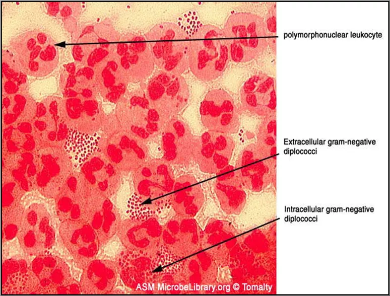
Morphology: In preparations made from gonorrheal pus , the gonococcus occurs in pairs or intracellular diplococcus resembling a pairs of kidney or beans. The long axis of the cocci in pairs are parallel and not in line. Morphologically the gonococcus is identical with the meningococcus .It is non capsulated, non spore forming and non motile. Cultural characters: Primary cultivation of the gonococcus on laboratory media is difficult, not only because the organism is fastidious in its growth requirements but also because it is highly susceptible to the toxic effects of a variety of substance commonly present in ordinary culture media. Most strains require an atmosphere containing 5-10% CO2to initiate development. A sufficient supply of moisture is essential for better growth. Best growth is obtained on blood and chocolate agar. On these media the colonies are 1-2mm, translucent, shining, viscid, convex, entire edge and slightly granular surface. Colonies of gonococcus are smaller than these of meningococcus and are more viscous and emulsify less readily. An effective selective medium for the cultivation of N. gonorrhoeae and N. meningitidis has been developed by Thayer and Martin (1966) which contains antibiotic Vancomycin, Colistin and Nystatin in the basic medium like chocolate agar. Vancomycin inhibits gram positive bacteria, colistin inhibits gram negative bacteria and nystatin for the inhibition of yeast. The medium permits the growth of pathogenic Neisseria while inhibiting saprophytic species such as N. sicca, N. catarrhalis, N. flava and also prevents the growth of bacterial contaminants encountered in cervical, vaginal, urethral and even rectal specimens. Colonies may be slow in development and growth may not appear for 2-3 days. Inoculation of material to be cultured should if possible be made directly from the patient on to suitable medium pre- warmed to 37 and the culture should be incubated at once.
. Biochemical reactions: The gonococcus is not very active biochemically . its attack on sugars is by oxidative methods . it ferments glucose and not maltose with out gas .catalase positive oxidase positive . Toxins: The toxicity of the gonococcus appears to be due to a lipopolysaccharide endotoxin. Failure to detect significant levels of antibodies has been related to the fact that the infection stimulates little antibody formation because of its local character . Classification: the gonococcus is divided into four types T1 , T2 , T3 , and T4 related to colonial appearance , autoagglotinatis virulence and other characters . T1 and T2 from small glistening , convex , brown colonies whose cells 1) autoagglutinate 2) virulent 3) posses pili which attack to epithelial cells 4) resistant to phagocytosis by polymorphonuclear cell. . 5) posses small on the cell surface often seen in association with pili and may be associated in some way with damage done to the host T3 and T4 from large colonies , flat , un pigmented whose cells luck pili and are a virulent . Pathogenicity: spherical structure found In male : urethritis , epididymitis , seminal vesiculitis prostatitis. In female : urithritis , cervicitis , endometritis , salpingitis , peritonitis . In children : vulvovaginitis , conjunctivitis . In new born : ophthalmia neonatorum. Laboratory examinations : The discharge from the gonococcal lesions is subjected to the following examinations: 1- microscopic examinations 2- cultural examination 3- oxidase reaction 4- biochemical reaction 5- immunofluorescent method 6- serodiagnosis
Neisseria meningitidis ( meningococcus) Morphology : gram negative oval or spherical diplococcus , with adjacent surfaces flattened . the long axis of the cocci in pairs are parallel and not in line as in the case of pneumococci . autolysis is a prominent feal of meningococi . non motile bacteria and non spore former. Groups A,C and D may from capsules . cultural character : strict aerobes and primary cultures are obtained better in the presence of 5-10 % CO2. highly fastidious optimum growth temperature is 35-37 . chocolate agar , blood agar , trypticase soya agar and starch casein hydrolysate agar are solid common media use for the propagation of these organisms. On chocolate and blood agar the colonies are moist , elevated smooth, semitransparent often 48hrs incubation , the centre of the colonies becomes elevated and more opaque while the periphery remains thin and transparent . in primary artificial culture , meningococcus dies with in 2-3 days , probably due to the formation of autolysin by the organisms , it is killed in a short time by drying and by exposure to dilute disinfectants. it dies with in few days at zero . Biochemical reaction : the meningococcus is not an active fomenter and ferments glucose and maltose with the production of acid and no gas, oxidase positive and catalase positive. Antigenic structure: It is divided into A,B,C,D,X,Y,Z,29E and W135 serogroups, Organisms in groups A,B and C are responsible for the great a majority of clinically recognized disease. Pathogenesis: The meningococci are strict parasites of man. Their natural Habitat is the nasopharynx and the organisms have a relatively low virulence for other species. This bacteria are actively toxigenic and potent products have been obtained from cultures. An endotoxin released by the autolysis of the bacteria is responsible for many signs of the disease . cerebrospinal meningitis and meningococcal septicaemia are the two types of meningococcal disease .
Laboratory diagnosis : 1- direct evidence by isolation of organisms . 2- indirect evidence by the identification of the specific antibodies which is not an important method for diagnosis of meningococcal infection . besides meningococcus other common bacteria producing pyogenic meningitis are pneumococcus haemophilus influenzae. The material for the demonstration of these organisms may be 1- cerebrospinal fluid 2- blood 3- skin lesions 4- nasopharyngeal swabs 1- cerebrospinal fluid : this is collected by lumbar puncture under aseptic precaution it may be divided in to 3 portion : one for physical and cytological , the second for biochemical and the third for bacteriological examinations. The C.S.F is subjected to the following examinations: A- physical examinations: C.S.F is clear but it became turbid if infection is present . presence of blood indicates bleeding . B- Cytological Examination : The total number of leucocytes is markedly increased and may account for thousand or more polymorphonuclear leucocytes per cmm. Leishman stain of the smear will reveal neutrophils .
Biochemical examination : Proteins are markedly increased whereas sugar is markedly reduced and chlorides are also reduced. D- Bacteriological examination: 1- smear examination 2- culture 3- immunofluorescent technique 2- Blood culture : the importance of blood cultures in the diagnosis of meningococcal septicaemia is obvious . in meningococcal meningitis , blood culture is positive in a proximately 50% of cases , if the culture is taken early in the course of the disease . 5-10ml blood is added to 100ml of tryptose phosphate infusion or casein hydrolysate broth , then incubated at 37 under 5-10% CO2 . subcultures are made every 48 h and continue for 8 days before discarded as negative .
Test Normal Acute bacterial meningitis Tuberculous meningitis Aseptic meningitis Appearance Clear Turbid or purulent Clear or opalescent Clear Colorless Total proteins 15-40mg/dl Markedly Moderately slightly Sugar 50-70mg/dl markedly Normal Chloride 700-740mg/dl Normal Cell count 0-3 polymorph- nuclears Lymphocytes Lymphocytes lymphocyte/c A few mm polymorphs. Culture Sterile C.S.F in meningitis


