Overview of Lymphoid Organs and Their Functions
Lymphocytes are crucial components of the lymphoid system, found in various lymphoid organs like the bone marrow, thymus, lymph nodes, and spleen. The primary, secondary, and tertiary lymphoid tissues play distinct roles in lymphocyte development, differentiation, and immune responses. Dysfunction of these organs can lead to immune deficiencies and diseases like DiGeorge Syndrome. Lymphatic circulation aids in transporting lymphocytes and antigens throughout the body.
Download Presentation

Please find below an Image/Link to download the presentation.
The content on the website is provided AS IS for your information and personal use only. It may not be sold, licensed, or shared on other websites without obtaining consent from the author. Download presentation by click this link. If you encounter any issues during the download, it is possible that the publisher has removed the file from their server.
E N D
Presentation Transcript
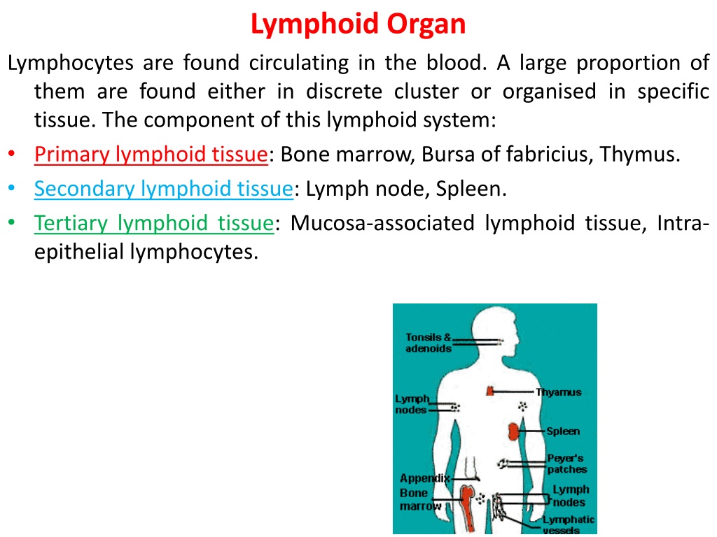
Lymphoid Organ Lymphocytes are found circulating in the blood. A large proportion of them are found either in discrete cluster or organised in specific tissue. The component of this lymphoid system: Primary lymphoid tissue: Bone marrow, Bursa of fabricius, Thymus. Secondary lymphoid tissue: Lymph node, Spleen. Tertiary lymphoid tissue: Mucosa-associated lymphoid tissue, Intra- epithelial lymphocytes.
Primary lymphoid tissue: Major sites of lymphopoiesis (lymphocyte differentiated from lymphoid stem cell, proliferate and mature to functional effector cells) and include: Bone marrow: all lymphocytes develop initially from haematopoietic stem cells in the bone marrow. Immature B cells remain in the bone marrow and develop in the mature cells. This processes are influenced by many factor including surface ligands and cytokines (particularly IL-7). Bursa of fabricius: The bursa is an epithelial and lymphoid organ that is found only in birds. It function differentiation of B-lymphocyte . Its composed of numerous lobes each has a cortical and medullary area. The cortex contain mostly large undifferentiated-lymphoid cells. While the medulla contains small, mature cells. include development and
Thymus: thymus in mammals is a bilobed organ, located in the thoracic cavity, the 2 lobes divided by trabeculae in to lobules, each of which has an outer cortex and an inner medulla. Epithelial cell in the cortex thymic nurse cells made up of sequamous cells that make and secret factors that attract T-cell precursors from the blood and promote subsequent maturation within thymus. These factor include: 1- Chemokine: called thymus-expressed cytokine (TECK ) 2- Hormones: thymulin, thymosin, and thymopoietine Nave cells enter the thymus from subcapsular sinus. They migrate through the cortex and medulla and undergo differentiation, express CD3 and either CD4 or CD8 and leave thymus as mature T cells. Thymus is relatively large and highly active at birth (22gm) reach its peak weight at puberty (35gm) and reach (6gm) of thymic tissue in adulthood.
If thymus surgically removed in neonates, they became severely immunodeficient and fail to thrive and babies without functional thymus lead to disease called Diageorge Syndrome. Adult have developed enough mature T cell that removal of thymus or reduction of its function has milder effect.
Secondary lymphoid tissue : are sites of accumulation and presentation of Ag to both virgin and memory lymphocyte populations. Lymphatic circulation: water and low molecular weight solutes leachout from blood vessel walls into the lower pressure intersitial space. Most of this fluid returns to blood stream through the walls of nearby venules, but a substantial amount dose not. Instead, this portion flows through the tissues,carring Ag and collected in a branching network of lymphatic vessels. Once the fluid enters these vessels it is know as lymph.
After passing throgh secondary lymphoid organ, lymph empties in to larger lymphatic vessels. Thoracic duct and right lymphatic duct which drain their contents in to right and left subclavian veins in the thorax. Lymph: interstitial fluid surrounding cells in tissue or organ low in protein content than blood plasma. Lymphatic vessels: (lymphocytes + dendritic)cell + lymph Blood vessels: (lymphocytes + RBC + monocyte + neutrophil + Basophil +eosinphil + M ) + plasma
Lymph node: the lymph nodes form part of a network which filters antigens from the interstitial tissue fluid and lymph during its passage from the periphery to the thoracic duct and the major collecting duct. Human lymph nodes are 2-10 mm in diameter and round or kidney shaped with blood vessels enter and leave the node from hilus. Its surrounded by a collagenous capsule. Radical trabeculae with reticular components. The lymph node consist of a B-cell area (cortex), a T-cell area (paracortex) and a central medulla containing T- cells, B- cells, abundant plasma cells and macrophages.lymph node cortex contain cellular aggregation called lymphoid follicles which composed of memory B-lymphocytes, smaller number of T-cell and follicular dendritic cell. fibers support cellular
Lymphoid follicles re of 2 types: Primary follicles: contain mature resting B-cells. Secondary follicles: with germinal center contain various stages of activation and blast transformation with numerous macrophage and occasional plasma cells may be seen. The paracortex contains specialized capillary vessels( high endothelial venules-HEV) that allow traffic of lymphocytes out of the circulation in to lymph node (lymphocyte traffic). The lymph that flows into a node may carry with it microorganism or other foreign matter from tissue. When such substance enters lymphocytes and macrophage in the node respond by activation as a result, some of the resident lymphocytes begins to proliferate, inflammatory mediators are released locally, blood flow to the node
Increases and node may become noticeably enlarged when infection develop. The swelling decrease when the infection ends.
Spleen: the spleen filters blood much as the lymph nodes filter lymph. Its located just below the diaphragm on the left side of the abdomen, the spleen weight 150gm in adult and enclused in a thin connective tissue capsule. Most of the spleen consist of red pulp and whit pulp.
Function of red pulp: Blood enters the spleen via the splenic artery, which divides into many arteries called central arteries. These arteries become thiner arterioles, which eventually enter the red pulp. The red pulp contains thin-walled blood vessels called venous sinusoid and in between these sinusoids are called splenic cords. Blood cells are emptied out of the arteries directly into the splenic cords. To re-enter the blood circulation, blood cells must transverse the splenic cords and enter the venous sinusoids. Venous sinusoids lined with macrophage and the splenic cords are full of macrophage. These macrophage recognize and phgocytose old or damaged red cells and pletelets preventing their re-entry in to the blood. Beside old red cells membranes are less elastic and unable to squeeze through the wall of endothelium of venous sinusoids to re-enter the blood stream.
Each arteriole is encased in lymphoid tissue that consist mainly of mature T- cells and is called the periarteriolar lymphoid sheath(PALS). Adjacent to PALS is the B-cell area containing primary follicles and secondary follicle with germinal centres. Between the whight pulp and the red pulp is the area called the marginal zone, which contains B- cells and macrophage.
1- Ag stimulates B cells at the junction of the marginal zone and primary follicle 2- and CD4 T cells in the PALS 3- the CD4 T cells migrateto b-cell area 4- Where CD4 T cell/B cells interaction take place, resulting in proliferation of B cells 5- and formation of some plasma cells in the same way as in the lymph node. The plasma cells migrate to the marginal zone and secrete antibody 6- other B cells and CD4 T cells enter primary follicles, leading to the development of the germinal centre
Tertiary lymphoid tissue: tissue in the body possess poorly organised collections of lymphoid cells. Such collections include mucosa- associated lymphoid tissue and intraepthelial lymphocytes(IEL). Mucosa-associated lymphoid tissue: diffuse lymphoid tissue found in sub mucosal regions and consititute the largest lymphoid organ, containing roughly half the lymphoid cell in the body including: Gut associated lymphoid tissue(GALT), Bronchus associated lymphoid tissue(BALT). GALT consist of peyers patches in the small intestine BALT consist of large collection of lymphocytes(majority B cells) found along the main bronchi in the lungs. At other sites, cells are organized in stable anatomic structures e.g. Tonsils (are nodular aggregates of macrophage and lymphoid cells located immediately beneath the stratified sequamous epithelium.
Intraepithelial lymphocytes: large number of lymphocytes are intrinsically associated with the epithelial surfaces of the body. Particularly reproductive tract, the lung and skin. these collection of lymphoid cells play a key role in the development of both local and systemic specific immune responses to antigens present at the body surface.


