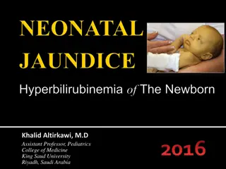
NEONATAL JAUNDICE
Neonatal jaundice, a common condition in newborns, results from elevated bilirubin levels. While often benign, high levels can lead to complications like kernicterus. Recognizing risk factors, limitations of clinical assessment, and appropriate evaluation and treatment are crucial in managing neonatal jaundice effectively.
Download Presentation

Please find below an Image/Link to download the presentation.
The content on the website is provided AS IS for your information and personal use only. It may not be sold, licensed, or shared on other websites without obtaining consent from the author.If you encounter any issues during the download, it is possible that the publisher has removed the file from their server.
You are allowed to download the files provided on this website for personal or commercial use, subject to the condition that they are used lawfully. All files are the property of their respective owners.
The content on the website is provided AS IS for your information and personal use only. It may not be sold, licensed, or shared on other websites without obtaining consent from the author.
E N D
Presentation Transcript
NEONATAL JAUNDICE D.Amani Elgadal
Objectives Understand etiology and pathophysiology of neonatal jaundice and kernicterus Identify high risk conditions Understand limits of clinical exam Describe appropriate evaluation Apply appropriate treatment to term and near term infants WWW.SMSO.NET
JAUNDICE Jaundice is the visible manifestation of increased level of bilirubin in the body. It is not a disease rather a symptom of diseases. In adults sclera appears jaundiced when serum bilirubin exceeds 2 mg/dl. Newborn skin >5 mg/dl.
BURDEN Important problem in the 1st week of life. Most neonates (60% Term and 80% Preterm) will have bilirubin > 5 mg/dl in the 1st week of life and become visibly jaundiced, vast majority being benign. Some of the term babies (8 to 9%) have levels exceeding 15 mg/dl in 1st 7 days of life.
BILIRUBIN End product of hemoglobin metabolism that is excreted in bile. In neonates -75% : from catabolism of circulating RBCs -25% :*from ineffective erythropoiesis (bone marrow) *from turnover of heme proteins & free heme( liver).
Hyperbilirubinemia -Direct( Conjugated) -Indirect( Un-conjugated) Conjugated Hyperbilirubinemia is present, * >20% of total bilirubin is conjugated * >2mg/dl is conjugated If neither criteria is met, hyperbilirubinemia is classified as Un-conjugated.
Neonatal jaundice Conjugated bilirubin Unconjugated bilirubin Hepatic Pathological Physiological Extra-hepatic Biliary atresia or bile duct obstruction Hemolytic Non-hemolytic *Infections *Intrinsic (Spherocytosis, elliptocytosis, G6PD deficiency, Pyruvate kinase deficiency, Sepsis, thalassemia) Breast milk jaundice Hep A/B TORCH Cephalohematoma Polycythemia Sepsis *Metabolic Urinary tract infection Sepsis Galactosemia Hypothyroidism Alpha-1- antitrypsin def *Extrinsic (Rh, ABO & minor group incompatibility) Gilbert's syndrome Crigler-Najjar syndrome Cystic fibrosis High GI obstruction
JAUNDICE APPEARING IN 1st day Hemolytic (Rh, ABO & minor group incompatibility, G6PD def. , spherocytosis, thalassemia), Neonatal sepsis. 2nd & 3rd days Physiological, Neonatal sepsis, Polycythemia, concealed hemorrhages (cephalhematoma, SAH, IVH) > 3 days (Prolonged) Neonatal sepsis, Hepatitis, Biliary atresia, Breast milk jaundice, metabolic disorders.
Prolonged Jaundice a) Direct Neonatal hepatitis (common) Extra-hepatic biliary atresia Breast milk jaundice Metabolic disorders Intra-hepatic biliary atresia
Prolonged Jaundice b) Indirect > Criggler Najjar Syndrome > Breast milk jaundice > Hypothyroidism > Pyloric stenosis > Ongoing hemolysis
Clinical classification: Physiological jaundice Pathological jaundice
Physiological jaundice First appears between 24-72 hours of age. Maximum intensity seen on 4-5th day in term and 7th day in preterm neonates. Does not exceed 15 mg/dl. Clinically undetectable after 14 days. No treatment is required but baby should be observed closely for signs of worsening jaundice.
Pathological jaundice Presence of any of the following signs denotes that the jaundice is pathological: Clinical jaundice detected before 24 hrs of age. Rise in serum total bilirubin by more than 5 mg/dl/ day (>5mg/dl on first day , 10 mg/dl on second day and 12- 13 mg/dl thereafter in term babies).
Pathological jaundice Serum bilirubin more than 15 mg/dl Clinical jaundice persisting beyond 14 days of life. Clay/white colored stool and/or dark urine staining the nappy yellow. Direct bilirubin >2 mg/dl at any time.
RISK FACTORS jaundice within first 24 hrs of life a sibling who was jaundiced as neonate premature infants and infants with low birth weight. Poor breastfeeding/suckling delayed passage of meconium deficiency of G6PD Infection Diabetic mother
COMPLICATIONS KERNICTERUS =bilirubin encephalopathy Brain damage caused by high levels of bilirubin (Bilirubin 25mg/dl) Early signs: Lethargy, poor feeding,, loss of Moro reflex. Late signs: Fever,high pitched cry, Bulging fontanel, paralysis of upward gaze, hypo/hypertonia, seizures, opisthotonos.
KERNICTERUS If significant brain damage occurs before treatment, a child can develop serious and permanent problems, such as: cerebral palsy (Permanent brain damage) hearing loss which can range from mild to severe. Death
APPROACH TO A CHILD WITH JAUNDICE
HISTORY HPI onset / duration color of stool/urine Associated symptoms: fever, poor feeding, convulsions
HISTORY PREGNANCY AND DELIVERY Term/preterm, Postnatal age in hours. Maternal illness/infection during pregnancy: diabetes; drug use, malaria, viral. Traumatic delivery, delayed cord clamping. Birth asphyxia, delay in meconium passage. Breast feeding.
HISTORY FAMILY HISTORY Family history of jaundice, liver disease Previous sibling with jaundice in the neonatal period. Anemia, splenectomy, or bile stones in family members or known heredity for hemolytic disorders
ON EXAMINATION Baby lethargic, poor feeding, temperature instability, with apnea: Sepsis Small for gestation: polycythemia Cataract, rash: TORCH infections Extra vascular bleed: Cephalhematoma Pallor: hemolysis
ON EXAMINATION Petechiae: sepsis, TORCH infections. Hepatosplenomegaly: Rh-isoimmunization, sepsis, TORCH infections. Dysmorphic features: congenital Hypothyroidism Look for evidence of kernicterus in deeply jaundiced NB (Lethargy and poor feeding, poor or absent Moro's, or convulsions)
HOW DO YOU LOOK FOR ICTERUS? Dermal staining :progresses from head to toe Examined in good day light skin of forehead, chest, abdomen, thigh, legs, palms, and soles. Blanched with digital pressure and the underlying color of the skin and subcutaneous tissue should be noted. Trans-cutaneous bilirubinometer ?!!!.
Clinical assessment of neonatal jaundice (Kramer s rule)
Diagnosis Lab Studies Total, conjugated & un- conjugated bilirubin. CBC + Retic. Peripheral blood film. Blood group. Direct coomb s test. Serum albumin * Osmotic Fragility test Radiology & US * TFT * LFTs * TORCH screening. * G6PD screening. 28
Clinical jaundice Measure bilirubin > 12 mg/dl & infant < 24hr old < 12 mg/dl & infant > 24hr old Direct Indirect Follow bilirubin level
Physiological jaundice Explain about benign nature of the disease Encourage to breastfeed frequently & exclusively Ask Mother to bring baby back if baby looks deep yellow or palms & soles have yellow staining.
Pathological jaundice 2 modalities of Treatment: PHOTOTHERAPY EXCHANGE TRANSFUSION
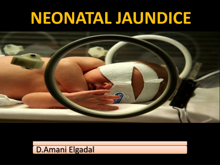
PHOTOTHERAPY ONLY if unconjugated hyperbilirubinemia. Single/multiple phototherapy Under blue-green light (460-490nm), insoluble bilirubin is converted into soluble isomers that can be excreted in urine & feces. To be effective, bilirubin must be present in skin; hence no role for prophylactic phototherapy
PHOTOTHERAPY The infant should be naked except for diaper , eye & gonads should be covered. distance between the skin and light source (35-50 cm) . when used spotlight , the infant is placed in center . turn infant every 2 hours.
PHOTOTHERAPY Ensure optimum breastfeeding as intermittent feeding sessions. routinely add 10-15% extra fluid. Monitor temperature of the baby every 2-4hrs Measure TSB every 12-24hrs. Phototherapy is discontinued if 2 TSB values 12hr apart < 10 mg/dl. Monitor for rebound bilirubin rise within 24hrs.
Guidelines for phototherapy in hospitalized infants of 35 or more weeks gestation Risk Factors: iso-immune hemolytic disease, G6PD def, asphyxia, significant lethargy, temp. instability, sepsis, acidosis or albumin < 3.0 g/dL
COMPLICATIONS Loose stools Skin rash hyperthermia Retinal damage Dehydration Bronze baby syndrome
Exchange transfusion Remove excess bilirubin & maternal antibodies. Fresh blood, O -ve (or infant BG Rh -ve) Umbilical venous route. Double Volume Exchange Transfusion (DVET) : 160-180ml/kg; is to be performed if TSB levels reach age-specific cut-offs or if the infant shows signs of bilirubin encephalopathy, irrespective of TSB levels.
Exchange transfusion Following exchange transfusion: maintain continuous multiple phototherapy TSB every 6-8 hrs after procedure. If baby shows signs of cardiac decompensation at birth, partial exchange transfusion with 50ml/kg of packed red cells should be done to quickly restore oxygen carrying capacity of blood.
Guidelines for exchange transfusion in infants of 35 or more weeks gestation Risk Factors: iso-immune hemolytic disease, G6PD def, asphyxia, significant lethargy, temp. instability, sepsis, acidosis or albumin < 3.0 g/dL
COMPLICATIONS Cardiac and respiratory disturbances. Shock due to inadequate replacement of blood. Infection. Clot formation.
Controversial Medications Phenobarbital: an inducer of hepatic bilirubin metabolism, has been used to enhance bilirubin metabolism. Several studies have shown that phenobarbital is effective in reducing mean serum bilirubin values during the first week of life.
Controversial Medications Intravenous immunoglobulin (IVIG): 500 mg/kg significantly reduce the need for exchange transfusions in infants with iso- immune hemolytic disease.
PREVENTION Ante-natal screening to detect Rh iso- immunization & prompt administration of Anti D after first obstetric event. Ensure adequate breast feeding. Educate parent about danger signs to ensure immediate checkup. Follow-up high risk babies( large cephal- hematoma, family history of jaundice) for 2-3 days of discharge.
