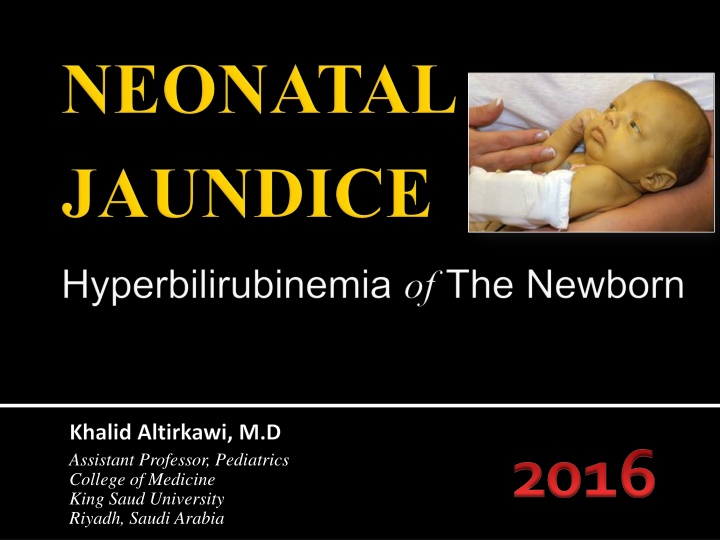
Neonatal Jaundice: A Guide for Medical Students
This presentation by Dr. Khalid Altirkawi, M.D., Assistant Professor of Pediatrics, aims to educate medical students on neonatal jaundice. The content covers metabolic pathways of bilirubin, types of jaundice, diagnostic tests, prevention, and treatment strategies. It provides valuable insights into the synthesis, transport, conjugation, and excretion of bilirubin, crucial for understanding neonatal jaundice.

Download Presentation

Please find below an Image/Link to download the presentation.
The content on the website is provided AS IS for your information and personal use only. It may not be sold, licensed, or shared on other websites without obtaining consent from the author.If you encounter any issues during the download, it is possible that the publisher has removed the file from their server.
You are allowed to download the files provided on this website for personal or commercial use, subject to the condition that they are used lawfully. All files are the property of their respective owners.
The content on the website is provided AS IS for your information and personal use only. It may not be sold, licensed, or shared on other websites without obtaining consent from the author.
E N D
Presentation Transcript
Khalid Altirkawi, M.D Assistant Professor, Pediatrics College of Medicine King Saud University Riyadh, Saudi Arabia 2016
This presentation is meant to guide students when rotating in the NICU. It is not to replace the recommended textbooks. Khalid Altirkawi, MD
By the end of this presentation, the student should be able to: Describe the metabolic pathways of bilirubin. Differentiate between physiological and pathological types of neonatal jaundice. Tell the appropriate diagnostic tests for evaluating jaundiced infants. Mention the strategies for prevention and treatment of neonatal jaundice.
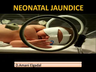
Neonatal Jaundice: A yellow discoloration of skin and sclera due to an increased concentration of serum bilirubin. Usually appears in the newborn infant when serum bilirubin concentration > 85 mol/L. Occurs in the majority of infants Term: >50%. Preterm: 80%
Synthesis From the breakdown of the heme molecule: The hemoglobin ... ... ... ... ... ... ... ... (80%) Nonerytheroid heme (cytochrome, myoglobin..) (20%) Bilirubin produced is Unconjugated (indirect). Lipid soluble (pass through BBB) but not water soluble.
Transport Unconjugated bilirubin is bound to albumin and carried via blood stream to liver. Hepatocytes uptake this Bilirubin and transport it intracellular where it is metabolized
Conjugation Bilirubin is conjugated to glucoronic acid by the enzyme glucoronyl transferase Conjugated bilirubin (Direct) is water-soluble (pass in urine) but not lipid soluble
Excretion Conjugated Bilirubin is excreted into the bile Under the effect of the intestinal flora Enzyme glucouronidase unconjugates it. Bilirubin urobilinogin stercobilin (~85%, in the stool) Urobilin (reabsorbed into blood) (~1% in the urine) Some is reabsorbed and transported back to liver (~15%, Enterohepatic circulation)
BILIRUBIN Metabolism Pathway
Unconjugated hyperbilirubinemia: 1. Defective conjugation 2. Increased production (e.g. hemolysis) 3. Increased bilirubin load (e.g. cephalhematoma) 4. Other unidentified mechanisms
Defective conjugation Physiological Jaundice Congenital hypothyroidism Enzyme deficiency (e.g. Crigler-Najjar syndrome)
Increased production (Hemolysis) Immune Iso-immune(hemolytic disease of the newborn) Auto-immune(Maternal auto-antibodies): e.g. SLE Non immune Inherited hemolytic anemias (e.g. hemoglobinopathies, G6PD, Spherocytosis) Others: Vitamin E deficiency, Drugs, Infections, sepsis, DIC.
Increased bilirubin load Extravascular blood collection Polycythemia Dehydration Increased enterohepatic circulation Paralytic ileus Hirschsprung s disease Intestinal stenosis or atresia
Uncertain mechanisms Breast milk jaundice Breast feeding jaundice Some ethnic groups
Conjugated hyperbilirubinemia Decreased excretion rate Obstructive lesions Congenital biliary atresia Tumors or cyst Inspissated bile Metabolic and endocrine abnormalities Hypothyroidism Galactosemia
Mixed hyperbilirubinemia Hepatocyte abnormalities Sepsis Intrauterine infections Neonatal hepatitis Hepatitis viruses TORCH Idiopathic hepatitis Hypoxic insult
Severe hemolysis Obstructive lesion Biliary atresia Neonatal hepatitis Prematurity Infection Hypothyroidism Breast milk jaundice Crigler-Najjar Physiological Dehydration Sepsis Cephalohematoma Polycythemia Crigler-Najjar
Within The 1st Week At Birth or Within 24h Hemolysis (Blood group incompatibility) Congenital infection (TORCH) Sepsis Physiological jaundice Congenital infection (TORCH) Sepsis Concealed hemorrhage Crigler-Najjar syndrome
After The 1st Week Persistent After 2nd Week Breast milk jaundice Sepsis Hepatitis Biliary atresia Hypothyroidism Galactosemia Inherited hemolytic anemia Neonatal hepatitis Biliary atresia Inspissated bile syndrome
Physiological Pathological 2nd-3rd day of life Onset At any time Level of Bilirubin Usually lower Usually higher Type of Bilirubin Unconjugated Any Slow increase (usually <85 mol/L/24h) May be faster (usually >85 mol/L/24h) Rate of increase Shorter Duration May be longer (7-10 days in the term & 14 days in the Preterm) Physical Exam and Lab. tests Normal, healthy infant Abnormal
Due to: Increased synthesis (excessive RBCs load) Relative immaturity of hepatic glucoronyl transferase Onset usually on 2nd-3rd day Disappear around day 7-10 Infant is usually healthy
Exacerbating Factors Prematurity Polycythemia Dehydration Breast feeding 50-65 mol/L than formula fed) (usually higher by Laboratory Rate of accumulation is < 85 mol/L/day
Treatment: Maintain sufficient feeding Serial measurements of serum bilirubin Look for pathological causes if bilirubin level exceeds the expected for age Phototherapy if needed
Indicated if bilirubin level exceeds a set point Exposure of infant to light with wave length around 450 nm Isomerization of bilirubin to nontoxic water soluble form Eyes should be protected Control for temperature and fluid balance is important
Unconjugated hyperbilirubineamia beyond 2nd week of life Disappears within 2 days of breast feeding discontinuation May take up to 3 months to resolve completely Due to (?) a substance in human milk that inhibits the activity of glucoronyl transferase Treatment: Reassurance after exclusion of other pathologies Stoppage of breast feeding is NOT recommended
May be related to decreased amount of milk consumed by the infant (breast-feeding failure ) More effective nursing may prevent early starvation in breastfed newborns and reduce the incidence of this type of jaundice
Rh isoimmunization Mother is Rh ve abd the fetus is Rh+ve First pregnancy usually goes with no fetal problems Mother is sensitized by fetal Rh+ve RBCs Antibodies to Rh+ve RBCs is formed and it is transmitted to the fetus during second pregnancy ~ 1/100 pregnancies and not in every Rh-ve mother May be prevented by Anti-D administered to the mother
ABO incompatibility (milder disease) The anti-A or Anti-B hemolysins (antibodies) present in a mother with blood group O is transferred to the fetus of A or B blood group This results in hemolysis (most transferred antibodies can be neutralized by the baby) 1 in every 200 births The first baby can be affected
Severe hemolysis manifestations Hydrops fetalis The most severe form Often still birth with severe anemia and massive edema and ascitis Icterus gravis neonatorum Jaundice in the first 24hr, not at birth Variable degree of hemolytic anemia Hepatosplenomegaly
Prevention Anti-D within 72 hrs after delivery of Rh+ve child or abortion. Management: Life support Establish diagnosis Early phototherapy Transfusion to restore normal hemoglobin level Exchange transfusion if needed.
Indicated in Kernictrus Cord bilirubin >85 mol/L or Cord hemoglobin <100 g/L Rate of bilirubin rise > 8.5 mol/L/hour Unconjugated bilirubin level > 340 mol/L at any age (at a lower level in presence of risk factor such as sepsis) Double volume exchange transfusion
Always pathological Direct bilirubin >34 mol/L Evaluation Serum bilirubin Liver function tests Cultures (blood, urine, .) Abdominal ultra sound Isotopic scan (scintegraphy) Liver biopsy
Neonatal hepatitis Biliary atresia Stool color Intermittent pale Persistent pale Serum Bilirubin Biphasic Mainly conjugated ALT, AST Marked increase Mild increase Alk. Phosphatase Mild increase Marked increase Uptake: Sluggish Uptake: Intact Liver scintegraphy Excretion of isotope: happens eventually Excretion of Isotope into intestine is absent Inflammatory Infiltration. Intact lobular architecture. Distorted lobular archit- ecture. Bile ductular proliferation or paucity. Liver biopsy Focal necrosis Perilobular edema & fibrosis
Treatment of chronic cholestasis Treatment of the cause Supportive Fat soluble vitamins Use of MCT-containing formula Attention to micronutrients as Ca, Ph, Zinc Surgical interference Biliary atresia: Kasai operation, Liver transplantation
Cause: Crossing of unconjugated bilirubin to the brain. The level at which injury happens depends on: Maturity/Postnatal age Binding capacity in the blood (S. Albumin) Associated risk factors (acidosis, infection, asphyxia )
Acute Encephalopathy Chronic Encephalopathy Three phases Hypotonia, lethargy, high pitched cry and poor sucking Extensors hypertonia, opithotonus, fever and seizures Hypotonia replaces hypertonia in a week Death may occur up the second phase Athetosis Sensorineural hearing deficit Limited upward gaze Dental dysplasia Intellectual deficit
Kernicterus Notice: Opithotonus position and fisted hands
Kernicterus: Yellowish staining of the brain tissue by bilirubin deposition Associated with evidence of neuronal injury Mostly affects basal ganglia, brain stem and cerebellar nuclei and anterior horn cells


")


")