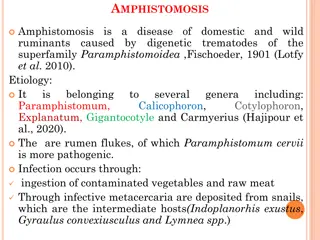
Myasthenia Gravis: Symptoms, Diagnosis, and Management
Myasthenia Gravis (MG) is a neuromuscular disorder characterized by muscle weakness and fatigability due to a decrease in acetylcholine receptors at neuromuscular junctions. It primarily affects individuals in two age peaks, with different symptoms and gender predominance. The cardinal feature is weakness, especially in cranial muscles, which leads to distinct clinical manifestations. Diagnosis involves assessing weakness and fatigability patterns. Treatment options aim to improve neuromuscular function and manage symptoms effectively.

Download Presentation

Please find below an Image/Link to download the presentation.
The content on the website is provided AS IS for your information and personal use only. It may not be sold, licensed, or shared on other websites without obtaining consent from the author.If you encounter any issues during the download, it is possible that the publisher has removed the file from their server.
You are allowed to download the files provided on this website for personal or commercial use, subject to the condition that they are used lawfully. All files are the property of their respective owners.
The content on the website is provided AS IS for your information and personal use only. It may not be sold, licensed, or shared on other websites without obtaining consent from the author.
E N D
Presentation Transcript
Myasthenia gravis (MG) is a neuromuscular disorder characterized fatigability of skeletal muscles. by weakness and The underlying defect is a decrease in the number of available acetylcholine receptors (AChRs) at neuromuscular junctions due to an antibody-mediated autoimmune attack.
The neuromuscular abnormalities in MG are brought about by an autoimmune response mediated by specific anti-AChR antibodies. The thymus appears to play a role in this process. The thymus is abnormal in ~75% of patients with MG; in ~65% the thymus is "hyperplastic," An additional 10% of patients have thymic tumors (thymomas). Muscle-like cells within the thymus (myoid cells), which bear AChRs on their surface, may serve as a source of autoantigen and trigger the autoimmune reaction within the thymus gland.
Epidemiology MG affects individuals in all age groups, but it has 2 peaks. The first peak, mainly in women, occurs between ages 10 and 40 years. The second peak has a male predominance and occurs from ages 50 to 75 years. Overall, women are affected more frequently than men, in a ratio of ~3:2.
Clinical Features The cardinal feature of MG is weakness, which has four general charecteristics; First fatigability. Second diurnal variation Third pattern of distribution . The cranial muscles, particularly the lids and extraocular muscles, are often involved early in the course of MG, Facial weakness produces a "snarling" expression when the patient attempts to smile. Weakness in chewing is most noticeable after prolonged effort, as in chewing meat. Speech may have a nasal quality caused by weakness of the palate.
In ~85% of patients, the weakness becomes generalized, affecting the limb muscles as well. The limb weakness in MG is often proximal and may be asymmetric. If weakness extraocular muscles for 3 years, it is likely that it will not become generalized, and these patients are said to have ocular MG. remains restricted to the Fourth Despite the muscle weakness, deep tendon reflexes and sensations are preserved.
Diagnosis The diagnosis is suspected on the basis of weakness and fatigability in the typical distribution described above, without loss of reflexes or impairment of sensation or other neurologic function.
AChR Antibodies and MuSK Antibodies Anti-AChR antibodies are detectable in the serum of ~85% of all myasthenic patients but in only about 50% of patients with weakness confined to the ocular muscles. The presence of anti-AChR antibodies is virtually diagnostic of MG, but a negative test does not exclude the disease. About half of the patients with negative results for anti- AChR Ab (seronegative MG) may have positive test results for antibody to muscle-specific kinase (MuSK)
Anticholinesterase Test Drugs that inhibit the enzyme AChE allow ACh to interact repeatedly with the limited number of AChRs, producing improvement in the strength of myasthenic muscles. Edrophonium (Tensilon) is used for diagnostic testing. An initial IV dose of 2 mg of edrophonium is given. If definite improvement occurs, the test is considered positive and is terminated. If there is no change, the patient is given an additional 8 mg IV.
Electrodiagnostic Testing Repetitive helpful diagnostic evidence of MG. Electric shocks are delivered at a rate of two or three per second to the appropriate nerves, and action potentials are recorded from the muscles. nerve stimulation often provides In normal individuals, the amplitude of the evoked muscle action potentials does not change at these rates of stimulation. However, in myasthenic patients there is a rapid reduction of >10 15% in the amplitude responses. of the evoked
Single-fiber electromyography (EMG) It involves inserting a fine wire electrode through your skin and into a muscle. In single- fiber EMGs, a single muscle fiber is tested. Most people find this test to be somewhat uncomfortable
Search for Associated Conditions Myasthenic patients have an increased incidence of several associated disorders. Thymic abnormalities which is detected by CT or MRI scanning of the anterior mediastinum. Hyperthyroidism occurs in 3 8% of patients and may aggravate the myasthenic weakness. Because of the association of MG with other autoimmune disorders, blood tests for RH factor and ANA should also be carried out. Chronic infection of any kind can exacerbate MG and should be sought carefully. PFT are valuable because of the frequency and seriousness of respiratory impairment in myasthenic patients.
Differential Diagnosis Lambert-Eaton myasthenic syndrome (LEMS) LEMS is a presynaptic disorder of the neuromuscular junction, caused by autoantibodies against calcium channels at the motor nerve terminals, result in impaired release of ACh from nerve terminals. Most patients with LEMS have an associated malignancy, most commonly small cell carcinoma of the lung, The proximal muscles of the lower limbs are most commonly affected, but other muscles may be involved as well. Cranial nerve findings, including ptosis of the eyelids and diplopia, occur in up to 70% of patients and resemble features of MG
Have depressed or absent reflexes, experience autonomic changes such as dry mouth and impotence, and have incremental rather than decremental responses on repetitive nerve stimulation.
Botulism Botulism is due to a potent bacterial toxin produced by Clostridium botulinum. The toxin interferes with the release of acetylcholine from the presynaptic neuromuscular junction. Patients present with bulbar weakness (e.g., diplopia, dysarthria, dysphagia), Weakness generalizes to the limbs and may result in respiratory failure. deep tendon reflexes are preserved early in the disease course, but diminished as the disease progresses. Autonomic findings include paralytic ileus, constipation, urinary retention, dilated or poorly reactive pupils, and dry mouth. The demonstration of toxin in serum by bioassay is definitive, but may be negative. incremental rather than decremental responses on repetitive nerve stimulation.
Treatment The most useful treatments for MG include; anticholinesterase medications immunosuppressive agents thymectomy plasmapheresis or intravenous immunoglobulin (IVIg).
Anticholinesterase Medications Anticholinesterase medication produces at least partial improvement in most myasthenic patients, although improvement is complete in only a few. Pyridostigmine (neostigmine) is the most widely used anticholinesterase drug. Overdosage with anticholinesterase medication may cause increased weakness and In some patients, muscarinic abdominal cramps, salivation, nausea). Atropine/diphenoxylate or loperamide is useful for the treatment of gastrointestinal symptoms. side effects (diarrhea,
Immunosuppression When symptomatic treatment with pyridostigmine is not effective, then treatment with immunosuppressant drugs is required. Immunosuppressant therapy includes corticosteroids (prednisone) azathioprine (Imuran) cyclophosphamide (Cytoxan) mycophenolate mofetil (CellCept).
Thymectomy Two separate issues should be distinguished: (1) surgical removal of thymoma, and (2) thymectomy as a treatment for MG. Surgical removal of a thymoma is necessary because of the possibility of local tumor spread. In the absence of a tumor, the available evidence suggests that up to 85% of patients experience improvement after thymectomy. It is the consensus that thymectomy should be carried out in all patients with generalized MG who are between the ages of puberty and at least 55 years.
Plasmapheresis Plasmapheresis has been used therapeutically in MG. Plasma, which contains antibodies, is mechanically separated from the blood cells, which are returned to the patient. the pathogenic Plasmapheresis produces a short-term reduction in anti-AChR antibodies, with clinical improvement in many patients. It is useful as a temporary mean in seriously affected patients or to improve the patient's condition prior to surgery (e.g., thymectomy).
Intravenous Immunoglobulin The indications for the use of IVIg are the same as those for plasma exchange: to produce rapid improvement to help the patient through a difficult period of myasthenic weakness or prior to surgery. This treatment has the advantages of not requiring special equipment or large-bore venous access, but it is costly. Improvement occurs in ~70% of patients, beginning during treatment, or within a week, and continuing for weeks to months. Adverse reactions are generally not serious but include headache, fluid overload, and rarely aseptic meningitis or renal failure.
Management of Myasthenic Crisis Myasthenic crisis is defined as an exacerbation of weakness sufficient to endanger life; it usually consists of respiratory failure caused by diaphragmatic and intercostal muscle weakness. Treatment should be carried out in intensive care units. The possibility that deterioration could be due to excessive anticholinesterase medication ("cholinergic crisis") is best excluded by temporarily stopping anticholinesterase drugs. The most common cause of crisis is intercurrent infection. This should be treated immediately. Early and effective antibiotic therapy, respiratory assistance, and pulmonary physiotherapy are essentials of the treatment program. As discussed above, plasmapheresis or IVIg is frequently helpful in hastening recovery.
The following factors may trigger or worsen exacerbations: Bright sunlight Surgery Immunization Emotional stress Menstruation Intercurrent illness (eg, viral infection) Medication
Drugs That May Exacerbate MG Antibiotics Aminoglycosides: e.g., streptomycin,tobramycin,kanamycin Quinolones: e.g., ciprofloxacin, levofloxacin,ofloxacin,gatifloxacin Macrolides: e.g., erythromycin, azithromycin, telithromycin Beta-blocking agents Local anesthetics and relatedagents Procaine, xylocainein large amounts Procainamide (for arrhythmias) Quinine derivatives . Quinine, quinidine, chloroquine, mefloquine(Lariam) Penicillamine phenytoin statins

 is a neuromuscular")
 is a neuromuscular")


")