Magnetic Resonance Imaging (MRI) in Radiology


MAGNETIC
RESONANCE
IMAGING
(MRI)
BY SAJID EJAZ

INT
R
ODUCTION
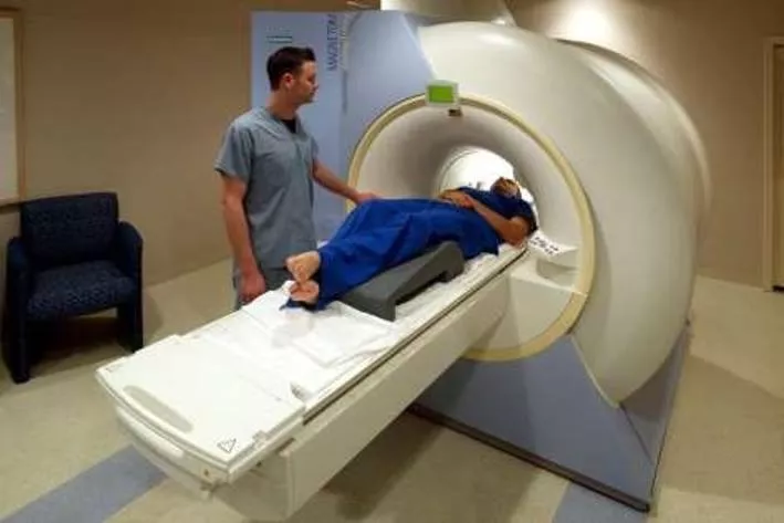
MRI is a
technique
in
radiology
which
uses magnetism,
radio
waves
and a
computer
to produce
images of body
structures.
Human body
is mainly
composed
of
fat
and
water,
which
makes
the
human body
composed
of
about 63%
hydrogen
Why
Are Protons
Important
to
MRI?
•
Positively charged.
•
Spin
about a
central
axis.
•
A moving
(spinning)
charge creates
a
magnetic
field.
•
The
straight
arrow
(vector) indicates
the
direction
of
the
magnetic
field.
USES
MRI
scans
are
used
as an
extremely
accurate
method of
detecting
disease
throughout
the
body.
Head
trauma,
Brain
Aneurysms,
Stroke,
Tumors
of
the
brain,
and
Spine
tumors,
are
all
abnormalities
that
are detected by
a
MRI
scan.
HIS
T
O
R
Y
The
first
MRI
image
was
published
in
1973.
The
first
studies
perform
on human
were
published
in
1997.
Created
by
Dr
Raymond
V.
Damadian,
Dr.Larry
Minkoff
and
Dr.Michael
Goldsmith.
In
2003,the
2003
nobel
prize
in
physiology
or
medicine
was awarded to
paul
C
Lauterbur
and
peter
mansfiled.
Made new mr
imaging
techniques.
Faster
and
more
effcinet
COMPONENTS
A magnet which
produces
a
very
powerful
uniform
magnetic
field.
Gradient
Magnets which
are
much
lower
in
strength.
RF
Coils
to
transmit
radio
frequency.
A
very powerful
computer
system,
which
translates
the
signals
transmitted
by
the
coils.
THE
MAGNET
The
most important
component
of
the MRI
scanner
is
the
magnet:
The
magnets
currently
used in
scanners
today
are
in
the
0.5 T to 2.0
T
range
(5,000
to 20,000-
gauss).
Higher values
are
used
for
research(up to
9T.
◦
While Earth
magnetic
field:
0.5-gauss
GRADIENT
MAGNET
These
magnets
have
a
much
lower
magnetic
field and
are
used
to
create
a
variable
field.
RF
COILS
T
r
a
n
smission
of
radio
frequency
PRINCIPAL
OF
MRI
Spinning
Atoms (hydrogen)
face
outside
magnetic
field.
Energy
Absorption
by
RF
Coils.
Resonance of
Hydrogen
atoms
Measured
by
RF
antenna
which
the
received
signal
is
sinusoidal
in
shape.
Imaging
by
The
computer
which
receives
mathematical
data,
which is
converted
through
the
use of
a
Fourier
transform into
an
image.
HOW
DOES
THE
MRI
WORK
Put
subject
in
big magnetic field.
Transmit
radio
waves into
subjects.
Receive radio
waves
that
has been
re-
transmitted
by
the
subject.
Store
the
data
&repeat
step
2.
Process
raw
data to
reconstruct
images.
Getting
high resolution
image.
When
placed
in a
large
magnetic field,
hydrogen
atoms
have
a
strong
tendency
to
align in the
direction
of
the
magnetic
filed
Inside the
bore
of
the
scanner,
the
magnetic field
runs
down
the
center
of
the tube in which the
patient
is
placed, so
the
hydrogen
protons
will line
up
in either
the
direction
of
the
feet
or
the
head.
The majority
will
cancel
each
other,
but
the
net
number of
protons
is
sufficient
to produce
an
image.
IMAGES
PROCESSING
DIAGRAM
BASIC
MRI
SCANS
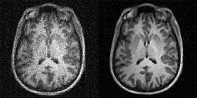
T1-weighted:
Differentiate
fat
from
water.
•
Water
is
darker,fat
is
brighter.
•
Provide good
grey matter/white
matter
contrast
in
brain.
T2-weighted:
Differentiate
fat
from
water.
•
Fat
shows
darker,and
water
lighter.
•
Good
for
imaging
edma.
•
Abnormal
accumulation
of
fluid beneath the
skin or in one or
more
cavities
of the
body.
A
D
V
AN
T
A
GES
Provide
very detailed
diagnostic
pictures
of
most
of
the
important
organs
and
tissues
in
your
body.
Ability
to
diagnose,
visualize,
and
evaluate
various
illnesses.
Are generally
painless.
Did not use
radiation
and
are
therefore
suitable
for
use
in children
and
pregnant
women.
DISADVANTAGES
Limited
availability.
It
is a
lengthy
procedure
“eg,
a
pituitary
gland
MRI
scan
can
take
up
to
30
minutes”.
MRI
scanning cannot be
performed
in the
presence
of
foreign
bodies
or
metallic
implants
“eg,
pacemakers”.
It is
relatively
expensive
compared
with other
forms
of
imaging.
Thank
you.....
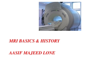
MRI is a powerful diagnostic tool utilizing magnetism, radio waves, and a computer to generate detailed images of body structures. Discover the importance of protons in MRI, its historical significance, key components, and working principles.

Download Presentation

Please find below an Image/Link to download the presentation.
The content on the website is provided AS IS for your information and personal use only. It may not be sold, licensed, or shared on other websites without obtaining consent from the author.If you encounter any issues during the download, it is possible that the publisher has removed the file from their server.
You are allowed to download the files provided on this website for personal or commercial use, subject to the condition that they are used lawfully. All files are the property of their respective owners.
The content on the website is provided AS IS for your information and personal use only. It may not be sold, licensed, or shared on other websites without obtaining consent from the author.
E N D
Presentation Transcript
MAGNETIC RESONANCE IMAGING (MRI) BY SAJID EJAZ
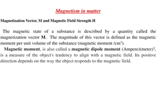
INTRODUCTION MRI is a technique in radiology which uses magnetism, radio waves and a computer to produce images of body structures. Human body is mainly composed of fat and water, which makes the human body composed of about 63% hydrogen Why Are Protons Important to MRI? Positively charged. Spin about a central axis. A moving (spinning) charge creates a magnetic field. The straight arrow (vector) indicates the direction of the magnetic field.
USES MRI scans are used as an extremely accurate method of detecting disease throughout the body. Head trauma, Brain Aneurysms, Stroke, Tumors of the brain, and Spine tumors, are all abnormalities that are detected by a MRI scan.
HISTORY The first MRI image was published in 1973. The first studies perform on human were published in 1997. Created by Dr Raymond V. Damadian, Dr.Larry Minkoff and Dr.Michael Goldsmith. In 2003,the 2003 nobel prize in physiology or medicine was awarded to paul C Lauterbur and peter mansfiled. Made new mr imaging techniques. Faster and more effcinet
COMPONENTS A magnet which produces a very powerful uniform magnetic field. Gradient Magnets which are much lower in strength. RF Coils to transmit radio frequency. A very powerful computer system, which translates the signals transmitted by the coils.
THE MAGNET The most important component of the MRI scanner is the magnet: The magnets currently used in scanners today are in the 0.5 T to 2.0 T range (5,000 to 20,000- gauss). Higher values are used for research(up to 9T. While Earth magnetic field: 0.5-gauss
GRADIENT MAGNET These magnets have a much lower magnetic field and are used to create a variable field.
RF COILS Transmission of radio frequency
PRINCIPAL OF MRI Spinning Atoms (hydrogen) face outside magnetic field. Energy Absorption by RF Coils. Resonance of Hydrogen atoms Measured by RF antenna which the received signal is sinusoidal in shape. Imaging by The computer which receives mathematical data, which is converted through the use of a Fourier transform into an image.
HOW DOES THE MRI WORK Put subject in big magnetic field. Transmit radio waves into subjects. Receive radio waves that has been re- transmitted by the subject. Store the data &repeat step 2. Process raw data to reconstruct images. Getting high resolution image.
When placed in a large magnetic field, hydrogen atoms have a strong tendency to align in the direction of the magnetic filed Inside the bore of the scanner, the magnetic field runs down the center of the tube in which the patient is placed, so the hydrogen protons will line up in either the direction of the feet or the head. The majority will cancel each other, but the net number of protons is sufficient to produce an image.
BASIC MRI SCANS T1-weighted: Differentiate fat from water. Water is darker,fat is brighter. Provide good grey matter/white matter contrast in brain. T2-weighted: Differentiate fat from water. Fat shows darker,and water lighter. Good for imaging edma. Abnormal accumulation of fluid beneath the skin or in one or more cavities of the body.
ADVANTAGES Provide very detailed diagnostic pictures of most of the important organs and tissues in your body. Ability to diagnose, visualize, and evaluate various illnesses. Are generally painless. Did not use radiation and are therefore suitable for use in children and pregnant women.
DISADVANTAGES Limited availability. It is a lengthy procedure eg, a pituitary gland MRI scan can take up to 30 minutes . MRI scanning cannot be performed in the presence of foreign bodies or metallic implants eg,pacemakers . It is relatively expensive compared with other forms of imaging.