Laboratory Diagnosis of Parasites: Methods and Clinical Specimens
Laboratory diagnosis of parasitic infections involves techniques such as demonstration of parasites, immunodiagnosis, and molecular biological methods. Clinical specimens for diagnosis include blood, urine, genital specimens, sputum, and tissue biopsy/aspiration. Chyluria, a condition with milky white urine, can be parasitic or non-parasitic. Immunodiagnosis includes skin tests and serological tests.
Download Presentation

Please find below an Image/Link to download the presentation.
The content on the website is provided AS IS for your information and personal use only. It may not be sold, licensed, or shared on other websites without obtaining consent from the author.If you encounter any issues during the download, it is possible that the publisher has removed the file from their server.
You are allowed to download the files provided on this website for personal or commercial use, subject to the condition that they are used lawfully. All files are the property of their respective owners.
The content on the website is provided AS IS for your information and personal use only. It may not be sold, licensed, or shared on other websites without obtaining consent from the author.
E N D
Presentation Transcript
LAB DIAGNOSIS OF PARASITES
LABORATORY DIAGNOSIS Laboratory diagnosis of parasitic infections can be carried out by * Demonstration of parasite * Immunodiagnosis * Molecular biological methods 2
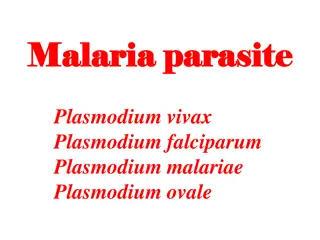
Demonstration of parasite CLINICAL SPECIMEN Blood PARASITES Plasmodium spp., Babesia spp. inside the RBCs L. donovani inside monocytes Trypomastigotes of T. b. gambiense, T.b.rhodesiense and T. cruzi and microfilaria of W. bancrofti and B. malayi in the blood Cysts/trophozoites: E. histolytica, G. lamblia, B. coli, I. belli Eggs: Many worms Larvae:S. stercoralis Adult worms: Taenia spp., A. lumbricoides, T. spiralis, E. vermicularis, hookworms Stool 3
CLINICAL SPECIMEN PARASITES Eggs of S. haematobium Trophozoites of T. vaginalis In the case of Chyluria cause by W.bancrofti, microfilaria in chyluria urine. Urine Trophozoites of T. vaginalis may be demonstrated in the vaginal and urethral discharge and in the prostatic secretions Genital specimens Trypomastigotes of T. b. gambiense and T.b.rhodesiense; Trophozoites of N. fowleri, Acanthamoeba spp., Ceresbrospinal fluid (CSF) Eggs of P. westermani Rarely migrating larvae of A. lumbricoides, S. stercoralis, A. duodenale, N. americanus Trophozoites of E. histolytica Sputum 4
CLINICAL SPECIMEN Tissue biopsy and aspiration PARASITES 1. Scolices and brood capsule in the fluid aspirate of Hydatid cyst 2. Amastigot L. donovani inside RES in the aspirate of spleen, bone marrow, liver, and lymph nodes. 3. Larvae of T. spiralis, T solium in muscle biopsy 4. Trophozoites of G. lamblia duodenum aspirate 5. Trophozoites of E.histolytica in pus aspirated from amoebic liver abcess and in necrotic tissue obtained from the based of the ulcers in the large intestine. Culture of parasites is particularly useful when the number of parasites in the specimens is too small. Culture It is useful in the detection of T. gondii and Babesia spp. in the clinical specimens. Animal Inoculation 5
Chyluria, also called chylous urine, is a medical condition involving the presence of chyle in the urine stream, which results in urine appearing milky white. The condition is usually classified as being either parasitic or non parasitic. a milky fluid containing fat droplets which drains from the lacteals of the small intestine into the lymphatic system during digestion.
Immunodiagnosis two types: Skin tests Serological tests. 7
Skin tests These tests are performed by intradermal injection of parasitic antigens and are read as under: 1. Immediate hyperensitivity reaction: It reveals erythema and induration after 30 minutes of injection. This reaction is seen in cases of hydatid disease, filariasis, schistosomiasis ascariasis, and strongyloidiasis 2. Delayed hypersensitivity reaction: It reveals erythema and induration after 48 hours of injection. This reaction is seen in cases of leishmaniasis, trypanosomiasis, toxoplasmosis and amoebiasis, 8
Serological test These tests detect antibodies or antigens in the patient serum and other clinical specimens TEST APPLICATION Toxoplasmosis, toxocariasis, leishmaniasis, Chagas diseases, malaria and schistosomiasis ELISA and RIA Indirect haemagglu- tination test Amoebiasis, hydatid disease, filariasis, cysticercosis, strongyloidiasis. Indirect fluorescent antibody test Amoebiasis, malaria, toxoplasmosis and schistosomiasis 10
TEST APPLICATION Complement fixation test Paragonimiasis, Chagas' disease and leishmaniasis Agglutination tests: Direct agglutination Visceral leishmaniasis 11
The latex agglutination test (LAT) utilizes a soluble antigen coated with latex particles .Upon addition of serum, a pattern of agglutination can be observed against a black surface that indicates that the serum sample is positive for parasites antibodies, this test is commercially available and easy to performe, but it can produce false-positive and false-negative results).
Tachyzoites of T. gondii can be cultured in laboratory animals such as mice, hamsters, guinea pigs and rabbits. All these animals are susceptible, but out-bread CD-1 mice or interferon-gamma gene knockout (KO) mice often used for tachyzoites production Tachyzoites grow in the peritoneal cavity of mice, sometimes producing ascites, and grow in most of the other host tissues after intraperitoneal inoculation (i.p) with tissue cysts, tachyzoites, or oocysts. Virulent strains such as, RH strain usually produce illness in mice and kill them within 1 to 2 weeks. Frequent, rapid passage of tachyzoites of low virulence may increase virulence .
Toxoplasma gondii tachyzoites will multiply in almost all mammalian cell lines in tissue cultures such as, Vero-monkey kidney cells, HeLa cell and human foreskin fibroblasts (HFF) which are extensively used. The yield of tachyzoites will vary with the cell line and the strain of T. gondii. Mouse- virulent strains rapidly destroy the cells while a virulent strains grow slowly causing minimal cell damage .
Indirect Immunofluorescent Antibody Test For detecting T. gondii slides were prepared from cell culture of T. gondii tachyzoites, processed through severe of washes and centrifugations, then cells were resuspended in saline to the desired concentration and added to multi-well IFAT slides . The actual IFAT process involves incubating serum on the prepared slide adding a species-specific fluorescent- labelled antibody and finally viewing the results under a fluorescent microscope.
Indirect Hemagglutination Test For the diagnosis of parasites , a soluble antigen from tachyzoites is coated on tanned red blood cells then agglutinated by immune serum. For the detection of T. gondii Ab, IHA detects antibodies later than the dye test, so acute and congenital infections are likely to be missed by this test). Complement Fixation Test Complement Fixation antibodies for T. gondii appear later than dye test antibodies and the test is positive during acute infection, but this largely depends on the antigenic preparation
Molecular biological methods These include DNA probes and polymerase chain reaction (PCR). 1. DNA probes DNA probe is a radio labelled or chromogenically labelled piece of single- stranded DNA complementary to a genome and unique to a particular parasitic strain, species and genus. Specific probe is added to the clinical specimen. If the specimen contains the parasitic DNA, probe will hybridize with it which can be detected. DNA probes are available for the detection of the infection with P. falciparum, W. Bancrofti, T. b. gambiense, T. b. rhodesiense, T. cruzi Onchocercaspp. segment of parasitic 17
2. Polymerase chain reaction (PCR) PCR is a DNA amplification system that allows molecular biologist to produce microgram quantities of DNA from picogram amounts of sorting material. It has been employed to detect faecal antigens for the diagnosis of intestinal amoebiasis, giardiasis and other intestinal parasitic infections.
Treatment of Parasitic Infection: The successful treatment includes Medical and Surgical measures. Adequate nutrition to build up general resistance. Specific chemotherapy, the successful chemotherapy depends upon the use of a drug that has a minimal toxic effect on the tissues of the host and lethal action on the parasite. New chemotherapeutic agents are being constantly developed, and therapeutic methods are continually undergoing revision.
Prevention of Parasitic Diseases: Prevention of parasitic diseases depends on: Establishment of barriers to the spread of parasites through the practical application of biological and epidemiological knowledge. Every parasite at some times in its life cycle is susceptible to special external measures, thus barriers such as sanitary excreta disposal may be established to break the weak links in the life cycle as departures of eggs, cysts etc.
The control of parasitic diseases include: 1- Reduction of sources of infection in human by therapeutic measures. 2- Establishment of biological barriers to transmission. 3- Education in personal prophylaxis to prevent the spread of infection and reduce the opportunities of exposure. 4- Sanitary control of water, food, living and waste disposal. 5- Destruction or control of reservoir hosts and vectors.
Community Participation Drugs: Method of controlling Parasitic diseases Health education of population at risk - Chemotherapy - Chemoprophylaxis Vector control Reservoir host control - Insecticides, - molluscicides, - Land development - Drainage, - Bush clearance - Destruction of animal hosts - Drug treatment of human hosts 22
LITERATURE 1. Arora, D. R. and B. Arora, 2007, Medical Parasitology, 2nd ed., CBS Publ. & New Dehli Distributor, 2. Markell, E. K., M. Voge, and D. T. John, 1992, Medical Parasitology, 7th ed., WB Saunders Co., Philadelphia 3. Peters, W. & H. M. Gilles, A Colour Atlas of Tropical Medicine & Parasitology, 3rd. ed., Wolfe Medical Publ. Ltd, Netherlands 24 maria immaculata iwo, sf itb


