Introduction to the Physics of Diagnostic X-Rays
The discovery of X-rays by W.C. Roentgen in 1895 was accidental, leading to a groundbreaking advancement in the field of radiology. X-ray photons are part of the electromagnetic spectrum and have applications in diagnostic radiology, radiation therapy, and nuclear medicine. The production of X-ray beams involves accelerating electrons towards a target to generate X-rays. Modern X-ray units consist of components similar to those found in color TV sets, with high potentials used to accelerate electrons. Understanding the physics behind X-rays is crucial for their safe and effective utilization in medical imaging.
Download Presentation

Please find below an Image/Link to download the presentation.
The content on the website is provided AS IS for your information and personal use only. It may not be sold, licensed, or shared on other websites without obtaining consent from the author.If you encounter any issues during the download, it is possible that the publisher has removed the file from their server.
You are allowed to download the files provided on this website for personal or commercial use, subject to the condition that they are used lawfully. All files are the property of their respective owners.
The content on the website is provided AS IS for your information and personal use only. It may not be sold, licensed, or shared on other websites without obtaining consent from the author.
E N D
Presentation Transcript
Part One: - Physics of Diagnostic X-Rays
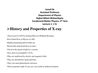
The X-ray photon is a member of the electromagnetic family that includes light of all types, radio waves, radar and television signals, and gamma rays. Like many important scientific breakthroughs, the discovery of X-rays was accidental. In the fall of 1895, W. C. Roentgen, a physicist at the University of Wurzburg in Germany, was studying cathode rays in his laboratory. He was using a fairly high voltage across a tube covered with black paper that had been evacuated to a low pressure. When he "excited" the tube with high voltage, he noticed that some crystals on a nearby bench glowed and that the rays causing this fluorescence could pass through solid matter. Within a few days Roentgen took the first X-ray film. Perhaps Roentgen suspected the dangers of X-rays; he did not X-ray his own hand, he X-rayed his wife's hand! Within a few weeks the news of his discovery had spread to all of the scientifically advanced countries.
The field of radiology has three major branches: - 1. Diagnostic Radiology. 2. Radiation Therapy. 3. Nuclear Medicine.
Production of X-ray beams A high-speed electron can convert some or all of its energy into an X-ray photon when it strikes an atom, and thus we need to speed up electrons to produce X-rays. Trying to speed up an electron in air is difficult since there are so many electrons on the atoms-about 4x10²⁰ in 1cm3; before an electron gets going it bumps into another one. It is thus necessary to eliminate most of the electrons, and this is done by using a glass bulb (X-ray tube) from which most of the atoms have been evacuated. For each 1 billion atoms in air only one atom remains in the evacuated X-ray tube, and the electrons can run unimpeded.
The main components of a modern X-ray unit are (1) a source of electrons-a filament, or cathode; (2) an evacuated space in which to speed up the electrons; (3) a high positive potential to accelerate the negative electrons; and (4) a target, or anode, which the electrons strike to produce X-ray. You may realize that the average home has a device that contains the same components-a color TV set. In color TVs, quite high voltages (~25kV) are used to accelerate the electrons. While a few years ago a number of defective color TVs did produce measurable amounts of X-rays. X-rays are not emitted from black-and white TVs because the very weak X-ray photons produced are absorbed in the glass walls of the tube.
Roentgen's X-rays were produced by electrons (cathode rays) from ionized gas in his cathode ray tube. In 1915, Coolidge invented an X-ray tube that produced electrons by "boiling" them off a red-hot filament, and the typical modern X-ray tube is a refinement of this design. In a modern X-ray tube the number of electrons accelerated toward the anode depends on the temperature of the filament, and the maximum energy of the X-ray photons produced is determined by the accelerating voltage-kilovolt peak (kVp). The kilovolt peak used for an X-ray study depends on the thickness of the patient and the type of study being done.
The intensity of the X-ray beam produced when the electrons strike the anode is highly dependent on the anode material. In general, the higher the atomic number (Z) of the target, the more efficiently X-rays are produced. The target material used should also have a high melting point since the heat produced when the electrons are stopped in the surface of the target is substantial. Most X-ray tubes have two filaments that can be interchanged to produce either a large or a small focal spot. The small focal spot produces less blurring of the X-ray image than the large focal spot, but it concentrates the heat on a smaller area of the target, increasing the chances of overheating and damage.
Many engineers found a way to increase the area on the target struck by electrons to avoid overheating without increasing the blurring of the X-ray image. This technique, called principle. Because of the angle of the target, typically 10oto 20o, the projected focal spot is smaller than the area struck by the electrons. years ago radiological line-focus the The second big breakthrough in designing anodes to avoid overheating was the development in 1930 by Bouwers of the rotating anode X-ray tube. The normal rotational rate of the anode is 3600 rpm, and the heat is spread over a large area as the anode rotates.
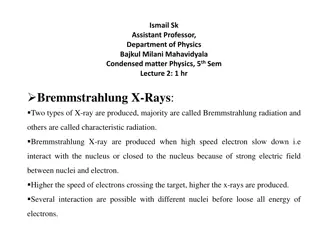
While the energy of most of the electrons striking the target is dissipated in the form of heat, the remaining few electrons produce useful X-rays. Many times one of these electrons gets close enough to the nucleus of a target atom to be diverted from its path and emits an X-ray photon that has some of its energy. X-rays produced in this way have a fancy German name, bremsstrahlung, which means "braking radiation".
Bremsstrahlung is also called white radiation since it is analogous to white light and has a range of wavelengths. The amount of bremsstrahlung produced for a given number of electrons striking the anode depend upon two factors: (1) the Z of the target-the more protons in the nucleus, the greater the acceleration of the electrons-and (2) the kilovolt peak-the faster the electrons, the more likely they will penetrate into the region of the nucleus.
Sometimes a fast electron strikes a K electron in a target atom and knocks it out of its orbit and free of the atom. The vacancy in the K shell is filled almost immediately when an electron from an outer shell of the atom falls into it, and in the process, a characteristicK X-ray photon is emitted. An X-ray photon emitted when an electron falls from the L level to the K level is called a K characteristic X-ray, and that emitted when an electron falls from the M shell to the K shell is called a K X-ray.
Since the energies of the electrons in the various shells of an atom are precisely determined by nature, an electron falling from an outer shell to an inner shell will always produce an X-ray with an energy characteristic of that atom. The spectrum of X-rays produced by a modern X-ray generator. The broad smooth curve is due bremsstrahlung, spikes represent characteristic X-rays. to the the the and
How X-rays are absorbed X-rays are not absorbed equally well by all materials; if they were, they would not be very useful in diagnosis. Heavy elements such as calcium are much better absorbers of X-rays than light elements such as carbon, oxygen, and hydrogen, and as a result, structures containing heavy elements, like the bones, stand out clearly. The soft tissues-fat, muscles, and tumors-all absorb about equally well and are thus difficult to distinguish from each other on an X-ray image . The attenuation of an X-ray beam is its reduction due to the absorption and scattering of some of the photons out of the beam. A simple method of measuring the attenuation of an X-ray beam is shown below.
A narrow beam of X-rays is produced with a collimator-a lead plate with a hole in it-and an X-ray detector measures the beam intensity. The unattenuated beam intensity is Io. As sheets of aluminum are introduced into the beam, the intensity Idecreases approximately exponentially. The lower energy (soft) X-rays are absorbed more readily than the higher energy (hard) X-rays.
The intensity of a monoenergetic X-ray beam would decrease exponentially. The exponential equation describing the attenuation curve for a monoenergetic X-ray beam is: - = e I I o Where e=2.718, x is the thickness of the attenuator, and is the linear attenuation coefficient of the attenuator. The linear attenuation coefficient is dependent on the energy of the X-ray photons; as the beam becomes harder, it decreases. The half-value layer (HVL) for an X-ray beam is the thickness of a given material that will reduce the beam intensity by one-half.
For a monenergetic X-ray beam, the second half-value layer equals the first half-value layer. The half-value layer is related to the linear attenuation coefficient by: - . 0 693 = HVL The mass attenuation coefficient m is used to remove the effect of density when comparing attenuation in several materials. The mass attenuation coefficient of a material is equal to the linear attenuation coefficient divided by the density of the materials. I=Ioe ( / )( x)=Ioe m( x) The quantity x is in grams per square centimeter and is sometimes called the area density; m is in square centimeters per gram.
* Calculate the percentage of X-ray beam absorbed by a bone of thickness 3cm and m/ of the bone to that X-ray energy is 0.2cm2/gm, and of the bone is 1.9gm/cm3. = I ( / )( ) m x I I e o = ( / )( ) m x e I o I . 1 . 1 = = = = 2 . 0 ( 9 . 1 )( ) 3 x 14 14 . 2 718 3 . 0 e e I o
Note that on a gram-for-gram basis, iodine is better absorber than lead from about 30 to about 90keV. This phenomenon is due to the Photoelectric Effect. The Photoelectric Effect is one way X-rays lose energy in the body. It occurs when the incoming X-ray photon transfers all of its energy to an electron which then escapes from the atom. The photoelectron uses some of its energy (the binding energy) to get away from the positive nucleus and spends the remainder ripping electrons off (ionizing) surrounding atoms.
The Photoelectric Effect is more apt to occur in the intense electric field near the nucleus than in the outer levels of the atoms, and it is more common in elements with high Z than in those with low Z. Of course, for a given electron to be librated its binding energy must be lower than the energy of the X-ray. When the energy of the X-ray is just slightly greater than the binding energy, the probability that the Photoelectric Effect will occur increases greatly, and this accounts for the sharp rises in the curve for iodine at 33keV and in the curve for lead at 88keV. These sharp rises are called K-edges.
Another important way X-rays lose energy in the body is by the Compton Effect. In 1922 A. H. Compton suggested that an X-ray photon can collide with a loosely bound outer electron much like a billiard ball collides with another billiard ball. At the collision, the electron receives part of the energy and the remainder is given to a Compton (scattered) photon, which then travels in a direction different from that of the original X-ray.
The energy transferred to the electron can be calculated in the same way as the energy transferred during a billiard ball collision by using the laws of conservation of energy and momentum. The X-ray has an effective mass m of E/c2 (from Einstein's famous equation E=mc2), and its momentum is E/c. We can calculate the energy equivalent of the electron mass to be 511keV, and the Compton Effect is most likely to occur when the X-ray has this energy. The number of Compton collisions depends only on the number of electrons per cubic centimeter, which is proportional to the density. A gram of bone has about the same number of electrons as 1g of water, and thus the number of Compton collisions will be about the same. However, since the Photoelectric Effect is more apt to occur in high Z materials than in low Z materials, the fraction of X-rays that lose energy by the Compton Effect is greatest in low Z elements.
Pair Production is the third major way X-rays give up energy. When a very energetic photon enters the intense electric field of the nucleus, it may be converted into two particles: an electron and a positron ( +), or positive electron. Providing the mass for the two particles requires a photon with energy of at least 1.02MeV, and the remainder of the energy over 1.02MeV is given to the particles as kinetic energy. The positron is a piece of antimatter. After it has spent its kinetic energy in ionization it does a death dance with an electron. Both then vanish, and their mass energy usually appears as two photons of 511keV each called annihilation radiation.
Since a minimum of 1.02MeV is necessary for Pair Production, this type of interaction is only important at very high energies. Because the intense electric field of the nucleus is involved, pair production is more apt to occur in high Z elements than in low Z elements. How are these interactions related to diagnostic radiology? You can see that Pair Production is of no use in diagnostic radiology because of the high energies needed and that the Photoelectric Effect is more useful than the Compton Effect because it permits us to see bones and other heavy materials such as bullets in the body. At 30keV bone absorbs X-rays about 8 times better than tissue due to the Photoelectric Effect. To make further use of the Photoelectric Effect radiologists often inject high Z materials, or contrast media, into different parts of the body.
Compounds containing iodine are often injected into the bloodstream to show the arteries, and an oily mist containing iodine is sometimes sprayed into the lungs to make the airways visible. Radiologists give barium compounds orally to see parts of the upper gastrointestinal tract (upper GI) and barium enemas to view the other end of the digestive system (lower GI). Since gases are poorer absorbers of X-rays than liquids and solids, it is possible to use air as a contrast medium. In a double-contrast study, barium and air are used separately to show the same organ. If the Photoelectric Effect did not exist and radiologists had to rely on the Compton Effect, X-rays would be much less useful because the Compton Effectdepends only on the density of the materials.
Making an X-ray image It is relatively easy to make an X-ray image, or roentgenogram-all that is needed is an X-ray source and a film wrapped in black paper on which to record the image. Unfortunately, X-ray cannot be focused to make a picture as with a camera. X-ray images are basically images of the shadows cast on film by the various structures in the body; they were once called skiagraphs, which is Greek for shadow graphs. To better understand the physical problems of recording sharp X-ray shadows, let us consider the problems of casting sharp shadows with visible light.
A large lightbulb produces a blurred shadow because the light from different parts of the bulb casts shadows in different places. The blurred edge of the shadow is called the penumbra, which means "next to the shadow". The width of the penumbra can be calculated from the dimensions of the lightbulb and the distances to the object and the paper. The penumbra can be reduced by using a smaller diameter lightbulb or by moving the object closer to the paper (moving the lightbulb further away also reduces the penumbra).
Another problem involved in casting a sharp shadow. The sediment in the water absorbs some of the light and scatters much of the light that is not absorbed. The problems involved in obtaining good X-ray shadows are analogous, and blurring can be reduced by using a small focal spot, positioning the patient as close to the film as possible (and increasing the distance between the X-ray tube and the film as much as possible), and reducing the amount of scattered radiation striking the film as much as possible. It is also necessary to avoid motion during the exposure, since motion causes blurring.
The nominal sizes of the focal spots on many X-ray units are 1mm (small focal spot) and 2mm (large focal spot). However, the focal spots are nearly always larger than the nominal sizes. In addition, a focal spot is generally not uniform and sometimes appears to be two spots close together. The actual size of the focal spot can be determined by several techniques. The physics approach is to make a pinhole image of the focal spot and calculate the size of the focal spot from the size of the image and the distances involved.
In another method of measuring the focal spot, a metal plate with patterns of openings of different sizes is placed 20cm above an X-ray film and an exposure is made. The penumbra prevents the smaller patterns from being resolved, and the smallest of the patterns that can be resolved indicates the focal spot size. An advantage of this technique is that it visually demonstrates the loss of detail due to the size of the focal spot.
While decreasing the size of the focal spot reduces the penumbra, it also necessitates lowering the power to avoid damaging the target. This reduces the intensity of the X-ray beam, requiring a longer exposure that usually results in blurring due to patient motion. While the patient is generally placed as close to the film as possible in order to reduce the penumbra, sometimes it is also possible to further reduce the penumbra by increasing the distance from the X-ray tube to the film.


