Hypertrophic Cardiomyopathy (HCM) in a Middle-Aged Female with Heart Failure
A 55-year-old female with a history of hypertension and heart failure presented with symptoms of dyspnea, chest pain, and a positive family history of cardiac disease. Exam findings included LVH on ECG and a systolic murmur at LLSB. This case highlights the importance of recognizing and managing hypertrophic cardiomyopathy (HCM) in patients with heart failure, describing its pathophysiological changes, clinical presentations, and physical examination findings.

Download Presentation

Please find below an Image/Link to download the presentation.
The content on the website is provided AS IS for your information and personal use only. It may not be sold, licensed, or shared on other websites without obtaining consent from the author.If you encounter any issues during the download, it is possible that the publisher has removed the file from their server.
You are allowed to download the files provided on this website for personal or commercial use, subject to the condition that they are used lawfully. All files are the property of their respective owners.
The content on the website is provided AS IS for your information and personal use only. It may not be sold, licensed, or shared on other websites without obtaining consent from the author.
E N D
Presentation Transcript
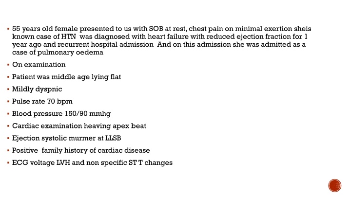
55 years old female presented to us with SOB at rest, chest pain on minimal exertion sheis known case of HTN was diagnosed with heart failure with reduced ejection fraction for 1 year ago and recurrent hospital admission And on this admission she was admitted as a case of pulmonary oedema On examination Patient was middle age lying flat Mildly dyspnic Pulse rate 70 bpm Blood pressure 150/90 mmhg Cardiac examination heaving apex beat Ejection systolic murmer at LLSB Positive family history of cardiac disease ECG voltage LVH and non specific ST T changes
Hypertrophic cardiomyopathies Dr. Mohammed adel F.I.B.M.C M.B.Ch.C University of Basra- Al zahraa teaching hospital
Hypertrophic cardiomyopathy (HCM) is a genetically determined heart muscle disease most often caused by mutations in one of several sarcomere genes that encode components of the contractile apparatus. HCM is characterized by left ventricular hypertrophy (LVH) of various morphologies, with a wide array of clinical manifestations and hemodynamic abnormalities Depending in part upon the site and extent of cardiac hypertrophy, patients with HCM can develop one or more of the following abnormalities: LV outflow obstruction Diastolic dysfunction Myocardial ischemia Mitral regurgitation In broad terms, the symptoms related to HCM can be categorized as those related to heart failure (HF), chest pain, or arrhythmias. For the majority of patients with HCM,
Pathophysiological changes in HOCM: Increase intraventricular pressure Prolong ventricular relaxation Increase myocardial wall stress Increase oxygen demand Decrease LVOT
Clinical presentation: Dyspnoea (90%), orthopnoea and PND Palpitation CHF Angina (70-80%) Syncope and presyncpe Sudden cardiac can be the first clinical manifestation
Physical examination: Carotid pulse (bifid ) JVP (prominent a wave ) Apical beat ( double or triple) Heart sound: S1 normal S2 usually split (reverse ) S3 indicate late stage S4 usually due hypertrophy Murmer: Medium pitch crescendo decrescendo LLSB and apex radiate to suprasternal notch. SAM with MR
Dynamic maneuverer: Murmur intensity increase with decrease preload (Valsalva, standing ) Murmur intensity decrease with increase preload (squatting , hand grip)
Diagnosis: ECG Echocardiography Cathetarization Cardiac imaging
ECG features of HCM Left ventricular hypertrophy with increased precordial voltages and non-specific ST segment and T- wave abnormalities Deep, narrow ( dagger-like ) Q waves in lateral (I, aVL, V5-6) +/- inferior (II, III, aVF) leads Other associated features may include: Left atrial enlargement( P mitrale ) left ventricular diastolic dysfunction may lead to compensatory left atrial hyertrophy Signs of WPW (short PR, delta wave) ECG features ofWolff-Parkinson-White (WPW) were seen in 33% of patients with HCM in one study, and at least one genetic mutation has been identified that is associated with both conditions Dysrhythmias:atrial fibrillation and supraventricular tachycardias are common; PACs, PVCs, VT Giant precordial T-wave inversions in apical HCM
ECHO FEATURES: Abnormal systolic motion of anterior leaflet of the mitral valve LVH Left atrial enlargement Diastolic dysfunction Small ventricular chamber size Septal wall hypertropthy Partial closure of the aortic valve in midsystole
Natural history Heart failure Only 15% develop NYHA3 systolic failure Only 3% develop end stage systolic heart failure Atrial fibrillation Develop in 30% in elder patient Endocarditis only 3-4% develop endocarditis Usually mitral valve effected Autonomic dysfunction develop in 25% of patients Poor prognosis
EVALUATION: Family screen Holter monitor Exercise test


 is a genetically")
 is a genetically")