Gout: Causes, Symptoms, and Management

GOUT
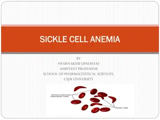
PATHOLOGY
excessive concentrations of uric acid and some purine bodies in
blood pre gout.
Whereas, the kidney is unable to separate this combination of
uric acid and purine bodies.
Then the uric acid salts accumulate in the blood.
PTO..
PATHOLOGY
Then, the crystal deposits in joints, tendons and surrounding
tissue , resulting in an attack of gout.
Gout may be confirmed by the presence of crystals in the joint
fluid .
Blood uric acid level may be normal during an attack.
PTO..
CLINICAL FEATURES
The disease is commonly seen in the first metatarsophalangeal
joint and metacarpophalangeal joints .
It can occur over the age between 30 -40 years.
The deposition of biurate of soda occur around the affected
joints ,
The joint changes include subchondral cysts , osteophytes
formation and in the later stages , reduction in the joint space.
SIGN AND SYMPTOMS :-
Symptoms can include joint pain, gout pain is often
intense , inflammation and redness .
The effected joint will often becomes
red,swallen,and tender to touch.
During the acute phase, the involved joint has signs
and symptoms of acute inflammation with a hard
and tender nodule known as ‘ chalk stones’ or
‘tophi’.
Fever often accompanies excrucitating night paiin,
but the involved joint is comparitively by painfree
during day.
FOOT
CLINICAL FEATURES
Fever often accompanies excruciating night pain , but the
involved joint is comparatively painfree during the day.
There will be decreased mobility as gout progresses , it may
limit the Range of motion .
If in case , gout occurs before the age of 30 there can be a
genetic metabolism disorder ,
( this condition is rare ).
PATHOPHYSIOLOGY :-
Gout is caused by disorders of purine metabolism resulting in
elivated levels of puric acid
->7mg /dl in men
->6mg /dl in women
Prolonged hyperuricemia leads to formation of monosodium
urate monohydrate crystals, the joint at the base of the big toe
is affected in about half of the cases , it may also result in ‘
tophi’.
It can also effect the kidney.
PREDISPOSING FACTOR OF GOUT
:-
1) gender –male > female ,
2) family history ,
3) Previous attack ,
4) obesity,
5) alchohal
6) diet Purine rich
7) dieuritics
8)renal insufficiency ,
9) rich diet ,
10) hypertension ,
11) diabetic conditions.
DIFFERENTIAL DIAGNOSIS :-
X-ray , which is generally normal ,( erosion is seen in chronic
stage ),
Joint aspiration –polarising light microscopy of crystals ,
A synovial fluid , gram stain and culture may be performed.
X-RAY
COMPLICATIONS OF GOUT:-
Gout ‘ tophi’ prsenting as nodules on the finger and helix of the
ear , tophi on the toe and ankle , tophus on the knee.
Gout complicated by rupture of tophi, the exudate of which
tested positiive for uric acid crystals.
COMPLICATIONS
Gout can present in multiple ways , although the most common
in a recurrent attack of acute inflammatory arthritis ( a red
tender hot , swollen joints ).
The metatarsal phalangeal joints at the base of big toe is
affected most often , accounting for half of the cases , other
joints such as the heels, knees, wrists , and fingers, may also be
effected .
Joint pain usually begins during night and peaks within 24 hours
of onset.
JOINTS AFFECTED
MANAGEMENT OF GOUT :-
There are three main goals of the medical management of gout
–
1) terminate acute attack
2) prevent re-occurance
3) correct , prevent furthur damage from hyperuricemia.
CHRONIC TOPHACEOUS GOUT :-
If gout is untreated,
Other joint involvement,
Formation of tophi-collection of crystals in soft tissues,
Bone erosions at joint –” punched-out”.
Erosion on x-ray.
CHRONIC TOPHACEOUS GOUT
CHRONIC TOPHACEOUS GOUT
PSEUDOGOUT
Pseudogout is usually present in the older age group and affects
knee and wrist joints.
It can be polyarticular with an evidence of calcification of the
cartilage ( chondro-calcinosis).
There is a deposition of calcium pyrophosphate crystals .
HAND
FOOT
TREATMENT :-
1) conservative treatment ,
2) physiotherapy treatment-
Conservative treatment :-1) NSAIDS,
2) colchicine ,( along with analgesics) , probencid,
3) glucocorticoids,
4) indomethacin 75-200 mg.
Lithium ionization is sometimes done in between the acute
attacks.
This iontophoresis forms soluble lithium urate in place of
insoluble sodium urate.
PHYSIOTHERAPY TREATMENT :-
1) ultrasound treatment ,( to reduce inflammation and pain ).
2) icing ( to calm the joint ) .. Cryotherapy in the form of crushed
ice packs .
3) strengthening to the muscles,
4) proprioception exercises – which assists in maintaining the
joint sense of position .
5) stretching exercises.
EXERCISES
T/T
Treatment plan will be the same in gout and psudogout .
AIM of treatment :- to give relaxation , to improve the range of
motion , to reduce the level of pain and inflammation .
Patient should be adviced to bring changes in sedantry lifestyle (
like in weight reduction ).
Thank you.
Gout is a condition caused by excessive uric acid levels in the blood, leading to crystal deposits in joints and tissues, resulting in intense pain, inflammation, and redness. Commonly seen in the first metatarsophalangeal joint, gout can present with joint changes and decreased mobility. Recognizing predisposing factors like gender, family history, obesity, and diet is vital for prevention and management.

Uploaded on Sep 17, 2024 | 1 Views
Download Presentation

Please find below an Image/Link to download the presentation.
The content on the website is provided AS IS for your information and personal use only. It may not be sold, licensed, or shared on other websites without obtaining consent from the author.If you encounter any issues during the download, it is possible that the publisher has removed the file from their server.
You are allowed to download the files provided on this website for personal or commercial use, subject to the condition that they are used lawfully. All files are the property of their respective owners.
The content on the website is provided AS IS for your information and personal use only. It may not be sold, licensed, or shared on other websites without obtaining consent from the author.
E N D
Presentation Transcript
PATHOLOGY excessive concentrations of uric acid and some purine bodies in blood pre gout. Whereas, the kidney is unable to separate this combination of uric acid and purine bodies. Then the uric acid salts accumulate in the blood. PTO..
PATHOLOGY Then, the crystal deposits in joints, tendons and surrounding tissue , resulting in an attack of gout. Gout may be confirmed by the presence of crystals in the joint fluid . Blood uric acid level may be normal during an attack. PTO..
CLINICAL FEATURES The disease is commonly seen in the first metatarsophalangeal joint and metacarpophalangealjoints . It can occur over the age between 30 -40 years. The deposition of biurate of soda occur around the affected joints , The joint changes include subchondral cysts , osteophytes formation and in the later stages , reduction in the joint space.
SIGN AND SYMPTOMS :- Symptoms can include joint pain, gout pain is often intense , inflammation and redness . The effected joint will often becomes red,swallen,and tender to touch. During the acute phase, the involved joint has signs and symptoms of acute inflammation with a hard and tender nodule known as chalk stones or tophi . Fever often accompanies excrucitating night paiin, but the involved joint is comparitively by painfree during day.
CLINICAL FEATURES Fever often accompanies excruciating night pain , but the involved joint is comparatively painfree during the day. There will be decreased mobility as gout progresses , it may limit the Range of motion . If in case , gout occurs before the age of 30 there can be a genetic metabolism disorder , ( this condition is rare ).
PATHOPHYSIOLOGY :- Gout is caused by disorders of purine metabolism resulting in elivated levels of puric acid ->7mg /dl in men ->6mg /dl in women Prolonged hyperuricemia leads to formation of monosodium uratemonohydrate crystals, the joint at the base of the big toe is affected in about half of the cases , it may also result in tophi . It can also effect the kidney.
PREDISPOSING FACTOR OF GOUT :- 1) gender male > female , 2) family history , 3) Previous attack , 4) obesity, 5) alchohal 6) diet Purine rich 7) dieuritics 8)renal insufficiency , 9) rich diet , 10) hypertension , 11) diabetic conditions.
DIFFERENTIAL DIAGNOSIS :- X-ray , which is generally normal ,( erosion is seen in chronic stage ), Joint aspiration polarising light microscopy of crystals , A synovial fluid , gram stain and culture may be performed.
COMPLICATIONS OF GOUT:- Gout tophi prsenting as nodules on the finger and helix of the ear , tophion the toe and ankle , tophus on the knee. Gout complicated by rupture of tophi, the exudate of which tested positiive for uric acid crystals.
COMPLICATIONS Gout can present in multiple ways , although the most common in a recurrent attack of acute inflammatory arthritis ( a red tender hot , swollen joints ). The metatarsal phalangeal joints at the base of big toe is affected most often , accounting for half of the cases , other joints such as the heels, knees, wrists , and fingers, may also be effected . Joint pain usually begins during night and peaks within 24 hours of onset.
MANAGEMENT OF GOUT :- There are three main goals of the medical management of gout 1) terminate acute attack 2) prevent re-occurance 3) correct , prevent furthur damage from hyperuricemia.
CHRONIC TOPHACEOUSGOUT :- If gout is untreated, Other joint involvement, Formation of tophi-collection of crystals in soft tissues, Bone erosions at joint punched-out . Erosion on x-ray.
PSEUDOGOUT Pseudogout is usually present in the older age group and affects knee and wrist joints. It can be polyarticular with an evidence of calcification of the cartilage ( chondro-calcinosis). There is a deposition of calcium pyrophosphate crystals .
TREATMENT :- 1) conservative treatment , 2) physiotherapy treatment- Conservative treatment :-1) NSAIDS, 2) colchicine ,( along with analgesics) , probencid, 3) glucocorticoids, 4) indomethacin 75-200 mg. Lithium ionization is sometimes done in between the acute attacks. This iontophoresis forms soluble lithium urate in place of insoluble sodium urate.
PHYSIOTHERAPY TREATMENT :- 1) ultrasound treatment ,( to reduce inflammation and pain ). 2) icing ( to calm the joint ) .. Cryotherapy in the form of crushed ice packs . 3) strengthening to the muscles, 4) proprioception exercises which assists in maintaining the joint sense of position . 5) stretching exercises.
T/T Treatment plan will be the same in gout and psudogout . AIM of treatment :- to give relaxation , to improve the range of motion , to reduce the level of pain and inflammation . Patient should be adviced to bring changes in sedantrylifestyle ( like in weight reduction ). Thank you.