Fluorescent DNA Nanotags: Advancements in Multiplex Biomolecule Labeling
This content explores the development and applications of fluorescent DNA nanotags for fast signal acquisition and high sensitivity in biomolecule labeling. The use of DNA scaffolds allows for precise positioning of fluorophores, enabling fine-tuning of emission wavelengths and efficient energy transfer pathways. The compact structure of DNA nanotags enhances brightness and photostability, while resisting degradation by nuclease enzymes. Overall, this technology enables the creation of bright, multichromophore assemblies termed DNA nanotags.
Uploaded on Sep 18, 2024 | 1 Views
Download Presentation

Please find below an Image/Link to download the presentation.
The content on the website is provided AS IS for your information and personal use only. It may not be sold, licensed, or shared on other websites without obtaining consent from the author.If you encounter any issues during the download, it is possible that the publisher has removed the file from their server.
You are allowed to download the files provided on this website for personal or commercial use, subject to the condition that they are used lawfully. All files are the property of their respective owners.
The content on the website is provided AS IS for your information and personal use only. It may not be sold, licensed, or shared on other websites without obtaining consent from the author.
E N D
Presentation Transcript
In the name of In the name of GOD GOD
Zeinab Mokhtari Fluorescent DNA Nanotags Based on a Self-Assembled DNA Tetrahedron ACSNANO VOL. 3 NO. 2 425 433 2009 11-Aug-2010
Fluorescent labeling of Fluorescent labeling of biomolecules biomolecules fast signal acquisition high sensitivity suitability for multiplex assaying by using fluorophores that emit at different wavelengths
Arrangement of multiple Arrangement of multiple dye molecules into arrays dye molecules into arrays improves the efficiency of light absorption provides efficient energy transfer pathways, allowing fine-tuning of the emission wavelength multichromophore multichromophore arrays arrays positioning the fluorophores at predefined spatial positions that prevent self-quenching but promote energy transfer
Oligonucleotides scaffolds for multiple covalent attachment of fluorescent dyes the phosphate backbone acts as a tunable spacer for the fluorophores energy transfer tags with multicolor labeling potential
DNA-templated multichromophore arrays with much higher extinction coefficients and efficient energy transfer capabilities relative to systems involving covalently attached dyes DNA nanostructures templates for the assembly of multiple intercalating dyes in a way which concentrated the fluorophores in a small, well-defined region
DNA nanotags The inherent steric constraints imposed by the DNA double helix restrict the placement of intercalating dyes to distances and orientations that prevent self-quenching, yet the compact structure of the nanotag allows efficient energy transfer to remote acceptor groups, resulting in bright multichromophore assemblies termed as DNA nanotags . loading with fluorescent intercalating dyes higher density of fluorophores without significantly compromised brightness improved photostability good resistance to degradation by nuclease enzymes
four oligonucleotide strands with partially complementary sequences can self-assemble into a tetrahedron consisting of 102 Watson-Crick base pairs. compact compact 3 3- -dimensional assembly dimensional assembly based on a tetrahedron ( based on a tetrahedron (TH) Nanotag Design TH) nanostructure nanostructure 8.5 nm hinges consisting of two unpaired nucleotides that edges : 17 base pairs in length The binding sites for the light-harvesting pintercalating dyes ensure sufficient flexibility to form the tetrahedron 8 intercalating chromophores bind to the 17 bp-long edge bisintercalating dimer of oxazole yellow: high affinity for double-stranded DNA, high extinction coefficient of 98900M-1cm-1, and fluorogenic properties
Nanotags Linear (1D) Branched 3-way junction (2D) Tetrahedron (3D) DNA (the compact nature of the TH)
Nanotag Assembly and Characterization neighbor exclusion principle one intercalator for every two base pairs due to steric constraints imposed by the DNA Saturation : half of the binding sites are occupied odd number of base pairs in each edge and the symmetry of the tetrahedron nanostructure only 4 bisintercalators bind per edge, giving 48 total intercalated chromophores. DNA nanostructures : thermal annealing process
Cooperative melting Cooperative melting transitions in the presence transitions in the presence of the dye of the dye a broad low temperature transition in the absence of YOYO-1 Intercalation of YOYO-1 increases the thermal stability of the DNA structures by approximately the same amount, regardless of the dimensionality of the nanostructure. The DNA-dye nanostructures are fully assembled at room temperature.
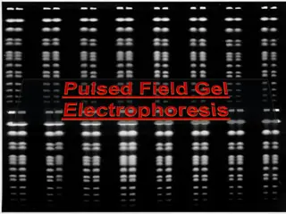
The impact of the intercalating dye on the TH nanostructure Nondenaturing gel electrophoresis The mobility of the TH is unchanged by intercalation of YOYO-1, whereas the linear and 3WJ DNA mobilities are retarded by the dye.
Optical Spectroscopy similarity among the three absorption spectra : comparable amounts of bound dye, regardless of the dimensionality of the DNA template. The unbound dye adopts a collapsed conformation in which the two chromophores are stacked into an H -dimer structure. Intercalation into DNA separates the chromophores and restores the monomer spectrum. HYPOCHROMIC EFFECT
significant differences in fluorescence intensities of the intercalator dye in the presence of the three nanostructures similar sequence composition (47-48% GC) for DNA templates The difference in fluorescence intensity is unlikely to be due to the known sequence dependence of fluorescence quantum yield for this dye. - one or more of the chromophores insert into the junction regions linking different helices in the 2- and 3-D structures not as constrained as a fully intercalated Dye lower quantum yield Intermediate weaker - traps for energy migrating through the assembly
Energy Transfer Experiments In flow cytometry and fluorescence microscopy, it is useful to have labels that fluoresce in different colors but that can be excited at the same wavelength, such that different populations can be distinguished while using the same light source. helix junction points multicolor labeling highly efficient ET from intercalated YOYO-1 to terminal acceptor dyes (Cy3 or WellRed-D2) attaching 1-4 Cy3 acceptor dyes to the 5 -termini of the DNA strands
The ET efficiency increases monotonically with the number of Cy3 acceptors, reaching a maximum of 95%. The compact nature of the TH, which localizes all intercalated dye molecules within a diameter of less than 10 nm, similar to the critical transfer distance of 7.3 nm for the YOYO-1/Cy3 pair.
energy migration among the cointercalated chromophores particularly within a given edge The TH acts as an antenna to harvest light with very strong efficiency and transfers the excitation to a lower energy acceptor dye at a nearby helix junction. TH nanotags with and without the Cy3 acceptor dyes can be used to label two different species of interest, while being excitable at the same wavelength.
Photostability Studies One critical parameter for effective use of fluorescent labels in imaging applications is the photostability of the fluorophores. Stable fluorescence intensity is desired for applications requiring long irradiations such as single molecule spectroscopy or fluorescence imaging of low abundance species. Cyanine dyes are known to undergo photobleaching via reaction with singlet oxygen. The TH nanotags were expected to minimize this reaction path by virtue of intercalation of the dyes into the DNA matrix, which should not only restrict access of molecular oxygen to the dye excited state, but also hinder reaction with any singlet oxygen that does get produced.
Comparison of the photostabilities of YOYO-1 intercalated in linear (1D) and TH (3D) nanostructures damage to the DNA template rather than to the dye The ability of the TH nanostructure to resist photodamage is a significant advantage relative to a linear DNA template.
Biochemical Stability Degradation by nuclease enzymes Releasing the dyes into solution and diminishing the fluorescence brightness of whatever was labeled by the nanotag Previous studies have shown that bisintercalating dyes improve the stability of a DNA duplex against endonuclease degradation, presumably because of the lengthening and unwinding of the duplex that occurs in response to intercalation.
monitoring the fluorescence intensity of the nanotags treated with endo- (DNase I) or exonuclease (Exo III) before or after loading with intercalator dyes protective effect of the intercalator dye
comparison for nanotags of different dimensionalities Meanwhile Exo III, which attacks the 3 end of the DNA strand and removes one nucleotide at a time, also exhibited lower activity against the TH than the 3WJ and comparable activity with the linear nanostructure, despite the fact that there are more 3 -termini in the TH than the 3WJ and linear nanostructures (4 versus 3 and 2, respectively). analyzing complex biological samples without having to purify or otherwise treat the sample more resistant because of the significantly shorter edges of the more compact TH nanostructure
DISCUSSION A compact 3D nanostructure provides a protective environment for the fluorophores, which leads to intense and stable optical signals. As the ratio of binding sites per unit volume of the nanostructure increases, brighter and smaller nanotags can result and find various applications. Because of the availability of well-established methods to functionalize DNA for conjugation to antibodies, proteins, peptides, or other nucleic acids, these nanotags can be utilized to label and track biomolecules. In addition to ease of synthesis and conjugation to biomolecules, having the ability to control both the number and the proximity of fluorophores on a 3D DNA template as opposed to being embedded irregularly in a matrix will be advantageous.


