DNA Transformation in Bacterial Cells
GENETIC ENGINEERING
College of Science/ biology department
fourth class
Assistant professor Dr. Munim Radwan Ali
Lecture seven
I
N
T
R
O
D
U
C
T
I
O
N
O
F
D
N
A
I
N
T
O
H
O
S
T
C
E
L
L
S
T
r
a
n
s
f
o
r
m
a
t
i
o
n
:
T
h
e
u
p
t
a
k
e
o
f
D
N
A
b
y
b
a
c
t
e
r
i
a
l
c
e
l
l
s
•
Transformation is the process of uptake of foreign DNA
(normally
plasmids)
by bacteria
.
•
In nature, transformation is probably
not a major
process by which
bacteria obtain genetic material. This is reflected by the fact that only a
few species (notably members of the genera
Bacillus
and
Streptococcus
)
can be transformed with ease in the laboratory, and studies have revealed
that these species possess sophisticated mechanisms for DNA binding and
uptake. Under normal circumstances, most bacterial species (including
E.
coli
) will only take up
limited amounts of DNA
. In order to transform these
species efficiently, the bacteria have to undergo some form of physical
and/or chemical treatment that will enhance their ability to take up DNA.
Cells that have undergone treatment are referred to as
competent cells
.
•
It was discovered in the early 1970s (cited by Old &
Primrose, 1994) that
E. coli
cells treated with solutions
containing
Ca2+ ions
were rendered susceptible to take
up
exogenous DNA
. The precise mechanism of this is not
understood. Ca2+ ions possibly cause the DNA to
precipitate on the outside of the cells
, or perhaps they
are responsible for some kind of
change in the cell wall
that improves DNA binding. Thus, treatment with Ca2+
ions affects
only
DNA binding, and not the actual uptake
of DNA .
•
Furthermore, when DNA is added to treated cells, it
remains attached to the exterior, and is not at this stage
transported into the cytoplasm. The actual movement of
the DNA into competent cells is stimulated by
briefly
rising the temperature to 42°C
(referred to as
heat
shock
) . This process induces enzymes involved in the
repair of DNA and other cellular components, which
allow the cell to recover from the unusual conditions of
the transformation process, and increases the efficiency.
After this, cells are incubated in a growth medium and
finally spread out on an agar plate and incubated until
single colonies of bacteria grow.
•
The quality of a given preparation of competent cells
may be measured by determining the
transformation
efficiency
. This can be defined
as the number of
colonies formed (on a selective plate) per microgram of
input DNA, where that DNA is a pure plasmid, most
commonly the vector to be used in a cloning
experiment
. Transformation efficiencies can range from
10
3
per μg for crude transformation protocols, to more
than 10
8
per μg for very carefully prepared competent
cells to be used in the construction of libraries. A
transformation efficiency of 10
5
per μg would be
adequate for a simple cloning experiment.
I
n
t
r
o
d
u
c
t
i
o
n
o
f
p
h
a
g
e
D
N
A
i
n
t
o
b
a
c
t
e
r
i
a
l
c
e
l
l
s
:
•
A recombinant DNA molecule, constructed with a phage vector, can
be introduced into a bacterial cell by transfection or
in vitro
packaging.
•
Transfection
: The process of transfection is equivalent to
transformation, the only difference being that phage DNA rather than
a plasmid is involved. As with transformation, the purified
recombinant phage molecule is added to competent
E. coli
cells and
DNA uptake is induced by heat shock .
•
In vitro
packaging :
Another method of introducing a
recombinant DNA molecule, constructed with a phage vector, is
in
vitro
packaging. To introduce such recombinants into the host cell,
they are first “packaged” into phage particles
in vitro
, and then the
bacteria are infected (transfected) with the packaged recombinant.
T
r
a
n
s
f
o
r
m
a
t
i
o
n
o
f
n
o
n
-
b
a
c
t
e
r
i
a
l
c
e
l
l
s
:
•
Ways of introducing DNA into yeast, fungi, animals and plants are also
needed if these organisms are to be used as hosts for gene cloning . The
uptake of DNA into eukaryotic cells is
more challenging
than bacterial
transformation, and the efficiency of the
process is much lower
. For
example, in yeast and plant cells the cell wall must be digested with
degradative
enzymes to yield fragile
protoplasts
, which may then take up
DNA quite readily. The cell walls are re-synthesized once the degrading
enzymes are removed. In contrast, animal cells in culture, which have no
cell wall, will take up DNA at low efficiency if it is precipitated on their
surface with calcium phosphate. Treating the cells with
a high voltage
,
which is believed to
open transient pores
in the cell membrane, may
increase the efficiency of the process.
•
This process is referred to as
electroporation
and can
also be used to introduce cloned genes into bacterial
cells. In addition, direct physical methods have been
used. DNA may be
microinjected
directly into the
cytoplasm or even the nucleus of individual animal or
plant cells in culture. Alternatively, DNA may be
introduced by the bombardment of the target cells with
metallic microprojectiles coated with DNA. This
technique is referred to as
biolistics
.
G
E
N
E
L
I
B
R
A
R
I
E
S
•
A gene library is a collection of different DNA sequences from
an organism each of which has been cloned into a vector for
ease of purification, storage and analysis. Gene libraries are
used for two main purposes: (i) the cloning of individual genes
(e.g. genes associated with certain genetic disorders), and (ii)
the construction of physical maps of genomes . There are
essentially two types of gene libraries that can be made
depending on the source of DNA used. If the DNA is genomic
DNA, the library is called
a genomic library
. If the DNA is a copy
of an mRNA population, that is cDNA, then the library is called
a
cDNA library
.
B
a
s
i
c
s
t
e
p
s
i
n
t
h
e
c
o
n
s
t
r
u
c
t
i
o
n
o
f
a
g
e
n
e
l
i
b
r
a
r
y
:
•
1. Extraction of genomic DNA of the organism of interest.
•
2. The extracted DNA is cut into gene-size pieces with restriction
enzymes.
•
3. The appropriate vector (for this purpose a plasmid) is cut with the
same restriction enzyme.
•
4. The gene-sized DNA and cut plasmids are combined into one test
tube. Some of the enzyme cut DNA pieces will combine with the cut
vectors and form recombinant DNA.
•
5. The recombinant plasmids are then transferred into bacteria using
either electroporation or heat shock (transformation).
•
6. The bacteria are grown on a culture dish and allowed to grow into
colonies. All the colonies on all the plates (cultures) are called a gene
library.
•
7. The gene library is then screened in order to discover which bacterial
colony is making copies of the one gene you are interested in. Library
screening identifies colonies which have that particular gene. Screening
can be based on detecting the DNA sequence of the cloned gene,
detecting a protein that the gene encodes, or the use of linked DNA
markers.
•
8. When the bacterial colony containing the desired gene is located, the
bacteria can be propagated to make millions of copies of the
recombinant plasmid that contains the gene. Furthermore, the plasmids
can be extracted for the next steps of genetic engineering, gene
modification, and transformation.
S
C
R
E
E
N
I
N
G
A
N
D
S
E
L
E
C
T
I
O
N
F
O
R
R
E
C
O
M
B
I
N
A
N
T
S
•
The art of cloning is to find the one particular transformed cell that contains
the cloning vector with the gene of interest (referred to as a recombinant
cell). It is thus most important to be able to
select recombinant cells from
transformed cells
(containing the vector without insert DNA) . If this goal
has been achieved the gene in question is said to have been cloned.
•
Although there are many different procedures by which the desired clone
can be obtained, all are variations of two basic concepts :
•
•
Direct selection
, which means that the cloning experiment is
designed in such a way that the clones obtained are the clones
containing the required gene. Almost invariably, selection occurs at
the plating-out stage. Direct selection is the method of choice, as it is
quick and usually unambiguous. However, it is not applicable to all
genes.
•
•
Identification of the clone from a gene library
, which entails an
initial “shotgun” cloning experiment, to produce a clone library
representing all or most of the genes present in the cell, followed by
analysis of the individual clones to identify the correct one.
•
Direct selection:
•
Most cloning vectors are designed so that the insertion of a DNA
fragment into the vector destroys the integrity of one of the genes
present on the molecule. This is referred to as
insertional inactivation
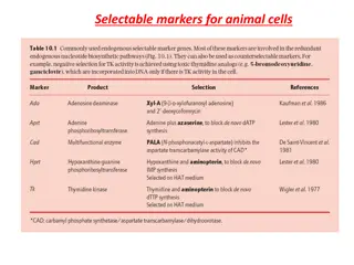
. Two examples will be discussed: (i) direct antibiotic resistance
screening, and (ii) blue-white colour screening.
D
i
r
e
c
t
a
n
t
i
b
i
o
t
i
c
r
e
s
i
s
t
a
n
c
e
s
c
r
e
e
n
i
n
g
•
Most cloning vectors are designed so that the insertion of a DNA
fragment into the vector destroys the integrity of one of the genes
present on the molecule (usually an antibiotic resistance gene). For
instance, if the vector carries an ampicillin resistance gene (
ampr
), it
will confer ampicillin resistance to the
E. coli
cells if the
transformation process was successful. This would mean that when
the clones are plated out on ampicillin-containing medium, only cells
containing a plasmid would be resistant to the antibiotic and,
therefore, be able to grow. However, this does not show whether the
cell contains the recombinant plasmid (with the inserted gene of
interest) or only the re-ligated vector (without the inserted gene of
interest).
B
l
u
e
-
w
h
i
t
e
c
o
l
o
u
r
s
c
r
e
e
n
i
n
g
•
This method also involves the insertional inactivation of a gene and
uses the production of a blue compound as an indicator . The gene is
lacZ
, which encodes the enzyme ß-galactosidase, and is under the
control of the
lac
promoter. If the host
E. coli
strain is expressing the
lac repressor, expression of a
lacZ
gene on the vector may be induced
using IPTG (isopropyl- ß-D-thiogalactopyranoside), and the expressed
enzyme can utilize the synthetic substrate X-gal (5-bromo-4-chlore-3-
indolyl- ß-D-galactopyranoside) to yield a blue product.
•
Insertion of a DNA fragment into the
lacZ
gene (insertional
inactivation of
lacZ
) in the production of a recombinant plasmid
would prevent the development of the blue colour. In this method,
the transformed cells are spread onto a plate containing an antibiotic
(to select for transformants in the usual way), IPTG and X-gal, to yield
a mixture of blue and white colonies. The white colonies have no
expressed ß-galactosidase and are hence likely to
contain the
inserted target fragment
. The blue colonies probably contain
religated vector.
I
d
e
n
t
i
f
i
c
a
t
i
o
n
o
f
t
h
e
c
l
o
n
e
f
r
o
m
a
g
e
n
e
l
i
b
r
a
r
y
:
•
Many different techniques are available for screening a library. The most
important approaches involve screening by nucleic acid hybridization
and screening by functional analysis. Nucleic acid hybridization requires
some prior knowledge of the DNA sequence either of the gene to be
cloned or of stretches of DNA in the vicinity of the gene to be cloned.
Functional screening involves the use of expression vectors that allow
cells containing the vector with the desired gene to express the
corresponding protein
. Under these circumstances, cells containing a
vector with the desired gene can be identified by means of
antibodies
directed against the protein.
N
u
c
l
e
i
c
a
c
i
d
h
y
b
r
i
d
i
z
a
t
i
o
n
•
Detection of an individual clone in a library can be achieved by
employing strategies of nucleic acid hybridization in which short
chemically synthesized labeled oligonucleotides (
probes
) are used to
detect complementary sequences in individual cells or phages
containing an insert. The success of colony or plaque hybridization
will depend on the availability of a DNA molecule that can be used as
probe.
This probe must share at least a part of the sequence of the
cloned gene
. If the gene itself is not available, it can be derived, for
example, from known protein sequences, or so-called degenerate
oligonucleotides (mixtures of oligonucleotides that differ from each
other by base substitutions at identical and/or different positions) can
be used.
•
The first step of a hybridization screening experiment involves
the transfer of the DNA in the plaque or colony to a nylon or
nitrocellulose membrane. The bacteria on the membrane are
lysed to release their DNA, and the DNA is denatured with alkali
to produce single strands that are bounded to the membrane by
heat treatment or UV irradiation. The membrane is then
immersed in a solution containing a nucleic acid probe (usually
radioactively labeled) and incubated to allow the probe to
hybridize to its complementary sequence. After hybridization,
the membrane is washed to remove unhybridized probe, and
regions where the probe has hybridized are visualized with
autoradiography .
F
u
n
c
t
i
o
n
a
l
s
c
r
e
e
n
i
n
g
•
The desired gene can also be identified by the activities of the
encoded gene product. In this case, one uses a so-called expression
library that has been established by cloning DNA (cDNA in the case of
eukaryotes) fragments into special cloning vectors allowing the
functional expression of cloned DNA fragments. Functional gene
products and hence the desired clones can then be detected either by
antibodies (
immunological screening
) or other
ligands
that specifically
recognize the encoded proteins or by exploiting a bioactivity of the
gene product, if known .
•
The procedure of immunological screening has
similarities to hybridization screening, discussed before,
though in this instance it is the protein encoded by the
DNA (or cDNA) rather than the DNA itself that is
detected on the membrane. The membrane is treated to
covalently attach the protein, and immersed in a
solution containing the antibodies. When the antibody
has bound to its epitope, it is detected by other
antibodies and/or chemicals that recognize it.
C
h
r
o
m
o
s
o
m
e
w
a
l
k
i
n
g
•
Chromosome walking
•
Often, a genomic clone
may not include all of
the sequence for a
particular gene so it is
necessary to isolate
overlapping clones
that cover the genomic
region of interest. This
process is known as
chromosome walking.
DNA transformation is a crucial process in genetic engineering, where foreign DNA is introduced into bacterial cells such as E. coli. This process, known as transformation, involves making the cells competent to uptake DNA through physical and chemical treatments. The uptake of DNA occurs after treating the cells with solutions containing Ca2+ ions, followed by a heat shock to facilitate the movement of DNA into the cytoplasm. Monitoring the transformation efficiency is important for evaluating the success of the process.

Uploaded on Sep 30, 2024 | 2 Views
Download Presentation

Please find below an Image/Link to download the presentation.
The content on the website is provided AS IS for your information and personal use only. It may not be sold, licensed, or shared on other websites without obtaining consent from the author.If you encounter any issues during the download, it is possible that the publisher has removed the file from their server.
You are allowed to download the files provided on this website for personal or commercial use, subject to the condition that they are used lawfully. All files are the property of their respective owners.
The content on the website is provided AS IS for your information and personal use only. It may not be sold, licensed, or shared on other websites without obtaining consent from the author.
E N D
Presentation Transcript
GENETIC ENGINEERING College of Science/ biology department fourth class Assistant professor Dr. Munim Radwan Ali Lecture seven
INTRODUCTION OF DNA INTO HOST CELLS INTRODUCTION OF DNA INTO HOST CELLS Transformation: The uptake of DNA by bacterial cells Transformation: The uptake of DNA by bacterial cells Transformation is the process of uptake of foreign DNA (normally plasmids) by bacteria. In nature, transformation is probably not a major process by which bacteria obtain genetic material. This is reflected by the fact that only a few species (notably members of the genera Bacillus and Streptococcus) can be transformed with ease in the laboratory, and studies have revealed that these species possess sophisticated mechanisms for DNA binding and uptake. Under normal circumstances, most bacterial species (including E. coli) will only take up limited amounts of DNA. In order to transform these species efficiently, the bacteria have to undergo some form of physical and/or chemical treatment that will enhance their ability to take up DNA. Cells that have undergone treatment are referred to as competent cells .
It was discovered in the early 1970s (cited by Old & Primrose, 1994) that E. coli cells treated with solutions containing Ca2+ ions were rendered susceptible to take up exogenous DNA. The precise mechanism of this is not understood. Ca2+ ions possibly cause the DNA to precipitate on the outside of the cells, or perhaps they are responsible for some kind of change in the cell wall that improves DNA binding. Thus, treatment with Ca2+ ions affects only DNA binding, and not the actual uptake of DNA .
Furthermore, when DNA is added to treated cells, it remains attached to the exterior, and is not at this stage transported into the cytoplasm. The actual movement of the DNA into competent cells is stimulated by briefly rising the temperature to 42 C (referred to as heat shock) . This process induces enzymes involved in the repair of DNA and other cellular components, which allow the cell to recover from the unusual conditions of the transformation process, and increases the efficiency. After this, cells are incubated in a growth medium and finally spread out on an agar plate and incubated until single colonies of bacteria grow.
The quality of a given preparation of competent cells may be measured by determining the transformation efficiency. This can be defined as the number of colonies formed (on a selective plate) per microgram of input DNA, where that DNA is a pure plasmid, most commonly the vector to be used in a cloning experiment. Transformation efficiencies can range from 103 per g for crude transformation protocols, to more than 108 per g for very carefully prepared competent cells to be used in the construction of libraries. A transformation efficiency of 105 per g would be adequate for a simple cloning experiment.
Introduction of phage DNA into bacterial cells: Introduction of phage DNA into bacterial cells: A recombinant DNA molecule, constructed with a phage vector, can be introduced into a bacterial cell by transfection or in vitro packaging. Transfection: The process of transfection is equivalent to transformation, the only difference being that phage DNA rather than a plasmid is involved. As with transformation, the purified recombinant phage molecule is added to competent E. coli cells and DNA uptake is induced by heat shock .
In vitro packaging : Another method of introducing a recombinant DNA molecule, constructed with a phage vector, is in vitro packaging. To introduce such recombinants into the host cell, they are first packaged into phage particles in vitro, and then the bacteria are infected (transfected) with the packaged recombinant.
Transformation of non Transformation of non- -bacterial cells: Ways of introducing DNA into yeast, fungi, animals and plants are also needed if these organisms are to be used as hosts for gene cloning . The uptake of DNA into eukaryotic cells is more challenging than bacterial transformation, and the efficiency of the process is much lower. For example, in yeast and plant cells the cell wall must be digested with degradative enzymes to yield fragile protoplasts, which may then take up DNA quite readily. The cell walls are re-synthesized once the degrading enzymes are removed. In contrast, animal cells in culture, which have no cell wall, will take up DNA at low efficiency if it is precipitated on their surface with calcium phosphate. Treating the cells with a high voltage, which is believed to open transient pores in the cell membrane, may increase the efficiency of the process. bacterial cells:
This process is referred to as electroporation and can also be used to introduce cloned genes into bacterial cells. In addition, direct physical methods have been used. DNA may be microinjected directly into the cytoplasm or even the nucleus of individual animal or plant cells in culture. Alternatively, DNA may be introduced by the bombardment of the target cells with metallic microprojectiles coated with DNA. This technique is referred to as biolistics .
GENE LIBRARIES GENE LIBRARIES A gene library is a collection of different DNA sequences from an organism each of which has been cloned into a vector for ease of purification, storage and analysis. Gene libraries are used for two main purposes: (i) the cloning of individual genes (e.g. genes associated with certain genetic disorders), and (ii) the construction of physical maps of genomes . There are essentially two types of gene libraries that can be made depending on the source of DNA used. If the DNA is genomic DNA, the library is called a genomic library. If the DNA is a copy of an mRNA population, that is cDNA, then the library is called a cDNA library.
Basic steps in the construction of a gene library: Basic steps in the construction of a gene library: 1. Extraction of genomic DNA of the organism of interest. 2. The extracted DNA is cut into gene-size pieces with restriction enzymes. 3. The appropriate vector (for this purpose a plasmid) is cut with the same restriction enzyme. 4. The gene-sized DNA and cut plasmids are combined into one test tube. Some of the enzyme cut DNA pieces will combine with the cut vectors and form recombinant DNA. 5. The recombinant plasmids are then transferred into bacteria using either electroporation or heat shock (transformation).
6. The bacteria are grown on a culture dish and allowed to grow into colonies. All the colonies on all the plates (cultures) are called a gene library. 7. The gene library is then screened in order to discover which bacterial colony is making copies of the one gene you are interested in. Library screening identifies colonies which have that particular gene. Screening can be based on detecting the DNA sequence of the cloned gene, detecting a protein that the gene encodes, or the use of linked DNA markers. 8. When the bacterial colony containing the desired gene is located, the bacteria can be propagated to make millions of copies of the recombinant plasmid that contains the gene. Furthermore, the plasmids can be extracted for the next steps of genetic engineering, gene modification, and transformation.
SCREENING AND SELECTION FOR RECOMBINANTS SCREENING AND SELECTION FOR RECOMBINANTS The art of cloning is to find the one particular transformed cell that contains the cloning vector with the gene of interest (referred to as a recombinant cell). It is thus most important to be able to select recombinant cells from transformed cells (containing the vector without insert DNA) . If this goal has been achieved the gene in question is said to have been cloned. Although there are many different procedures by which the desired clone can be obtained, all are variations of two basic concepts :
Direct selection, which means that the cloning experiment is designed in such a way that the clones obtained are the clones containing the required gene. Almost invariably, selection occurs at the plating-out stage. Direct selection is the method of choice, as it is quick and usually unambiguous. However, it is not applicable to all genes. Identification of the clone from a gene library, which entails an initial shotgun cloning experiment, to produce a clone library representing all or most of the genes present in the cell, followed by analysis of the individual clones to identify the correct one.
Direct selection: Most cloning vectors are designed so that the insertion of a DNA fragment into the vector destroys the integrity of one of the genes present on the molecule. This is referred to as insertional inactivation . Two examples will be discussed: (i) direct antibiotic resistance screening, and (ii) blue-white colour screening.
Direct antibiotic resistance screening Direct antibiotic resistance screening Most cloning vectors are designed so that the insertion of a DNA fragment into the vector destroys the integrity of one of the genes present on the molecule (usually an antibiotic resistance gene). For instance, if the vector carries an ampicillin resistance gene (ampr), it will confer ampicillin resistance to the E. coli cells if the transformation process was successful. This would mean that when the clones are plated out on ampicillin-containing medium, only cells containing a plasmid would be resistant to the antibiotic and, therefore, be able to grow. However, this does not show whether the cell contains the recombinant plasmid (with the inserted gene of interest) or only the re-ligated vector (without the inserted gene of interest).
Blue Blue- -white white colour colour screening screening This method also involves the insertional inactivation of a gene and uses the production of a blue compound as an indicator . The gene is lacZ, which encodes the enzyme -galactosidase, and is under the control of the lac promoter. If the host E. coli strain is expressing the lac repressor, expression of a lacZ gene on the vector may be induced using IPTG (isopropyl- -D-thiogalactopyranoside), and the expressed enzyme can utilize the synthetic substrate X-gal (5-bromo-4-chlore-3- indolyl- -D-galactopyranoside) to yield a blue product.
Insertion of a DNA fragment into the lacZ gene (insertional inactivation of lacZ) in the production of a recombinant plasmid would prevent the development of the blue colour. In this method, the transformed cells are spread onto a plate containing an antibiotic (to select for transformants in the usual way), IPTG and X-gal, to yield a mixture of blue and white colonies. The white colonies have no expressed -galactosidase and are hence likely to contain the inserted target fragment. The blue colonies probably contain religated vector.
Identification of the clone from a gene library: Identification of the clone from a gene library: Many different techniques are available for screening a library. The most important approaches involve screening by nucleic acid hybridization and screening by functional analysis. Nucleic acid hybridization requires some prior knowledge of the DNA sequence either of the gene to be cloned or of stretches of DNA in the vicinity of the gene to be cloned. Functional screening involves the use of expression vectors that allow cells containing the vector with the desired gene to express the corresponding protein. Under these circumstances, cells containing a vector with the desired gene can be identified by means of antibodies directed against the protein.
Nucleic acid hybridization Nucleic acid hybridization Detection of an individual clone in a library can be achieved by employing strategies of nucleic acid hybridization in which short chemically synthesized labeled oligonucleotides (probes) are used to detect complementary sequences in individual cells or phages containing an insert. The success of colony or plaque hybridization will depend on the availability of a DNA molecule that can be used as probe. This probe must share at least a part of the sequence of the cloned gene. If the gene itself is not available, it can be derived, for example, from known protein sequences, or so-called degenerate oligonucleotides (mixtures of oligonucleotides that differ from each other by base substitutions at identical and/or different positions) can be used.
The first step of a hybridization screening experiment involves the transfer of the DNA in the plaque or colony to a nylon or nitrocellulose membrane. The bacteria on the membrane are lysed to release their DNA, and the DNA is denatured with alkali to produce single strands that are bounded to the membrane by heat treatment or UV irradiation. The membrane is then immersed in a solution containing a nucleic acid probe (usually radioactively labeled) and incubated to allow the probe to hybridize to its complementary sequence. After hybridization, the membrane is washed to remove unhybridized probe, and regions where the probe has hybridized are visualized with autoradiography .
Functional screening Functional screening The desired gene can also be identified by the activities of the encoded gene product. In this case, one uses a so-called expression library that has been established by cloning DNA (cDNA in the case of eukaryotes) fragments into special cloning vectors allowing the functional expression of cloned DNA fragments. Functional gene products and hence the desired clones can then be detected either by antibodies (immunological screening) or other ligands that specifically recognize the encoded proteins or by exploiting a bioactivity of the gene product, if known .
The procedure of immunological screening has similarities to hybridization screening, discussed before, though in this instance it is the protein encoded by the DNA (or cDNA) rather than the DNA itself that is detected on the membrane. The membrane is treated to covalently attach the protein, and immersed in a solution containing the antibodies. When the antibody has bound to its epitope, it is detected by other antibodies and/or chemicals that recognize it.
Chromosome walking Chromosome walking Chromosome walking Often, a genomic clone may not include all of the sequence for a particular gene so it is necessary to isolate overlapping clones that cover the genomic region of interest. This process is known as chromosome walking.