Enterotomy in Dogs and Cats: Anatomy and Procedure
The small intestine anatomy in carnivores like dogs and cats, including the structure, length, and components. Details on enterotomy indications, procedure, and closure techniques for foreign bodies and biopsies.
Download Presentation

Please find below an Image/Link to download the presentation.
The content on the website is provided AS IS for your information and personal use only. It may not be sold, licensed, or shared on other websites without obtaining consent from the author. Download presentation by click this link. If you encounter any issues during the download, it is possible that the publisher has removed the file from their server.
E N D
Presentation Transcript
Enterotomy Dr.Alaa Ahmed Ibrahim
Antomy carnivore s intestine is approximately five times the length of its trunk applies reasonably well to dogs and cats The small intestine measures between 1.0 and 1.5 m in cats and between 2 and 5 m in dogs The small intestine is composed of the duodenum, jejunum, and ileum and therefore extends from the pylorus to the ileocolic junction The duodenum is the relatively fixed, short proximal part of the small intestine. the jejunum, the longest portion of the small intestines
The root of the mesentery includes the cranial mesenteric artery, intestinal lymphatics, and large mesenteric plexus of nerves that supply the small intestine. Cranial mesenteric artery arises beneath the first lumbar vertebra and anastomoses proximally with a branch of the celiac artery the cranial mesenteric artery divides into 12 to 15 major branches
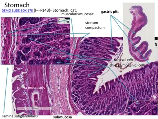
The small intestine is structurally composed of four layers: the mucosa, submucosa, muscularis, and serosa
Enterotomy indication Foreign bodies Biopsy samples A longitudinal incision is made in the antimesenteric border of the bowel immediately distal to the foreign object. This ensures the suture line is placed in healthy bowel that has not undergone pressure necrosis or distension from presence of the foreign body. The enterotomy is closed longitudinally in a single layer, appositional pattern with simple interrupted or continuous sutures. When full-thickness intestinal biopsies are needed for histopathologic evaluation, a longitudinal elliptical incision is made in the antimesenteric border of the bowel
longitudinal closure of the defect compromises the lumen, the defect is closed transversely in a single layer, appositional, simple interrupted pattern


