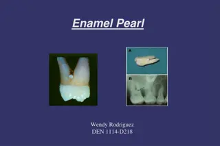
Enamel: Structure, Formation, and Properties

E
n
a
m
e
l
Enamel is formed on the tooth while
the tooth is developing within the
gum, before interrupts into the
mouth. Once fully formed, it does not
contain blood vessels or nerves.
Embryologically, enamel is formed by
ameloblast cells, which originate
from the ectoderm.
Ameloblasts have short extensions
toward the dentinoenamel junction
(DEJ) and these are called 
Tomes'
processes
.
Chemically:
- It is the hardest substance in the human
body.
-
It contains the highest percentage of
inorganic matter (minerals);95%-98%  by
weight.
-
The primary mineral is 
hydroxyapatite
(
crystalline
 
calcium phosphate
).
- The organic content of about 1-2% and
water.
- Enamel does not contain 
collagen
, as
found in other hard tissues such as
dentin and 
bone
, but it does contain two
unique classes of proteins:
amelogenins
 and 
enamelins
.
 
Enamel
O
n
c
e
 
i
t
 
i
s
 
m
a
t
u
r
e
,
 
e
n
a
m
e
l
 
i
s
 
a
l
m
o
s
t
 
t
o
t
a
l
l
y
w
i
t
h
o
u
t
 
t
h
e
 
s
o
f
t
e
r
 
o
r
g
a
n
i
c
 
m
a
t
t
e
r
.
 
E
n
a
m
e
l
 
i
s
a
v
a
s
c
u
l
a
r
 
a
n
d
 
h
a
s
 
n
o
 
n
e
r
v
e
 
s
u
p
p
l
y
 
w
i
t
h
i
n
 
i
t
a
n
d
 
i
s
 
n
o
t
 
r
e
n
e
w
e
d
,
 
h
o
w
e
v
e
r
,
 
i
t
 
i
s
 
n
o
t
 
a
s
t
a
t
i
c
 
t
i
s
s
u
e
 
a
s
 
i
t
 
c
a
n
 
u
n
d
e
r
g
o
 
m
i
n
e
r
a
l
i
z
a
t
i
o
n
c
h
a
n
g
e
s
.
Structurally
T
h
e
 
b
a
s
i
c
 
u
n
i
t
 
o
f
 
e
n
a
m
e
l
 
i
s
 
c
a
l
l
e
d
t
h
e
 
e
n
a
m
e
l
 
r
o
d
,
 
w
h
i
c
h
 
i
s
 
a
b
o
u
t
 
4
8
 
μ
m
 
i
n
 
d
i
a
m
e
t
e
r
.
 
A
n
 
e
n
a
m
e
l
 
r
o
d
,
f
o
r
m
a
l
l
y
 
c
a
l
l
e
d
 
a
n
 
e
n
a
m
e
l
 
p
r
i
s
m
,
i
s
 
a
 
t
i
g
h
t
l
y
 
p
a
c
k
e
d
 
m
a
s
s
 
o
f
h
y
d
r
o
x
y
a
p
a
t
i
t
e
 
c
r
y
s
t
a
l
s
 
i
n
 
a
n
o
r
g
a
n
i
z
e
d
 
p
a
t
t
e
r
n
.
 
I
n
 
c
r
o
s
s
s
e
c
t
i
o
n
,
 
i
t
 
i
s
 
b
e
s
t
 
c
o
m
p
a
r
e
d
 
t
o
 
a
k
e
y
h
o
l
e
,
 
w
i
t
h
 
t
h
e
 
t
o
p
,
 
o
r
 
h
e
a
d
,
o
r
i
e
n
t
e
d
 
t
o
w
a
r
d
 
t
h
e
 
c
r
o
w
n
 
o
f
 
t
h
e
t
o
o
t
h
,
 
a
n
d
 
t
h
e
 
b
o
t
t
o
m
,
 
o
r
 
t
a
i
l
,
o
r
i
e
n
t
e
d
 
t
o
w
a
r
d
 
t
h
e
 
r
o
o
t
 
o
f
 
t
h
e
t
o
o
t
h
.
 
 
T
h
e
 
a
r
e
a
 
a
r
o
u
n
d
 
t
h
e
e
n
a
m
e
l
 
r
o
d
 
i
s
 
k
n
o
w
n
a
s
 
i
n
t
e
r
r
o
d
 
e
n
a
m
e
l
.
I
n
t
e
r
r
o
d
 
e
n
a
m
e
l
 
h
a
s
 
t
h
e
s
a
m
e
 
c
o
m
p
o
s
i
t
i
o
n
 
a
s
e
n
a
m
e
l
 
r
o
d
;
 
h
o
w
e
v
e
r
 
t
h
e
c
r
y
s
t
a
l
 
o
r
i
e
n
t
a
t
i
o
n
 
i
s
d
i
f
f
e
r
e
n
t
 
i
n
 
e
a
c
h
.
 
T
h
e
b
o
r
d
e
r
 
w
h
e
r
e
 
t
h
e
 
c
r
y
s
t
a
l
s
o
f
 
e
n
a
m
e
l
 
r
o
d
s
 
a
n
d
c
r
y
s
t
a
l
s
 
o
f
 
i
n
t
e
r
r
o
d
e
n
a
m
e
l
 
m
e
e
t
 
i
s
 
c
a
l
l
e
d
t
h
e
 
r
o
d
 
s
h
e
a
t
h
.
Physical prosperities:
1
-
 
C
o
l
o
r
The normal color of E varies from light
yellow to grayish (bluish) white.
At the edges of teeth where there is no
dentin underlying the enamel, the color
sometimes has a slightly blue tone.
 Since enamel is 
semitranslucent
, the
color of dentin, the thickness of enamel,
and any material underneath the enamel
strongly affects the 
appearance
 of a tooth.
The enamel on primary teeth has a
more opaque crystalline form and thus
appears whiter than on permanent teeth.
2
.
 
T
h
i
c
k
n
e
s
s
I
t
 
i
s
 
t
h
i
c
k
 
a
t
 
t
h
e
 
i
n
c
i
s
a
l
 
e
d
g
e
 
a
n
d
 
c
u
s
p
 
t
i
p
o
f
 
m
o
l
a
r
s
 
a
n
d
 
p
r
e
m
o
l
a
r
s
 
(
2
-
2
.
5
 
m
m
)
 
a
n
d
e
n
d
s
 
c
e
r
v
i
c
a
l
l
y
 
a
s
 
k
n
i
f
e
 
e
d
g
e
.
H
o
w
e
v
e
r
,
 
e
n
a
m
e
l
 
t
h
i
c
k
n
e
s
s
 
d
e
c
r
e
a
s
e
s
 
i
n
p
i
t
s
 
a
n
d
 
f
i
s
s
u
r
e
 
a
r
e
a
s
 
o
f
 
o
c
c
l
u
s
a
l
 
s
u
r
f
a
c
e
s
o
f
 
m
o
l
a
r
s
 
a
n
d
 
p
r
e
m
o
l
a
r
s
 
w
h
e
r
e
 
d
e
e
p
i
n
v
a
g
i
n
a
t
i
o
n
s
 
o
c
c
u
r
.
T
h
e
s
e
 
f
i
s
s
u
r
e
s
 
a
c
t
 
a
s
 
f
o
o
d
 
a
n
d
 
b
a
c
t
e
r
i
a
l
t
r
a
p
s
 
t
h
a
t
 
m
a
y
 
p
r
e
d
i
s
p
o
s
e
 
d
e
n
t
a
l
 
c
a
r
i
e
s
.
N
o
r
m
a
l
l
y
,
 
o
c
c
l
u
s
a
l
 
g
r
o
o
v
e
s
 
s
e
r
v
e
 
a
n
i
m
p
o
r
t
a
n
t
 
f
u
n
c
t
i
o
n
 
a
s
 
a
n
 
e
s
c
a
p
e
 
w
a
y
 
f
o
r
m
o
v
e
m
e
n
t
 
o
f
 
t
h
e
 
f
o
o
d
 
t
o
 
t
h
e
 
f
a
c
i
a
l
 
a
n
d
l
i
n
g
u
a
l
 
s
u
r
f
a
c
e
s
 
d
u
r
i
n
g
 
m
a
s
t
i
c
a
t
i
o
n
.
3.Hardness & Brittleness
Enamel is as hard as steel. It's the
hardest substance of the human
body.
 However, enamel may wear
because of attrition of friction
contact against opposing enamel
or harder restorative.
Heavy occlusal wear is
demonstrated when rounded
cuspal contacts re ground to flat
facets.
 
Depending on the factors
such as; bruxism,
malocclusion, age, diet, and
bad habits, cusps may be
completely lost and enamel
abraded away so that dentin
is exposed.
Therefore cavity outline form
should be designed so that
the margins of restorations be
away of critical stress area of
occlusal contact.
Bad Habits


4
.
 
S
o
l
u
b
i
l
i
t
y
:
Enamel is soluble when exposed to acid
medium.
The high mineral content of enamel,
amakes it susceptible to a
demineralization process which often
occurs as 
dental caries
.
The mouth contains a great number
and variety of 
bacteria
, and
when 
sucrose
, coats the surface of the
mouth, some bacteria interact with it
and form 
lactic acid
, which decreases
the pH in the mouth.
Then, the hydroxyapatite crystals of
enamel demineralize, allowing for
greater bacterial invasion deeper into
the tooth.
Fluoride effect
5
.
 
P
e
r
m
e
a
b
i
l
i
t
y
Although enamel is a very hard and dense structure,
it is permeable to certain ions and molecules.
The route of passage  through hypomineralized and
rich in organic content such as rod sheaths, enamel
cracks, and other defects.
It appears that water plays an important role as a
transporting medium.
Enamel permeability decreases with age because of
the changes in the enamel matrix. This decrease is
referred to as enamel maturation.
T
h
a
n
k
 
y
o
u


Dentin or Dentine
It forms the largest portion of
tooth structure, extending
almost the full length of the
tooth. Externally dentin is
covered by enamel on the
anatomical crown and by
cementum on the root.
Dentin is a bone-like matrix that is
porous and yellow material.
It is made up of 75% inorganic
materials, 20% organic materials
and 5% water.
Because it is softer than enamel, it
decays more rapidly and is
subject to severe cavities if not
properly treated.
T
y
p
e
s
 
o
f
 
d
e
n
t
i
n
The most recently formed layer of dentin is always on the pulpal surface.
This unmineralized zone of dentin is immediately next to the cell bodies
of the odontoblasts is called 
predentin
.
Dentin formation begins at areas subadjacent to the cusp tip or incisal
ridge and gradually spread to the apex of the root. The dentin which forms
the initial shape of the tooth is called 
primary dentin.
 
The dentinal tubules are
small canals that extend
from the DEJ or DCJ to the
pulp.
Each tubule contains the
cytoplasmic cell process
(Tomes' fiber) of
odontoblasts.
Each dentinal tubule is
lined with a layer of
peritubular
 dentin, which
is much more mineralized
than the surrounding
intertubular
 dentin.
S
e
c
o
n
d
a
r
y
 
d
e
n
t
i
n
It grows much more slowly
than primary dentin, but
maintains its incremental
aspect of growth.
It has a similar structure to
primary dentin,
Its deposition is mainly in
the pulp horns and on the
floor of the pulp chamber.
 It is the growth of this
dentin that causes the
decrease in the size of the
pulp chamber with age.
 
Tertiary Dentin- (irregular)
Is formed in response to
moderate-level irritants,.
In about 15 days,
mesenchymal cells of the
pulp are converted or
differentiated to secondary
odontoblasts, and form
irregularly organized
tubules.
 The rate of formation and
the thickness and
organization of reparative
dentin depend on the
intensity and duration of
the stimulus. 
 
D
e
n
t
i
n
 
p
e
r
m
e
a
b
i
l
i
t
y
Dentin permeability is directly related to
its protective function.
When the external cap of enamel and
cementum is lost from the dentinal
tubules, the exposed tubules becomes
conduits between the pulp and the
external oral environment.
such capillary action, differential
thermal expansion and diffusion of
various fluids through permeable
dentinal tubules to reach the pulp,
putting the tooth at risk for pulpal
inflammation and sensitivity.
D
e
n
t
i
n
 
s
e
n
s
i
t
i
v
i
t
y
the most accepted theory of pain
transmission is the
hydrodynamic theory
.
This theory accounts for pain
transmission by the small, rapid
movements of fluid that occur
within the dentinal tubules.
Because many tubules contain
mechanoreceptor never ending
near the pulp.
Small fluid movement in the
tubules arising from cutting,
drying, pressure changes,
osmotic shifts, or changes in
temperature accounts for the
majority of pain transmission.
D
e
n
t
i
n
 
i
s
 
d
i
s
t
i
n
g
u
i
s
h
e
d
 
f
r
o
m
 
e
n
a
m
e
l
b
y
1
.
 
C
o
l
o
r
:
 
d
e
n
t
i
n
 
i
s
 
n
o
r
m
a
l
l
y
y
e
l
l
o
w
 
w
h
i
t
e
 
a
n
d
 
s
l
i
g
h
t
l
y
 
d
a
r
k
e
r
t
h
a
n
 
e
n
a
m
e
l
.
2
.
 
H
a
r
d
n
e
s
s
:
 
d
e
n
t
i
n
 
i
s
 
s
o
f
t
e
r
t
h
a
n
 
e
n
a
m
e
l
,
 
s
h
a
r
p
 
e
x
p
l
o
r
e
r
t
e
n
d
s
 
t
o
 
c
a
t
c
h
 
a
n
d
 
h
o
l
d
 
i
n
d
e
n
t
i
n
.
3
.
 
R
e
f
l
e
c
t
a
n
c
e
:
 
d
e
n
t
i
n
 
m
o
r
e
o
p
a
q
u
e
 
a
n
d
 
d
u
l
l
,
 
l
e
s
s
 
r
e
f
l
e
c
t
i
v
e
t
o
 
l
i
g
h
t
 
t
h
a
n
 
e
n
a
m
e
l
,
 
w
h
i
c
h
a
p
p
e
a
r
 
s
h
i
n
y
.
4
.
 
S
o
u
n
d
:
 
w
h
e
n
 
m
o
v
i
n
g
 
a
n
e
x
p
l
o
r
e
r
 
t
i
p
 
o
v
e
r
 
t
h
e
 
t
o
o
t
h
,
e
n
a
m
e
l
 
s
u
r
f
a
c
e
s
 
p
r
o
v
i
d
e
 
a
s
h
a
r
p
e
r
,
 
h
i
g
h
e
r
 
 
p
i
t
c
h
e
d
 
s
o
u
n
d
t
h
a
n
 
d
e
n
t
i
n
 
s
u
r
f
a
c
e
s
.
 
T
h
e
 
p
u
l
p
 
The pulp cavity is divided into:
the 
coronal
 
pulp
 located in the
pulp chamber of the crown
portion of the tooth
 the 
radicular pulp 
located in
the canals of the root portion of
the tooth.
 The radicular pulp is
continuous with the periapical
tissues by connecting through
the apical foramen of the root.
Accessory canals may extend
from the pulp canals laterally
through the root dentin to the
periodontal tissues.
T
h
e
 
p
u
l
p
 
i
s
 
a
 
s
p
e
c
i
a
l
i
z
e
d
 
o
r
g
a
n
 
s
e
r
v
i
n
g
f
o
u
r
 
f
u
n
c
t
i
o
n
s
:
1- 
Formative of developmental
; is the production of primary
and secondary dentin by the odontoblasts.
2- 
Nutritive
; supplies nutriments and moisture to the dentin
through the blood vascular supply to the odontoblasts and their
processes.
3- 
Sensory or protective
; provide sensory nerve fibers within
the pulp to mediate the sensation of pain.
4- 
Defensive or reparative
; is related primarily to its response
to irritation. Such irritants can cause the degeneration or death
of the odontoblastic processes and corresponding odontoblasts
and the formation by the pulp of replacement odontoblasts
(from undifferentiated mesenchymal cells) that lay down
irregular or reparative dentin.
:
C
o
m
p
o
s
i
t
i
o
n
 
o
f
 
t
h
e
 
p
u
l
p
 
From innermost to outermost:
1. 
Pulpal core
, which is in the center of the pulp chamber with many cells
and an extensive vascular supply.
2. 
Cell rich zone
; which contains fibroblasts and undifferentiated
mesenchymal cells.
3. 
Cell free zone
; which is rich in both capillaries and nerve networks.
4. 
Odontoblastic layer
; outermost layer which contains odontoblasts and
lies next to the predentin and mature dentin.
Cells found in the dental pulp include 
fibroblasts
 (the principal
cell), 
odontoblasts
,defence cells
like 
histiocytes
macrophage
granulocytes
mast cells
 and 
plasma cells
.
 
Thank you
Slide Note
Embed
Share

Enamel is the hardest substance in the human body, formed by ameloblast cells during tooth development. Chemically, it consists mostly of inorganic matter, primarily hydroxyapatite. Mature enamel lacks organic matter, is avascular, and not renewed. Its structural unit is the enamel rod, surrounded by interrod enamel. Enamel's physical properties include color variations and thickness differences across teeth.

  • Enamel
  • Tooth development
  • Hydroxyapatite
  • Enamel structure
  • Dental health

Uploaded on Oct 01, 2024 | 0 Views


Download Presentation

Please find below an Image/Link to download the presentation.

The content on the website is provided AS IS for your information and personal use only. It may not be sold, licensed, or shared on other websites without obtaining consent from the author. Download presentation by click this link. If you encounter any issues during the download, it is possible that the publisher has removed the file from their server.

E N D

Presentation Transcript


  1. Enamel Enamel is formed on the tooth while the tooth is developing within the gum, before interrupts into the mouth. Once fully formed, it does not contain blood vessels or nerves. Embryologically, enamel is formed by ameloblast cells, which originate from the ectoderm. Ameloblasts have short extensions toward the dentinoenamel junction (DEJ) and these are called Tomes' processes.

  2. Chemically: - It is the hardest substance in the human body. -It contains the highest percentage of inorganic matter (minerals);95%-98% by weight. -The primary mineral is hydroxyapatite (crystalline calcium phosphate). - The organic content of about 1-2% and water. - Enamel does not contain collagen, as found in other hard tissues such as dentin and bone, but it does contain two unique classes of proteins: amelogenins and enamelins.

  3. Enamel Once it is mature, enamel is almost totally without the softer organic matter. Enamel is avascular and has no nerve supply within it and is not renewed, however, it is not a static tissue as it can undergo mineralization changes.

  4. Structurally The basic unit of enamel is called theenamel rod, which is about 4 8 m in diameter. An enamel rod, formally called an enamel prism, is a tightly packed mass of hydroxyapatite crystals in an organized pattern. In cross section, it is best compared to a keyhole, with the top, or head, oriented toward the crown of the tooth, and the bottom, or tail, oriented toward the root of the tooth.

  5. The area around the enamel rod is known as interrod enamel. Interrod enamel has the same composition as enamel rod; however the crystal orientation is different in each. The border where the crystals of enamel rods and crystals of interrod enamel meet is called the rod sheath.

  6. Physical prosperities: 1- Color The normal color of E varies from light yellow to grayish (bluish) white. At the edges of teeth where there is no dentin underlying the enamel, the color sometimes has a slightly blue tone. Since enamel is semitranslucent, the color of dentin, the thickness of enamel, and any material underneath the enamel strongly affects theappearance of a tooth. The enamel on primary teeth has a more opaque crystalline form and thus appears whiter than on permanent teeth.

  7. 2. Thickness It is thick at the incisal edge and cusp tip of molars and premolars (2-2.5 mm) and ends cervically as knife edge. However, enamel thickness decreases in pits and fissure areas of occlusal surfaces of molars and premolars where deep invaginations occur. These fissures act as food and bacterial traps that may predispose dental caries. Normally, occlusal grooves serve an important function as an escape way for movement of the food to the facial and lingual surfaces during mastication.

  8. 3.Hardness & Brittleness Enamel is as hard as steel. It's the hardest substance of the human body. However, enamel may wear because of attrition of friction contact against opposing enamel or harder restorative. Heavy occlusal wear is demonstrated when rounded cuspal contacts re ground to flat facets.

  9. Depending on the factors such as; bruxism, malocclusion, age, diet, and bad habits, cusps may be completely lost and enamel abraded away so that dentin is exposed. Therefore cavity outline form should be designed so that the margins of restorations be away of critical stress area of occlusal contact.

  10. Bad Habits

  11. 4. Solubility: Enamel is soluble when exposed to acid medium. The high mineral content of enamel, amakes it susceptible to a demineralization process which often occurs as dental caries. The mouth contains a great number and variety of bacteria, and when sucrose, coats the surface of the mouth, some bacteria interact with it and form lactic acid, which decreases the pH in the mouth. Then, the hydroxyapatite crystals of enamel demineralize, allowing for greater bacterial invasion deeper into the tooth.

  12. Fluoride effect

  13. 5. Permeability Although enamel is a very hard and dense structure, it is permeable to certain ions and molecules. The route of passage through hypomineralized and rich in organic content such as rod sheaths, enamel cracks, and other defects. It appears that water plays an important role as a transporting medium. Enamel permeability decreases with age because of the changes in the enamel matrix. This decrease is referred to as enamel maturation.

  14. Thank you

  15. Dentin or Dentine It forms the largest portion of tooth structure, extending almost the full length of the tooth. Externally dentin is covered by enamel on the anatomical crown and by cementum on the root. Dentin is a bone-like matrix that is porous and yellow material. It is made up of 75% inorganic materials, 20% organic materials and 5% water. Because it is softer than enamel, it decays more rapidly and is subject to severe cavities if not properly treated.

  16. Types of dentin The most recently formed layer of dentin is always on the pulpal surface. This unmineralized zone of dentin is immediately next to the cell bodies of the odontoblasts is called predentin. Dentin formation begins at areas subadjacent to the cusp tip or incisal ridge and gradually spread to the apex of the root. The dentin which forms the initial shape of the tooth is called primary dentin.

  17. The dentinal tubules are small canals that extend from the DEJ or DCJ to the pulp. Each tubule contains the cytoplasmic cell process (Tomes' fiber) of odontoblasts. Each dentinal tubule is lined with a layer of peritubular dentin, which is much more mineralized than the surrounding intertubular dentin.

  18. Secondary dentin It grows much more slowly than primary dentin, but maintains its incremental aspect of growth. It has a similar structure to primary dentin, Its deposition is mainly in the pulp horns and on the floor of the pulp chamber. It is the growth of this dentin that causes the decrease in the size of the pulp chamber with age.

  19. Tertiary Dentin- (irregular) Is formed in response to moderate-level irritants,. In about 15 days, mesenchymal cells of the pulp are converted or differentiated to secondary odontoblasts, and form irregularly organized tubules. The rate of formation and the thickness and organization of reparative dentin depend on the intensity and duration of the stimulus.

  20. Dentin permeability Dentin permeability is directly related to its protective function. When the external cap of enamel and cementum is lost from the dentinal tubules, the exposed tubules becomes conduits between the pulp and the external oral environment. such capillary action, differential thermal expansion and diffusion of various fluids through permeable dentinal tubules to reach the pulp, putting the tooth at risk for pulpal inflammation and sensitivity.

  21. Dentin sensitivity the most accepted theory of pain transmission is the hydrodynamic theory. This theory accounts for pain transmission by the small, rapid movements of fluid that occur within the dentinal tubules. Because many tubules contain mechanoreceptor never ending near the pulp. Small fluid movement in the tubules arising from cutting, drying, pressure changes, osmotic shifts, or changes in temperature accounts for the majority of pain transmission.

  22. Dentin is distinguished from enamel by 1. Color: dentin is normally yellow white and slightly darker than enamel. 2. Hardness: dentin is softer than enamel, sharp explorer tends to catch and hold in dentin. 3. Reflectance: dentin more opaque and dull, less reflective to light than enamel, which appear shiny. 4. Sound: when moving an explorer tip over the tooth, enamel surfaces provide a sharper, higher pitched sound than dentin surfaces.

  23. The pulp The pulp cavity is divided into: the coronalpulp located in the pulp chamber of the crown portion of the tooth the radicular pulp located in the canals of the root portion of the tooth. The radicular pulp is continuous with the periapical tissues by connecting through the apical foramen of the root. Accessory canals may extend from the pulp canals laterally through the root dentin to the periodontal tissues.

  24. : The pulp is a specialized organ serving four functions: 1- Formative of developmental; is the production of primary and secondary dentin by the odontoblasts. 2- Nutritive; supplies nutriments and moisture to the dentin through the blood vascular supply to the odontoblasts and their processes. 3- Sensory or protective; provide sensory nerve fibers within the pulp to mediate the sensation of pain. 4- Defensive or reparative; is related primarily to its response to irritation. Such irritants can cause the degeneration or death of the odontoblastic processes and corresponding odontoblasts and the formation by the pulp of replacement odontoblasts (from undifferentiated mesenchymal cells) that lay down irregular or reparative dentin.

  25. Composition of the pulp From innermost to outermost: 1. Pulpal core, which is in the center of the pulp chamber with many cells and an extensive vascular supply. 2. Cell rich zone; which contains fibroblasts and undifferentiated mesenchymal cells. 3. Cell free zone; which is rich in both capillaries and nerve networks. 4. Odontoblastic layer; outermost layer which contains odontoblasts and lies next to the predentin and mature dentin. Cells found in the dental pulp include fibroblasts (the principal cell), odontoblasts,defence cells like histiocytes, macrophage, granulocytes, mast cells and plasma cells.

  26. Thank you

More Related Content

giItT1WQy@!-/#giItT1WQy@!-/#giItT1WQy@!-/#giItT1WQy@!-/#giItT1WQy@!-/#giItT1WQy@!-/#giItT1WQy@!-/#giItT1WQy@!-/#giItT1WQy@!-/#giItT1WQy@!-/#giItT1WQy@!-/#giItT1WQy@!-/#giItT1WQy@!-/#