Degenerative Spine Disease and Disc Degeneration


Degenerative
Spine
Disease
Dr.Demet Demircioğlu

Problem
Statement
2002
survey
26%
americans
–
low
back pain
and
14%
-
neck
pain
890
million
office
visits
due
to
back
pain
2005
-
JAMA
-
$86
billion
health
expenditures in
spine
related
problems
Anatomy
and
Physiology
of
Spine
Degeneration
Kirkaldy-
Willis
three-joint
complex
Theory
spine
–
at
each
level
composed
of
three
joints
complex
that
are
affected
in
degenerative
process
This
comprise
of
intervertebral
disc
and
two
zygapophyseal
joints
(dorsal articulating
joints)
Degeneration
of
any
one
joint
leads to
degeneration
of
the
other
two,
initiating
a
casacade
that
leads to
spinal
degenerative
disease
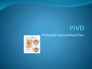
Pathology..
Disc
Degeneration..
Components
of
disc
nucleus
pulposus
semigelatinous
structure
situated
near
the
center
remnant
of
notochord;
composed
of
mucopolysaccharide
+
salt
+
water
annulus fibrosis
multilayered
circular
structures
that
surrounds
the
pulposus
composed
of
fibrocartilaginous
lamellae;
stiffer
than
nucleus
cartilagenous
end
plates
Mechanism
of
disc
degeneration
Part
of
natural
aging
process
Repetitive
loading
results
in
forces
that
foster
degeneration
Aging
dessication
collagen
and
proteoglycans
are
replaced
with
fibrous
tissue
Continued
Axial
pressure
less
compliant
annulus
develops
circumferential
tears
most
frequently
in
dorsolateral
aspect
tears
enlarge
and
develop
into
radial
tears
herniation
of
nucleus
pulposus
Relative
dorsal
location
of
nucleus pulposus
and
presence
of
posterior
longitudinal
ligament
lead
to
classical
dorsolateral
disc
herniation
Circumferential
bulge
of
annulus
due
to
annular
tear
loss
of
disc
height
and
osteophyte
formation
at
the
attachment of
the
annulus
to
vertebral
body
narrowing
of
central
canal
and
neural
foramen
Dorsal
joint
degeneration
Articulating
facets
from
superior
and
inferior
vertebral
segments
Joints
–
composed
of cartilage,
synovial
membrane
and
capsule
Aging
synovial
reaction,
fibrillation
of
articular
cartilage,
osteophyte
formation
laxity
of
joint
capsule
Leads
to
subluxation
of
joint
Osteophyte
spinal
canal and
lateral
recess
stenosis
Combine
Three-joint
complex
Degeneration
Individual
aging
process
of
disc
and
dorsal
facet
joints
are
interlaced
to
contribute
to
the clinical manifestation
of
spondylosis
Disc
degeneration
loss
of
disc
height
subluxation
of
dorsal
joints
This
compounds
to
natural
process
of facet
joint
degeneration
Subluxation
of
rostral vertebral
body
ventrally
with
respect
to the
caudal
vertebral
body
(spondylolisthesis)
This
results
in
further
narrowing
of
neural
foramina
lateral
nerve root
entrapment
Three
stages
of
Degenerative
Spine
Disease
Dysfunction
stage
Destabilization
stage
Restabilization
stage
Dysfunction
stage..
Characterized by
synovial
reaction
in
dorsal
joint
and
small
tears
in
the
intervertebral
discs
Minor
or
absent
clinical
symptoms
that
are
best
treated
conservatively
Destabilization
Stage..
Kirkaldy-Willis
defines
this
stage
as
greater
degeneration
in the
three-joint
complex,
manifesting
as
laxity
and
subluxation
in the
dorsal
joints and progressive
disc
degeneration
Abnormal
spinal
motion
Natural
mobility of
the
spine
lost
Compounded
by
advanced
disc
degeneration
and
disc
height
reduction
lead
to
spondylolisthesis
Rx
–
core
strengthening
and
flexibility
program
to
stabilize
and
normalize
dysfunctional
motion
segment
Restabilization
Stage
Instability
is
reduced
via
osteophyte
formation
secondary
to
a
prior
increased
joint laxity
and
loss
of
disc
interspace
height
Resolution
of
symptoms
can
occur
due
to
gradually
decreased
spinal
motion
There
may
be
radiculopathy
from
spinal
nerve
entrapment
or
claudication
symptoms
from
central
canal and
lateral
recess
stenosis
CT
Myelography
effective
alternative
to
MRI
for
assessing
neural
elements,
central
or
foraminal
stenosis
MRI
gold
standard
for
evaluation
spinal
canal
stenosis
soft
tissue
surrounding
spinal
canal,
discs,
ligamentum
flavum
and
facet
joints
visualized
hypertrophy
of
PLL,
ligamentum
flavum
and
facet
hypertrophy
can
be
localized
specifically
Discography
more
invasive
diagnostic
strategy
can
be
done
if
clinical
presentation
does
not match
the
findings
in
other
imaging
modality
useful
to
identify
and
characterize
disease
involves
injecting
contrast
material
into
the
disc
in
question
normal
cervical
disc
tolerates
0.2-0.5
ml fluid
whereas
a
degenerated
disc
can
accept
0.5-1.5
ml
fluid
if
the
pain produced
following
contrast
injection
is
concordant
with
the
typical
pain
experienced
by
the
patient
pathological
Treatment
Options
Non
operative
management
natural
course
of
spinal
stenosis
–
47%
patients
with
neurogenic
claudication
and
radiculopathy
symptomatic
improvement
without
intervention
Reason
–
progressive
disc
dehydration
shrinking
of
disk
decrease
in
root
compression
Medical
symptomatic
relief
to
reduction
in
inflammation
NSAIDs
Narcotics
to
supplement
masks
degenerative
process
until
it
progresses
and
improves
spontaneously
muscle
relaxants
–
shows
some
benefit
Systemic
oral
steroids
–
anti
inflammatory
effects
may
reduce
nerve root
irritation
Physical
reconditioning
via
physical
therapist
–
strengthen
core
muscles
Flexibility
exercise to
help
preserve
normal
motion
Epidural
steroid
injections
–
short
term
benefit
Facet
joint
injection
(long
acting anesthetic
and
steroids)
33%
patients
reported
>50%
pain
relief
–
result
consistent
with
placebo
also;
hence
controversial
key
to
efficacy
is
proper
selection
of patients
with
facet
syndrome
Facet
syndrome
defined
as
pain
in
the
hips
and
buttocks
area,
cramping
thigh
pain,
and
back
stiffness
that
is
worse
in
the
morning,
without
lower
extremity
paresthesia
Spinal
Manipulative
Therapy
(SMT)
by
chiropractors,
physical
therapists
and
osteopathic
physicians
3
main types
of
manipulations
–
therapeutic
massage,
mobilization
and
manipulative
procedures
Hypothesis
–
neck
or
back
pain
is
caused
by
either
a
limited
range
of
motion
or
abnormal dorsal
intervertebral
joint
motion
SMT
“resets”
the
joint
by
extending
the
joint
beyond
the
passive
range
of
motion,
into the
“paraphysiologic
range
of
motion”
Operative
Treatment
May
correlate
with
the
extent
of
disease
progression
Discectomy
ventral
or
dorsal approach
Cervical
Spine:
ventral
approach
often utilized in
cervical
spine
Ventral
–
performed through
a
paramedian
incision
requires
little
muscle
splitting
low
amount
of
postoperative
pain and
morbidity
complications
dysphagia secondary to
retraction
of
esophagus
(1-79%
cases)
damage
to
recurrent
laryngeal
n.
vocal
cord
palsy
improves with
time
Dorsal
approach
in
cervical
spine
effective
in
eliminating
unilateral
n.
root
compression
foraminotomies
with
or
without
discectomy
requires
muscle
splitting
variable
post
operative
pain
simple
foraminotomies
and
discectomies
do
not
require
fusion
minimally
invasive
techniques
to
reduce
amount
of
muscle
dissection
shown
to
have
up
to
97%
success
rate
alleviating
radiculopathy
symptoms
Lumbar
spine
categorized
as
anterior
and
posterior
approach
posterior
approach
more
often
utilized
involves
unilateral
muscle
dissection
exposing
the
lamina
hemilaminectomy
removal
of
herniated
disc
The
Spine
Patient
Outcomes
Research
Trial
(SPORT)
–
prospective,
randomized
trial
evaluating
lumbar
discectomies
against
nonoperative
treatment
conclusion
–
patients
undergoing
lumbar
discectomies
enjoyed
reduction
of pain,
improvements
in
physical
functioning,
and
a
greater
improvement
in
their
disability
index
than
conservative
mgmt
grp.
Laminectomy
Decompress
spine
via
the
removal
of
lamina and
spinous
process
Applied
for
multilevel reduction
of
spinal
canal
stenosis
Effective
in
cervical canal
stenosis
with
spondolytic
myelopathy
and
ossification
of
posterior
longitudinal
ligament
Development
of
postoperative
kyphosis
in
14-47%
cases
This
led to
use
of
cervical
laminectomy
combined
with
fusion
and
laminoplasty
decreased
incidence
of
postoperative
kyphosis
Laminectomy
in
lumbar
spine
involves
removal
of
lamina and
medial
facetectomy to eliminate
lateral
recess
stenosis
Laminoplasty
Detachment
of
lamina
on
only
one
side
by
creating
a
trough,
and
thinning
the
lamina
on
the
contralateral
side
to
allow
for
“hinging”
at
the
attached
lamina
site
Detached
lamina
elevated
and
secured
using
small
bone
graft
to
maintain
the
decompressed
state
Preservation
of
posterior
element
effectively
decompress
the
spinal
canal without
the
consequences
of
fusion
such
as
loss
of
range
of
motion
and
adjacent
segment
degeneration
Laminoplasty
–
27%
improvement
in
preventing
the
incidence
of
postoperative
kyphosis
Fu
s
ion
Debated
topic
None
of
the
study
provides
class I
evidence
to
indicate
clear
benefit
Consideration
to
perform
fusion
is
based
on
the
need
to
create stability
in
an
unstable
region
of
the
spine
A
review
of
13
class II
and
III studies
comparing
outcome
of
anterior
cervical
discectomies
with
or
without
fusion
performed
by
Matz
et.
Al.
Demonstrated
no
clinically
significant
advantage
of
including
fusion
Although
no class I
or
II
evidence
to
support
the
use
of
cervical
laminectomy
with
fusion,
there
is
class
III
evidence
that
fusion
reduces
postoperative
kyphosis
A
great
deal
controversy
regarding
fusion
in
lumbar
spine
Indicated
typically
in
Kirkaldy-Willis
second
stage,
where
maximum
destabilization
is
present
Lumbar
fusion can
be used
to
augment
the
transition
of
the
second
to
third
stage
of
restabilization.
Autograft
bone
is
used
either
in
the
dorsolateral
spaces
or
the
interspaces
to
facilitate
bony
fusion
while
the
construct
immobilizes
the
spinal
segment.
There
are
no
clear data
to
support
the
presumption
that
fusion
results
in
better
outcomes
compared
to
simple
laminectomy
alone.
Oswestry disability
Index
(ODI)
Modic
change
–
vertebral
body
marrow
change
Thank
you!!!
Degenerative spine disease is a prevalent condition affecting millions, leading to substantial healthcare costs. The anatomy and physiology of spine degeneration involve the three-joint complex theory. Pathologically, disc degeneration impacts the nucleus pulposus and annulus fibrosis, contributing to disc herniation. Mechanistically, aging and repetitive loading play key roles in disc degeneration. The process involves the development of circumferential tears, herniation, and dorsal joint degeneration, ultimately causing spinal degenerative disease.

Download Presentation

Please find below an Image/Link to download the presentation.
The content on the website is provided AS IS for your information and personal use only. It may not be sold, licensed, or shared on other websites without obtaining consent from the author.If you encounter any issues during the download, it is possible that the publisher has removed the file from their server.
You are allowed to download the files provided on this website for personal or commercial use, subject to the condition that they are used lawfully. All files are the property of their respective owners.
The content on the website is provided AS IS for your information and personal use only. It may not be sold, licensed, or shared on other websites without obtaining consent from the author.
E N D
Presentation Transcript
Degenerative Spine Disease Dr.Demet Demircio lu
Problem Statement 2002survey 26% americans low back pain and 14% - neck pain 890 million office visits due to back pain 2005- JAMA - $86 billion health expenditures in spine relatedproblems
Anatomy and Physiology of SpineDegeneration Kirkaldy-Willis three-joint complex Theory spine at each level composed of three joints complex that are affected in degenerative process This comprise of intervertebral disc and two zygapophysealjoints (dorsal articulating joints) Degeneration of any one joint leads to degeneration of the other two, initiating a casacade that leads to spinal degenerative disease
Pathology.. Disc Degeneration.. Components of disc nucleus pulposus semigelatinous structure situated near the center remnant of notochord; composed of mucopolysaccharide + salt + water annulus fibrosis multilayered circular structures that surrounds the pulposus composed of fibrocartilaginous lamellae; stiffer thannucleus cartilagenous end plates
Mechanism of discdegeneration Part of natural aging process Repetitive loading results in forces that foster degeneration Aging fibrous tissue dessication collagen and proteoglycans are replaced with
Continued Axial pressure less compliant annulus develops circumferential tears most frequently in dorsolateral aspect tears enlarge and develop into radial tears herniation of nucleus pulposus Relative dorsal location of nucleus pulposus and presence of posterior longitudinal ligament lead to classical dorsolateral disc herniation Circumferential bulge of annulus due to annular tear and osteophyte formation at the attachment of the annulus to vertebral body narrowing of central canal and neural foramen loss of disc height
Dorsal joint degeneration Articulating facets from superior and inferior vertebral segments Joints composed of cartilage, synovial membrane andcapsule Aging formation synovial reaction, fibrillation of articular cartilage, osteophyte laxity of jointcapsule Leads to subluxation of joint Osteophyte spinal canal and lateral recess stenosis
Combine Three-joint complex Degeneration Individual aging process of disc and dorsal facet joints are interlaced to contribute to the clinical manifestation of spondylosis Disc degeneration loss of discheight subluxation of dorsaljoints This compounds to natural process of facet jointdegeneration Subluxation of rostral vertebral body ventrally with respect to the caudal vertebral body (spondylolisthesis) This results in further narrowing of neural foramina entrapment lateral nerve root
Three stages of Degenerative SpineDisease Dysfunction stage Destabilization stage Restabilization stage
Dysfunction stage.. Characterized by synovial reaction in dorsal joint and small tears in the intervertebral discs Minor or absent clinical symptoms that are best treated conservatively
Destabilization Stage.. Kirkaldy-Willis defines this stageas greater degeneration in the three-joint complex, manifesting as laxity and subluxation in the dorsal joints and progressive disc degeneration Abnormal spinal motion Natural mobility of the spine lost Compounded by advanced disc degeneration and disc height reduction lead to spondylolisthesis Rx core strengthening and flexibility program to stabilize and normalize dysfunctional motion segment
Restabilization Stage Instability is reduced via osteophyte formation secondary to a prior increased joint laxity and loss of disc interspaceheight Resolution of symptoms can occur due to gradually decreasedspinal motion There may be radiculopathy from spinal nerve entrapment or claudication symptoms from central canal and lateral recess stenosis
CT Myelography effective alternative to MRI for assessing neural elements, central or foraminal stenosis MRI gold standard for evaluation spinal canal stenosis soft tissue surrounding spinal canal, discs, ligamentum flavum and facet joints visualized hypertrophy of PLL, ligamentum flavum and facet hypertrophy can be localized specifically
Discography more invasive diagnostic strategy can be done if clinical presentation does not match the findings in other imaging modality useful to identify and characterize disease involves injecting contrast material into the disc in question normal cervical disc tolerates 0.2-0.5 ml fluid whereas a degenerated disc can accept 0.5-1.5 ml fluid if the pain produced following contrast injection is concordant with the typical pain experienced by the patient pathological
TreatmentOptions Non operativemanagement natural course of spinal stenosis 47% patients with neurogenic claudication and radiculopathy symptomatic improvement without intervention Reason progressive disc dehydration root compression shrinking of disk decrease in
Medical symptomatic relief to reduction in inflammation NSAIDs Narcotics to supplement progresses and improvesspontaneously muscle relaxants shows some benefit Systemic oral steroids anti inflammatory effects may reduce nerve root irritation masks degenerative process until it Physical reconditioning via physical therapist strengthen core muscles Flexibility exercise to help preserve normal motion
Epidural steroid injections short term benefit Facet joint injection (long acting anesthetic and steroids) 33% patients reported >50% pain relief result consistent with placebo also; hence controversial key to efficacy is proper selection of patients with facetsyndrome Facet syndrome defined as pain in the hips and buttocks area, cramping thigh pain, and back stiffness that is worse in the morning, without lower extremity paresthesia
Spinal Manipulative Therapy (SMT) by chiropractors, physical therapists and osteopathic physicians 3 main types of manipulations therapeutic massage, mobilization and manipulative procedures Hypothesis neck or back pain is caused by either a limited range of motion or abnormal dorsal intervertebral joint motion SMT resets the joint by extending the joint beyond the passive range of motion, into the paraphysiologic range of motion
Operative Treatment May correlate with the extent of disease progression Discectomy ventral or dorsal approach Cervical Spine: ventral approach often utilized in cervical spine Ventral performed through a paramedian incision requires little muscle splitting postoperative pain and morbidity complications dysphagia secondary to retraction of esophagus low amount of (1-79%cases) damage to recurrent laryngeal n. improves with time vocal cord palsy
Dorsal approach in cervical spine effective in eliminating unilateral n. root compression foraminotomies with or without discectomy requires muscle splitting variable post operative pain simple foraminotomies and discectomies do not require fusion minimally invasive techniques to reduce amount of muscledissection shown to have up to 97% success rate alleviatingradiculopathy symptoms
Lumbar spine categorized as anterior and posterior approach posterior approach more often utilized involves unilateral muscle dissection exposing the lamina hemilaminectomy removal of herniated disc The Spine Patient Outcomes Research Trial (SPORT) prospective, randomized trial evaluating lumbar discectomies against nonoperative treatment conclusion patients undergoing lumbar discectomies enjoyed reduction of pain, improvements in physical functioning, and a greater improvement in their disability index than conservative mgmt grp.
Laminectomy Decompress spine via the removal of lamina and spinous process Applied for multilevel reduction of spinal canal stenosis Effective in cervical canal stenosis with spondolytic myelopathy and ossification of posterior longitudinal ligament Development of postoperative kyphosis in 14-47%cases This led to use of cervical laminectomy combined with fusion and laminoplasty decreased incidence of postoperative kyphosis Laminectomy in lumbar spine involves removal of lamina and medial facetectomy to eliminate lateral recess stenosis
Laminoplasty Detachment of lamina on only one side by creating a trough, and thinning the lamina on the contralateral side to allow for hinging at the attached lamina site Detached lamina elevated and secured using small bone graft to maintain the decompressed state Preservation of posterior element canal without the consequences of fusion such as loss of range of motion and adjacent segment degeneration effectively decompress the spinal Laminoplasty 27% improvement in preventing the incidence of postoperative kyphosis
Fusion Debated topic None of the study provides class I evidence to indicate clearbenefit Consideration to perform fusion is based on the need to create stability in an unstable region of thespine A review of 13 class II and III studies comparing outcome of anterior cervical discectomies with or without fusion performed by Matz et. Al. Demonstrated no clinically significant advantage of includingfusion Although no class I or II evidence to support the use of cervical laminectomy with fusion, there is class III evidence that fusion reduces postoperative kyphosis
A great deal controversy regarding fusion in lumbarspine Indicated typically in Kirkaldy-Willis second stage, where maximum destabilization is present Lumbar fusion can be used to augment the transition of the secondto third stage of restabilization. Autograft bone is used either in the dorsolateral spaces or the interspaces to facilitate bony fusion while the construct immobilizes the spinal segment. There are no clear data to support the presumption that fusion results in better outcomes compared to simple laminectomy alone.


 by chiropractors, physical")


")