Comprehensive Guide on Effective Communication Skills
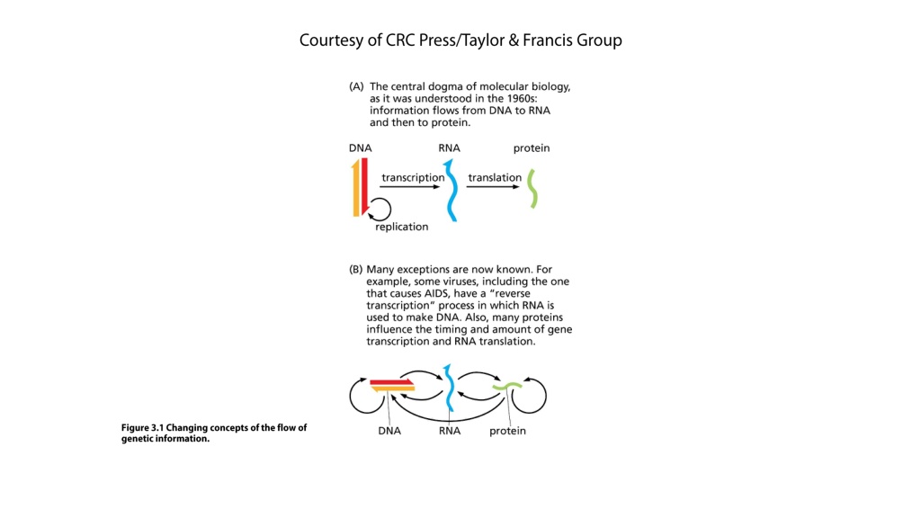

Courtesy of CRC Press/Taylor & Francis Group
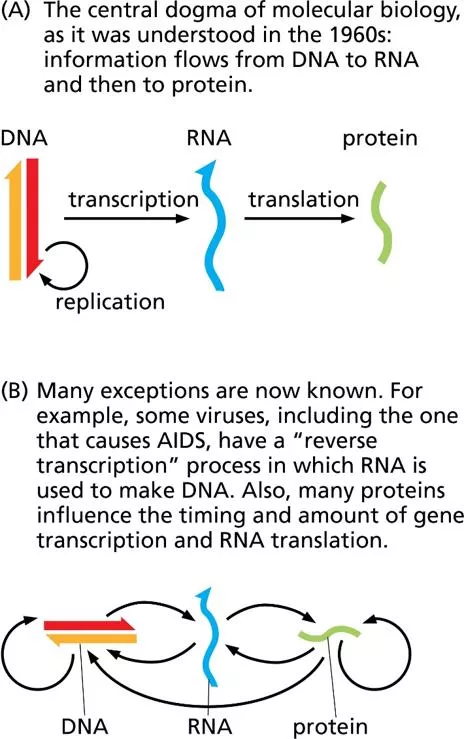
Figure 3.1 Changing concepts of the flow of
genetic information.
Courtesy of CRC Press/Taylor & Francis Group
Figure 3.2 The structure of RNA.
The molecule as a whole is usually single-stranded, but short portions of some RNA molecules can base-pair with other portions of the same molecule.
Courtesy of CRC Press/Taylor & Francis Group
Figure 3.3 The transcription
of DNA into RNA.
Courtesy of CRC Press/Taylor & Francis Group
Figure 3.4 Translation of an mRNA into protein.
The two subunits of a ribosome (shown in light and dark green) bind to the mRNA and move along it, interpreting the genetic code to link
amino acids together into a protein chain. The protein chain elongates as the ribosome moves along, adding one amino acid at a time.
(A)
In the electron micrograph, notice that the mRNA
strand appears as a very thin straight line. Both here and in the schematic diagram,
(B)
notice that multiple ribosomes can be engaged in different stages of translation of the same mRNA at one
time, leading to synthesis of multiple protein molecules.
Courtesy of CRC Press/Taylor & Francis Group
Figure 3.5 The structure of tRNA and its role in translation. (A)
tRNA molecules make internal base pairs to form a cloverleaf structure with the three-letter anticodon at the tip of the middle
“leaf” and a “stem” that becomes attached to the amino acid corresponding to the mRNA codon that the anticodon recognizes. In the example shown here, the mRNA codon UUC matches the
tRNA anticodon AAG, and this tRNA is attached to the amino acid phenylalanine (abbreviated Phe) that is specified by the UUC codon.
(B)
The ribosome (containing ribosomal proteins and
rRNA) holds the tRNA in place on the mRNA. Here, the tRNA cloverleaf is shown in a more schematic structure for simplicity.
Courtesy of CRC Press/Taylor & Francis Group
Table 3.1 The genetic code (coding dictionary)
Courtesy of CRC Press/Taylor & Francis Group
Figure 3.6 Steps in translation.
Courtesy of CRC Press/Taylor & Francis Group
Figure 3.7 Examples of two types of mutations.
The DNA sequences shown are the template strands from which the mRNA codons are copied.
Courtesy of CRC Press/Taylor & Francis Group
Figure 3.8 Human female and male karyotypes.
Courtesy of CRC Press/Taylor & Francis Group
Figure 3.9 The relationship (simplified) between the
SRY
gene and sexual development in humans.
The SRY protein is also known as testisdetermining factor (TDF).
Courtesy of CRC Press/Taylor & Francis Group
Figure 3.10 Inheritance of red-green color blindness,
a sex-linked recessive trait.
Courtesy of CRC Press/Taylor & Francis Group
Figure 3.11 Two variations in human X and Y karyotypes.
Courtesy of CRC Press/Taylor & Francis Group
Figure 3.12 Abnormal meiosis during egg
production, showing how certain chromosomal
variations may arise.
(Nondisjunction can occur
during either the first or second division of meiosis;
in either case, the resulting chromosomal
configurations are as shown here.)
Courtesy of CRC Press/Taylor & Francis Group
Figure 3.13 Down syndrome.
Courtesy of CRC Press/Taylor & Francis Group
Figure 3.14 An example of simple
Mendelian inheritance in humans.
Albinism, a recessive trait, arises in
most cases from matings between
heterozygotes, although it could
also arise from matings between a
heterozygous person and someone
who is homozygous recessive.
Courtesy of CRC Press/Taylor & Francis Group
Figure 3.15 Biochemical
pathways for three inborn errors
of metabolism: phenylketonuria,
alkaptonuria, and albinism.
Courtesy of CRC Press/Taylor & Francis Group
Figure 3.16 Microsatellite DNA marker alleles can be distinguished by the distance they travel during electrophoresis.
Courtesy of CRC Press/Taylor & Francis Group
Figure 3.17 Using DNA markers to establish a linkage between a DNA region and an inherited phenotype.
Courtesy of CRC Press/Taylor & Francis Group
Figure 3.18 Techniques for prenatal detection of genetic conditions.
Courtesy of CRC Press/Taylor & Francis Group
Figure 3.19 The polymerase chain reaction (PCR).
Courtesy of CRC Press/Taylor & Francis Group
Figure 3.20 Two ways of assessing risk for a recessive trait with single-gene, simple Mendelian inheritance.
Slide Note
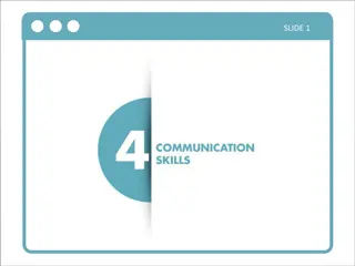
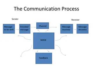
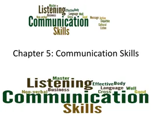
Enhance your communication skills by learning the importance of effective communication in various aspects of life. Discover practical tips and techniques to improve your verbal, non-verbal, and written communication skills.

Download Presentation

Please find below an Image/Link to download the presentation.
The content on the website is provided AS IS for your information and personal use only. It may not be sold, licensed, or shared on other websites without obtaining consent from the author. Download presentation by click this link. If you encounter any issues during the download, it is possible that the publisher has removed the file from their server.
E N D