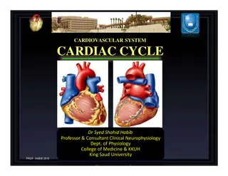
Cardiac Auscultation: Heart Sounds and Murmurs

Cardiovascular Block
Physiology
Heart Sounds and Murmurs

Intended learning outcomes (ILOs)
List the standard positions of stethoscope placement for cardiac auscultation.
Distinguish between the 1
st
, 2
nd
, 3
rd
and 4
th
heart sounds.
Explain physiological splitting of the 2
nd
heart sound and depict the pathophysiology of
fixed and paradoxical splitting of the 2
nd
heart sound.
Define and classify cardiac murmurs and list cause of heart murmurs.
Outline how heart murmurs are described and graded.
Outline the haemodynamic changes and murmurs in conditions of:
Aortic stenosis
Aortic regurgitation
Mitral stenosis
Mitral regurgitation
Mitral valve prolapse
Ventricular septal defect
Patent ductus arteriosus
After reviewing the PowerPoint presentation and the associated learning resources,
the student should be able to:
Learning Resources
Learning Resources
•
Guyton and Hall, Textbook of Medical Physiology; 13
th
Edition;
Unit III-Chapter 9
and Unit IV-Chapter 23.
Linda Costanzo, Physiology, 5
th
Edition; Chapter 4.
•
Ganong’s Review of Medical Physiology; 25
th
Edition; Section
V; Chapter 30.
Events of the cardiac cycle and heart sounds
Events of the cardiac cycle and heart sounds
The heart sounds are detected
over anterior chest wall by:
Auscultation: using
stethoscope
Phonocardiography:
Phonocardiogram is a graphic
recording of the heart sounds.
It involves picking up the sonic
vibrations from the heart
through a highly sensitive
microphone. Such waves are
then converted into electrical
energy and fed into a
galvanometer, where they are
recorded on paper.
Heart sounds
•
Normally, only S
1
and S
2
are
heard with the stethoscope.
•
S
3
and S
4
are detectable by
phonocardiogram.
•
Occasionally S
4
is heard in
normal individuals.
•
S
3
is often heard about one
third of the way through
diastole in children and in many
normal young individuals.
Auscultation of the Heart
•
Generally the sounds produced by each valve is best
heard over a particular region of the chest.
•
Pulmonary area:
•
2
nd
- 3
rd
Lt intercostal space, near the sternum.
•
Aortic area:
•
2
nd
- 3
rd
Rt intercostal space, near the sternum.
•
Mitral area:
•
5
th
Lt intercostal space crossing mid-clavicular
line (apex), or
•
9 cm (2.5-3 in) from sternum.
•
Tricuspid area:
•
Left lower sternal border.
In general both the first and the second heart
sounds can be heard at all areas.
Heart sounds; S
1
•
S
1
is caused by vibrations set
up by the sudden closure of the
AV valves at the start of
ventricular systole
.
•
S
1
is
a slightly prolonged, low
“lub” sound.
•
S
1
has a duration of about
0.15 s and a frequency of 25–45
Hz.
•
S
1
is soft when the heart rate
is low, because the ventricles
are well filled with blood and
the leaflets of the AV valves
float together before systole.
Heart sounds; S
2
•
S
2
is caused by vibrations
associated with closure of the
aortic and pulmonary valves
just after the end of
ventricular systole, and at the
beginning of the isometric
relaxation phase.
•
S
2
is a shorter, high-pitched
“dup” sound.
•
S
2
lasts about 0.12 s, with a
frequency of 50 Hz.
•
S
2
is loud and sharp when
the diastolic pressure in the
aorta or pulmonary artery is
elevated, causing the
respective valves to shut
briskly at the end of systole.
Physiological splitting of S
2
During inspiration, the
aortic valve closes before
pulmonary valve
→
reduplication (physiologic
splitting of S
2
.
The increased venous
return to the right side of
the heart delays closure of
the pulmonary valve. The
right ventricle has more
blood than usual to eject
and it thus takes more time.
No splitting of the second
heart sound is normally
seen during expiration.
Fixed splitting of S
2
Splitting of S
2
is heard both during
inspiration and expiration, with
the aortic valve closing before the
pulmonary valve.
This is heard in cases of ASD.
Wide splitting of S
2
A split in the second heart sound during inspiration may become wider and the split may also
be seen during expiration if:
1.
There is a delay in the closing of the pulmonic valve (as would be seen in right bundle
branch block due to delay in right ventricular depolarization and contraction).
2. The aortic valve closes earlier than normal (this is seen with either mitral regurgitation
or ventricular septal defect).
Paradoxical (reversed) splitting of S
2
Reversed (
paradoxical
) splitting of
the second heart sound is typically
heard during expiration, with the
pulmonary valve closing before the
aortic valve. No splitting is apparent
during inspiration, since the
pulmonary valve is closing earlier
(relative to the aortic valve) than
normal.
This may be caused by the following:
Delayed onset of left ventricular
systole (example:
left bundle
branch block
).
Prolonged left ventricular systole
(examples:
aortic stenosis
,
severe
hypertension
,
left-sided
congestive heart failure
).
Early onset of right ventricular
systole (example:
Wolff-
Parkinson White syndrome
).
Heart sounds; S
3
•
S
3
is heard about one third of
the way through diastole in
children and many normal
young individuals.
•
S
3
coincides with the period
of rapid ventricular filling and
occurs during transition
between rapid filling and slow
filling of ventricle.
•
It is probably due to
vibrations set up by the inrush
of blood.
•
S
3
is a
soft, low-pitched.
•
Duration
≈ 0.05 sec.
•
Best heard at the mitral area.
Heart sounds; S
4
•
S
4
is caused by
oscillations of the
ventricles during atrial
contraction.
•
Recorded during ‘atrial
systole’.
•
S
4
is usually not audible
.. (very low pitch).
•
Duration
≈
0.04 sec.
•
? heard in elderly.
•
Best heard at Mitral
area.
Significance of heart sounds
Important for diagnosis
of abnormal heart
sounds (murmurs)
What makes sounds
and noise in the heart
Closure of the valves:
Atrio-ventricular = (S
1
)
Semilunar = (S
2
)
Increased intra-cardiac
hemodynamics:
Blood striking the left
ventricle, e.g., S
3
, S
4
.
Murmurs:
Increased flow across
normal valves.
Turbulent flow through an
abnormal valve.
Turbulent flow through
septal defect.
Murmurs are longer than heart sounds
Causes of murmurs
Innocent murmurs: common in children and young adults.
Physiological murmurs:
Associate with increased blood flow across normal valves: e.g.
•
Pregnancy
•
Hyperthyroidism
•
Anemia
•
Fever
•
Children
Pathological murmurs:
Turbulent flow through abnormal valves, or septal defect.. ? Congenital: e.g.
The most common abnormalities of the valves are:
Stenosis (narrowing): the valve does not open properly.
Insufficiency (the valve fails to close completely, and hence causing backflow
or leaks of the blood across the insufficient valve. Valvular insufficiency is also
known as Regurgitation or Incompetency).
A combination of Stenosis and Insufficiency.
How to describe heart murmurs
Timing (systolic or diastolic)
Shape
Location
Radiation
Intensity (grade)
Pitch
Quality
1. Timing of heart murmurs
Murmurs are described according to their position in the cardiac cycle:
Systolic murmurs: are further classified into:
Early systolic murmurs.
Mid systolic murmurs (ejection systolic murmurs; ESM).
Late systolic murmurs.
Pansystolic (holosystolic murmurs).
Diastolic: are further classified into:
Early diastolic murmurs.
Mid diastolic murmurs.
Late diastolic murmurs.
Continuous:
2. Shape of heart murmurs
Crescendo (
increasing intensity
).
Decrescendo (
decreasing
intensity
).
Crescendo-decrescendo
(Diamond-shaped); (
increasing
then immediate decreasing
intensity
).
Plateau (uniform); the intensity of
the murmur remains uniform
throughout.
3. Location of maximum intensity of heart murmurs
Determined by the site where the murmur originates
e.g. A, P, T, M listening areas
4. Radiation of heart murmurs
Reflects intensity of the murmur & direction of blood flow
5. Intensity of heart murmurs
Graded on a 6 point according to Levine scale:
Grade 1: Grade I/VI
Grade 2: Grade II/VI
Grade 3: Grade III/VI
Grade 4: Grade IV/VI
Grade 5: Grade V/VI
Grade 6: Grade VI/VI
5. Intensity (grades) of heart murmurs
Graded on a 6 point according to Levine scale:
A thrill is a slight palpable
vibration felt by the hand
over the chest wall
6. Pitch (frequency) of heart murmurs
High, medium, low
7. Quality of heart murmurs
Blowing, harsh, rumbling, musical
8. Others
Variation with respiration
Murmurs increasing with expiration originate with left side (aortic
or mitral) valves, while murmurs increasing in intensity with
inspiration originate with tricuspid or pulmonary valves.
Variation with position of patient
Variation with special maneuvers
Valsalva maneuver decreases the intensity and duraion of most
murmurs.
Pathophysiology of systolic murmurs
Systolic murmurs are derived from harsh &
↑
turbulence in
blood flow during ventricular systole.
They are associated with:
↑
flow across a normal valve.
↑
flow into a dilated great vessel.
↑
flow across a narrowed ventricular outflow tract or
stenosis of the aortic valve or pulmonary valve.
↑
flow across an incompetent AV valve, e.g., mitral and
tricuspid regurgitation.
↑
flow across a VSD.
Mid-systolic murmurs (ejection systolic murmurs; ESM)
Most common kind of heart murmur.
Usually crescendo-decrescendo.
They may be:
Innocent:
In children & young adults.
Physiological:
As result of increased intra-cardiac hemodynamics,, as in
cases of anemia, pregnancy, fever & hyperthyroidism.
Pathological:
Secondary to structural cardiovascular abnormalities, e.g.,
Aortic/pulmonary stenosis, hypertrophic cardiomyopathy &
mitral valve prolapse.
Aortic stenosis; AS
Narrowing of the aortic outflow tract causes
obstruction of flow from the LV into ascending aorta.
This causes a mid-systolic murmur; ejection systolic
murmur.
Best heard at the aortic area, and radiates along
carotid arteries.
The ESM of AS has a crescendo-decrescendo contour
and a gap between the end of the audible sound and
S
2
.
It is harsh, loud, and may have associated thrill,
“ejection click”.
AS may be associated with:
Older age as a result of wear and tear of the
aortic valve in the elderly,
Bicuspid aortic valve,
Scarring of the aortic valve due to rheumatic
fever as a child or young adult.
•
The narrow orifice of the
aortic valve increases
resistance of the aortic valve
and slows the rate at which
SV is ejected.
•
Ventricular systolic
pressure increases to
overcome the increased
resistance of the aortic valve.
•
Thus, there is a pressure
gradient between the left
ventricle and aorta during
ejection.
•
AS may result in concentric
hypertrophy of the LV.
Aortic stenosis; AS
Reversed splitting of S
2
in aortic stenosis
Diagram showing the reverse paradoxical splitting in aortic stenosis with
different levels of severity.
Mitral valve prolapse
In mitral valve prolapse, there is
bulging of one or both mitral
valve leaflets into LA during LV
systole.
This causes a mid- late systolic
murmur.
Best heard at the apex.
Characterized by mid systolic
click.
Mitral valve prolapse may be
seen in ~5% normal population,
asymptomatic, ? sudden death.
Pan-systolic murmurs (holosystolic murmurs)
Pan-systolic murmurs are pathological murmur.
They begin immediately with S1 & continue up to S2.
Heard with:
Mitral/tricuspid regurgitation.
Ventricular septal defect (VSD).
Mitral regurgitation; MR
An incompetent mitral valve allows blood to
regurgitate from the left ventricle to the left
atrium throughout ventricular systole. Thus,
•
Left atrial volume and pressure are
increased during ventricular systole.
•
Left ventricular volume and pressure are
increased during diastole, but there is NO
pressure gradient between the LA and the
LV.
This results in a holosystolic murmur.
The sound is of reasonably constant intensity
throughout the ejection period.
Best heard at apex, radiates to left axilla.
Soft, high-pitched, blowing.
May be associated with MV prolapse, MV
myxomatous degeneration, MI, rheumatic heart
disease, cardiomyopathy, endocarditis.
left ventricular
pressure
left atrial
pressure
S
4
S
1
S
2
S
3
Ventricular septal defect; VSD
Summary of systolic murmurs
Diastolic murmurs almost always indicate heart disease.
There are two basic types:
Early decrescendo diastolic murmurs:
This murmur signifies regurgitant flow through an
incompetent semilunar valve, e.g., aortic/pulmonary
regurgitation (incompetency).
Rumbling mid- or late diastole diastolic murmurs:
This murmur suggests stenosis of an AV valve, e.g.,
mitral/tricuspid stenosis.
Aortic regurgitation; AR
•
The aortic valve does not close properly at the
beginning of diastole.
•
As a result, there is retrograde flow of blood from
the aorta into the ventricle during diastole.
•
The amount of the blood regurgitated into the left
ventricle may be as much as 60-70% of the amount
ejected during systole.
•
Thus, there is:
•
Decreased aortic diastolic pressure.
•
increased left ventricular and aortic systolic
pressures.
•
Increased aortic pulse pressure.
•
AR causes an early diastolic murmurs; decrescendo
murmur.
•
Best heard at the 2
nd
– 4
th
left intercostal space
with the patient sitting up, leaning forward, at end
expiration.
•
High pitched, loud blowing. It wanes with time as
aortic pressure falls.
•
May be associated with aortic root degeneration,
rheumatic heart disease, VSD, w/aortic valve
prolapse (kids).
Mitral stenosis; MS
•
Narrowing of the mitral valve orifice
impairs emptying of the left atrium
into the left ventricle during diastole.
•
Left a
trial pressure greatly exceeds
left ventricular pressure when the
stenotic valve is open. T
his generates a
pressure gradient between the left
atrium and the left ventricle during
filling.
•
Thus, pressure and volume can be
dramatically elevated in the left
atrium.
•
However, in most cases of MS, LV
pressure curve is normal, and similarly,
the
aortic pressure curve is also
normal.
Mitral stenosis; MS
•
MS results in a diastolic murmur.
•
The murmur is
often heard with
an opening snap (OS) This gives the
murmur a decreschendo-
creschendo profile.
•
It begins early after the OS. Its
timing is thus mid-diastolic or pre-
systolic.
•
It is
best heard at the apex.
•
It is
a low-frequency (
low pitched)
blowing sound and thus
heard with
the bell of the stethoscope.
•
MS may be associated with
rheumatic fever.
Summary of diastolic murmurs
Failure of closure of the ductus arteriosus between
pulmonary artery & aorta results in a continuous
murmur.
Best heard at upper left sternal border.
Machine-like.
May be associated with left to right shunt, cyanosis.
Intended learning outcomes (ILOs)
List the standard positions of stethoscope placement for cardiac auscultation.
Distinguish between the 1
st
, 2
nd
, 3
rd
and 4
th
heart sounds.
Explain physiological splitting of the 2
nd
heart sound and depict the pathophysiology
of fixed and paradoxical splitting of the 2
nd
heart sound.
Define and classify cardiac murmurs and list cause of heart murmurs.
Outline how heart murmurs are described and graded.
Outline the haemodynamic changes and murmurs in conditions of:
Aortic stenosis
Aortic regurgitation
Mitral stenosis
Mitral regurgitation
Mitral valve prolapse
Ventricular septal defect
Patent ductus arteriosus
After reviewing the PowerPoint presentation and the associated learning resources,
the student should be able to:
Thank You
Thank You
Delve into the intricacies of cardiac auscultation with a focus on heart sounds and murmurs. Learn about stethoscope placement, heart sound differentiation, physiological splitting, cardiac murmur classification, and hemodynamic changes in various heart conditions. Explore essential learning outcomes and resources to deepen your understanding.

Download Presentation

Please find below an Image/Link to download the presentation.
The content on the website is provided AS IS for your information and personal use only. It may not be sold, licensed, or shared on other websites without obtaining consent from the author.If you encounter any issues during the download, it is possible that the publisher has removed the file from their server.
You are allowed to download the files provided on this website for personal or commercial use, subject to the condition that they are used lawfully. All files are the property of their respective owners.
The content on the website is provided AS IS for your information and personal use only. It may not be sold, licensed, or shared on other websites without obtaining consent from the author.
E N D
Presentation Transcript
Cardiovascular Block Physiology Heart Sounds and Murmurs
Intended learning outcomes (ILOs) After reviewing the PowerPoint presentation and the associated learning resources, the student should be able to: List the standard positions of stethoscope placement for cardiac auscultation. Distinguish between the 1st, 2nd, 3rd and 4th heart sounds. Explain physiological splitting of the 2nd heart sound and depict the pathophysiology of fixed and paradoxical splitting of the 2nd heart sound. Define and classify cardiac murmurs and list cause of heart murmurs. Outline how heart murmurs are described and graded. Outline the haemodynamic changes and murmurs in conditions of: Aortic stenosis Aortic regurgitation Mitral stenosis Mitral regurgitation Mitral valve prolapse Ventricular septal defect Patent ductus arteriosus
Learning Resources Guyton and Hall, Textbook of Medical Physiology; 13th Edition; Unit III-Chapter 9 and Unit IV-Chapter 23. Linda Costanzo, Physiology, 5th Edition; Chapter 4. Ganong s Review of Medical Physiology; 25th Edition; Section V; Chapter 30.
Events of the cardiac cycle and heart sounds The heart sounds are detected over anterior chest wall by: Auscultation: using stethoscope Phonocardiography: Phonocardiogram is a graphic recording of the heart sounds. It involves picking up the sonic vibrations from the heart through a highly sensitive microphone. Such waves are then converted into electrical energy and fed into a galvanometer, where they are recorded on paper.
Heart sounds Normally, only S1 and S2 are heard with the stethoscope. S3 and S4 are detectable by phonocardiogram. Occasionally S4 is heard in normal individuals. S3 is often heard about one third of the way through diastole in children and in many normal young individuals.
Auscultation of the Heart Generally the sounds produced by each valve is best heard over a particular region of the chest. Pulmonary area: 2nd - 3rd Lt intercostal space, near the sternum. Aortic area: 2nd - 3rd Rt intercostal space, near the sternum. Mitral area: 5th Lt intercostal space crossing mid-clavicular line (apex), or 9 cm (2.5-3 in) from sternum. Tricuspid area: Left lower sternal border. In general both the first and the second heart sounds can be heard at all areas.
S1 is caused by vibrations set up by the sudden closure of the AV valves at the start of ventricular systole. Heart sounds; S1 S1 is a slightly prolonged, low lub sound. S1 has a duration of about 0.15 s and a frequency of 25 45 Hz. S1 is soft when the heart rate is low, because the ventricles are well filled with blood and the leaflets of the AV valves float together before systole.
S2 is caused by vibrations associated with closure of the aortic and pulmonary valves just after the end of ventricular systole, and at the beginning of the isometric relaxation phase. Heart sounds; S2 S2 is a shorter, high-pitched dup sound. S2 lasts about 0.12 s, with a frequency of 50 Hz. S2 is loud and sharp when the diastolic pressure in the aorta or pulmonary artery is elevated, causing the respective valves to shut briskly at the end of systole.
Physiological splitting of S2 During inspiration, the aortic valve closes before pulmonary valve reduplication (physiologic splitting of S2. The increased venous return to the right side of the heart delays closure of the pulmonary valve. The right ventricle has more blood than usual to eject and it thus takes more time. No splitting of the second heart sound is normally seen during expiration.
Fixed splitting of S2 Splitting of S2 is heard both during inspiration and expiration, with the aortic valve closing before the pulmonary valve. This is heard in cases of ASD.
Wide splitting of S2 A split in the second heart sound during inspiration may become wider and the split may also be seen during expiration if: 1.There is a delay in the closing of the pulmonic valve (as would be seen in right bundle branch block due to delay in right ventricular depolarization and contraction). 2. The aortic valve closes earlier than normal (this is seen with either mitral regurgitation or ventricular septal defect).
Reversed (paradoxical) splitting of the second heart sound is typically heard during expiration, with the pulmonary valve closing before the aortic valve. No splitting is apparent during inspiration, since the pulmonary valve is closing earlier (relative to the aortic valve) than normal. Paradoxical (reversed) splitting of S2 This may be caused by the following: Delayed onset of left ventricular systole (example: left bundle branch block). Prolonged left ventricular systole (examples: aortic stenosis, severe hypertension, left-sided congestive heart failure). Early onset of right ventricular systole (example: Wolff- Parkinson White syndrome).
S3 is heard about one third of the way through diastole in children and many normal young individuals. Heart sounds; S3 S3 coincides with the period of rapid ventricular filling and occurs during transition between rapid filling and slow filling of ventricle. It is probably due to vibrations set up by the inrush of blood. S3 is a soft, low-pitched. Duration 0.05 sec. Best heard at the mitral area.
S4 is caused by oscillations of the ventricles during atrial contraction. Heart sounds; S4 Recorded during atrial systole . S4 is usually not audible .. (very low pitch). Duration 0.04 sec. ? heard in elderly. Best heard at Mitral area.
Significance of heart sounds Important for diagnosis of abnormal heart sounds (murmurs)
Closure of the valves: What makes sounds and noise in the heart Atrio-ventricular = (S1) Semilunar = (S2) Increased intra-cardiac hemodynamics: Blood striking the left ventricle, e.g., S3, S4. Murmurs: Increased flow across normal valves. Turbulent flow through an abnormal valve. Turbulent flow through septal defect. Murmurs are longer than heart sounds
Causes of murmurs Innocent murmurs: common in children and young adults. Physiological murmurs: Associate with increased blood flow across normal valves: e.g. Pregnancy Hyperthyroidism Anemia Fever Children Pathological murmurs: Turbulent flow through abnormal valves, or septal defect.. ? Congenital: e.g. The most common abnormalities of the valves are: Stenosis (narrowing): the valve does not open properly. Insufficiency (the valve fails to close completely, and hence causing backflow or leaks of the blood across the insufficient valve. Valvular insufficiency is also known as Regurgitation or Incompetency). A combination of Stenosis and Insufficiency.
How to describe heart murmurs Timing (systolic or diastolic) Shape Location Radiation Intensity (grade) Pitch Quality
1. Timing of heart murmurs Murmurs are described according to their position in the cardiac cycle: Systolic murmurs: are further classified into: Early systolic murmurs. Mid systolic murmurs (ejection systolic murmurs; ESM). Late systolic murmurs. Pansystolic (holosystolic murmurs). Diastolic: are further classified into: Early diastolic murmurs. Mid diastolic murmurs. Late diastolic murmurs. Continuous:
2. Shape of heart murmurs Crescendo (increasing intensity). Decrescendo (decreasing intensity). Crescendo-decrescendo (Diamond-shaped); (increasing then immediate decreasing intensity). Plateau (uniform); the intensity of the murmur remains uniform throughout.
3. Location of maximum intensity of heart murmurs Determined by the site where the murmur originates e.g. A, P, T, M listening areas 4. Radiation of heart murmurs Reflects intensity of the murmur & direction of blood flow
5. Intensity of heart murmurs Graded on a 6 point according to Levine scale: Grade 1: Grade I/VI Grade 2: Grade II/VI Grade 3: Grade III/VI Grade 4: Grade IV/VI Grade 5: Grade V/VI Grade 6: Grade VI/VI
5. Intensity (grades) of heart murmurs Graded on a 6 point according to Levine scale: A thrill is a slight palpable vibration felt by the hand over the chest wall
6. Pitch (frequency) of heart murmurs High, medium, low 7. Quality of heart murmurs Blowing, harsh, rumbling, musical 8. Others Variation with respiration Murmurs increasing with expiration originate with left side (aortic or mitral) valves, while murmurs increasing in intensity with inspiration originate with tricuspid or pulmonary valves. Variation with position of patient Variation with special maneuvers Valsalva maneuver decreases the intensity and duraion of most murmurs.
Pathophysiology of systolic murmurs Systolic murmurs are derived from harsh & turbulence in blood flow during ventricular systole. They are associated with: flow across a normal valve. flow into a dilated great vessel. flow across a narrowed ventricular outflow tract or stenosis of the aortic valve or pulmonary valve. flow across an incompetent AV valve, e.g., mitral and tricuspid regurgitation. flow across a VSD.
Mid-systolic murmurs (ejection systolic murmurs; ESM) Most common kind of heart murmur. Usually crescendo-decrescendo. They may be: Innocent: In children & young adults. Physiological: As result of increased intra-cardiac hemodynamics,, as in cases of anemia, pregnancy, fever & hyperthyroidism. Pathological: Secondary to structural cardiovascular abnormalities, e.g., Aortic/pulmonary stenosis, hypertrophic cardiomyopathy & mitral valve prolapse.
Narrowing of the aortic outflow tract causes obstruction of flow from the LV into ascending aorta. Aortic stenosis; AS This causes a mid-systolic murmur; ejection systolic murmur. Best heard at the aortic area, and radiates along carotid arteries. The ESM of AS has a crescendo-decrescendo contour and a gap between the end of the audible sound and S2. It is harsh, loud, and may have associated thrill, ejection click . AS may be associated with: Older age as a result of wear and tear of the aortic valve in the elderly, Bicuspid aortic valve, Scarring of the aortic valve due to rheumatic fever as a child or young adult.
Aortic stenosis; AS The narrow orifice of the aortic valve increases resistance of the aortic valve and slows the rate at which SV is ejected. Ventricular systolic pressure increases to overcome the increased resistance of the aortic valve. Thus, there is a pressure gradient between the left ventricle and aorta during ejection. AS may result in concentric hypertrophy of the LV.
Reversed splitting of S2 in aortic stenosis Diagram showing the reverse paradoxical splitting in aortic stenosis with different levels of severity.
Mitral valve prolapse In mitral valve prolapse, there is bulging of one or both mitral valve leaflets into LA during LV systole. This causes a mid- late systolic murmur. Best heard at the apex. Characterized by mid systolic click. Mitral valve prolapse may be seen in ~5% normal population, asymptomatic, ? sudden death.
Pan-systolic murmurs (holosystolic murmurs) Pan-systolic murmurs are pathological murmur. They begin immediately with S1 & continue up to S2. Heard with: Mitral/tricuspid regurgitation. Ventricular septal defect (VSD).
An incompetent mitral valve allows blood to regurgitate from the left ventricle to the left atrium throughout ventricular systole. Thus, Left atrial volume and pressure are increased during ventricular systole. Left ventricular volume and pressure are increased during diastole, but there is NO pressure gradient between the LA and the LV. Mitral regurgitation; MR This results in a holosystolic murmur. The sound is of reasonably constant intensity throughout the ejection period. left ventricular pressure Best heard at apex, radiates to left axilla. Soft, high-pitched, blowing. left atrial pressure May be associated with MV prolapse, MV myxomatous degeneration, MI, rheumatic heart disease, cardiomyopathy, endocarditis. S4 S1 S2 S3
Diastolic murmurs Diastolic murmurs almost always indicate heart disease. There are two basic types: Early decrescendo diastolic murmurs: This murmur signifies regurgitant flow through an incompetent semilunar valve, e.g., aortic/pulmonary regurgitation (incompetency). Rumbling mid- or late diastole diastolic murmurs: This murmur suggests stenosis of an AV valve, e.g., mitral/tricuspid stenosis.
Aortic regurgitation; AR The aortic valve does not close properly at the beginning of diastole. As a result, there is retrograde flow of blood from the aorta into the ventricle during diastole. The amount of the blood regurgitated into the left ventricle may be as much as 60-70% of the amount ejected during systole. Thus, there is: Decreased aortic diastolic pressure. increased left ventricular and aortic systolic pressures. Increased aortic pulse pressure. AR causes an early diastolic murmurs; decrescendo murmur. Best heard at the 2nd 4th left intercostal space with the patient sitting up, leaning forward, at end expiration. High pitched, loud blowing. It wanes with time as aortic pressure falls. May be associated with aortic root degeneration, rheumatic heart disease, VSD, w/aortic valve prolapse (kids).
Mitral stenosis; MS Narrowing of the mitral valve orifice impairs emptying of the left atrium into the left ventricle during diastole. Left atrial pressure greatly exceeds left ventricular pressure when the stenotic valve is open. This generates a pressure gradient between the left atrium and the left ventricle during filling. Thus, pressure and volume can be dramatically elevated in the left atrium. However, in most cases of MS, LV pressure curve is normal, and similarly, the aortic pressure curve is also normal.
Mitral stenosis; MS MS results in a diastolic murmur. The murmur is often heard with an opening snap (OS) This gives the murmur a decreschendo- creschendo profile. It begins early after the OS. Its timing is thus mid-diastolic or pre- systolic. It is best heard at the apex. It is a low-frequency (low pitched) blowing sound and thus heard with the bell of the stethoscope. MS may be associated with rheumatic fever.
Continuous murmurs Patent ductus arteriosus; PDA Failure of closure of the ductus arteriosus between pulmonary artery & aorta results in a continuous murmur. Best heard at upper left sternal border. Machine-like. May be associated with left to right shunt, cyanosis.
Intended learning outcomes (ILOs) After reviewing the PowerPoint presentation and the associated learning resources, the student should be able to: List the standard positions of stethoscope placement for cardiac auscultation. Distinguish between the 1st, 2nd, 3rd and 4th heart sounds. Explain physiological splitting of the 2nd heart sound and depict the pathophysiology of fixed and paradoxical splitting of the 2nd heart sound. Define and classify cardiac murmurs and list cause of heart murmurs. Outline how heart murmurs are described and graded. Outline the haemodynamic changes and murmurs in conditions of: Aortic stenosis Aortic regurgitation Mitral stenosis Mitral regurgitation Mitral valve prolapse Ventricular septal defect Patent ductus arteriosus

")


 splitting of")


 of heart murmurs")
 of heart murmurs")

")


")


")