Assessment and Diagnostic Tests in Musculoskeletal System Pain
Pain in musculoskeletal conditions varies, such as bone pain described as dull and deep, muscular pain as soreness, fracture pain as sharp, and joint pain worsens with movement. Specific assessments by nurses include checking body alignment, joint symmetry, signs of inflammation, pressure sources, and skin tension. Questions to ask about altered sensations like paresthesias involve abnormal feelings, comparison with unaffected areas, onset, worsening, and associated pain. Factors affecting musculoskeletal health include occupation, exercise, alcohol and tobacco use, diet, health conditions, genetics, and trauma history.
Download Presentation

Please find below an Image/Link to download the presentation.
The content on the website is provided AS IS for your information and personal use only. It may not be sold, licensed, or shared on other websites without obtaining consent from the author.If you encounter any issues during the download, it is possible that the publisher has removed the file from their server.
You are allowed to download the files provided on this website for personal or commercial use, subject to the condition that they are used lawfully. All files are the property of their respective owners.
The content on the website is provided AS IS for your information and personal use only. It may not be sold, licensed, or shared on other websites without obtaining consent from the author.
E N D
Presentation Transcript
University of Basrah College of Nursing Fundamentals of Nursing Department Second stage Adult Nursing (II) Adult Nursing (II) 2 2nd Stage nd Stage (Theory) ) Lecture Lecture 1 1 (Theory Assessment and diagnostic test in musculoskeletal system - Dr. Dr. Mohamad Mohamad Abdulridha Abdulridha - M.B.CH.B F.I.C.M.S (ORTHO) Orthopedic Surgeon M.B.CH.B F.I.C.M.S (ORTHO) Orthopedic Surgeon -
Pain most patients with diseases and traumatic conditions or disorders of the muscles, bones, and joints experience pain. Pain 1.Bone pain is typically described as a dull, deep ache that is boring in nature. This pain is not typically related to movement and may interfere with sleep. 2. Muscular pain is described as soreness or aching and is referred to as muscle cramps
3. Fracture pain is sharp and piercing and is relieved by immobilization. Sharp pain may also result from bone infection with muscle spasm or pressure on a sensory nerve. 4.Joint pain is felt around or in the joint and typically worsens with movement
Specific assessments that the nurse should make regarding the pain include the following: Is the body in proper alignment? Are the joints symmetrical or are bony deformities present? Is there any inflammation or arthritis, swelling, warmth, tenderness, or redness? Is there pressure from traction, bed linens, a cast, or other appliances? Is there tension on the skin at a pin site?
Paresthesias nerves or by circulatory impairment Questions that the nurse should ask regarding altered sensations include the following: 1.Is the patient experiencing abnormal sensations, such as burning, tingling, or numbness? 2.If the abnormal sensation involves an extremity, how does this feeling compare to sensation in the unaffected extremity? 3.When did the condition begin? Is it getting worse? 4.Does the patient also have pain Paresthesias may be caused by pressure on
1.occupation (e.g., does the patients work require physical activity or heavy lifting?), 2. exercise patterns 3. alcohol consumption, tobacco use, 4.dietary intake (e.g., calcium and vitamin D). 5.Concurrent health conditions (e.g., diabetes, heart disease, chronic obstructive pulmonary disease, infection, and preexisting disability) 6. familial or genetic abnormalities 7. history of trauma or injury
1.Assess for other similarly affected family members in the past three generations. 2.Assess for the presence of other related genetic conditions (e.g., hematologic, cardiac, integumentary conditions). 3.Determine the age at onset (e.g., fractures present at birth such as osteogenesis imperfecta, hip dislocation present at birth in DDH, or early- onset osteoporosis).
1.Assess stature for general screening purposes (unusually short stature may be related to achondroplasia; unusually tall stature may be related to Marfan syndrome). 2. Assess for disease-specific skeletal findings (e.g., pectusexcavatum, scoliosis, long fingers [Marfan syndrome], 3.Assessment findings that could indicate a genetic musculoskeletal disorder include:e.g Enlarged hands or feet Flat feet or highly arched feet
Techniques of inspection and palpation are used to evaluate the patient s posture, gait, bone integrity, joint function, and muscle strength and size. In addition, assessing the skin and neurovascular status is an important part of a complete musculoskeletal assessment.
The normal curvature of the spine is convex through the thoracic portion and concave through the cervical and lumbar portions. kyphosis curvature of the thoracic spine (e.g., arthritis or disc degeneration), fractures related to osteoporosis, and injury or trauma 2. lordosis curvature of the lumbar spine e.g pregnancy 3. scoliosis the spine can be congenital, idiopathic e,g muscular dystrophy kyphosis, which is an increased forward lordosis, or swayback, an exaggerated scoliosis, which is a lateral curving deviation of
Gait is assessed by having the patient walk away from the examiner for a short distance. e.g unsteadiness or irregular movements, limping.
The bony skeleton is assessed for deformities and alignment. Symmetric parts of the body, such as extremities, are compared. Abnormal bony growths due to bone tumors may be observed. Shortened extremities, amputations, and body parts that are not in anatomic alignment are noted.
The articular system is evaluated by noting range of motion, deformity, stability, tenderness, swelling, effusion and nodular formation. Rhoumatoid arthritis soft nodule at long extensor tendon Gout hard nodule neat the joint
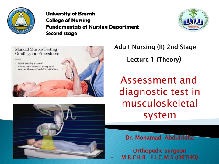
The muscular system is assessed by noting muscular strength and coordination, the size of individual muscles, and the patient s ability to change position. Muscle tone: palpating the muscle while passively moving the relaxed extremity Muscle strength: patient perform certain maneuvers with and without added resistance. Muscle girth: Measurements are taken at the maximum circumference of the extremity
X-ray to determine bone density, texture, erosion, and changes in bone relationships, Multiple xrays, with multiple views (e.g., anterior, posterior, lateral), are needed . Serial x-rays may be indicated to determine the status of the healing process. RELATED NURSING CARE :No special preparation needed for standard x-rays. If contrast medium is used, assess for allergy to shellfish, iodine, or contrast medium used in previous tests. If allergy is present, test will not be performed. Ask women if they are pregnant; x-rays should be avoided during the first trimester
may be performed with or without the use of oral or intravenous (IV) contrast agents, It may be used to visualize and assess tumors, severe trauma to the chest, abdomen, pelvis, head, or spinal cord. It is also used to identify the location and extent of fractures in areas that are difficult to evaluate (e.g., acetabulum) RELATED NURSING CARE No special preparation is needed.
PURPOSE AND DESCRIPTION Used in diagnosis and evaluation of avascular necrosis, osteomyelitis, tumors, disk abnormalities, and tears in ligament or cartilage. Uses radio waves and magnetic fields; Gadolinium dye may be injected to increase visualization of bony or muscular structures. Close mri and open type RELATED NURSING CARE Assess for metallic implants or metal on clothing (metallic implants, such as clips on aneurysms, pacemakers, or shrapnel, will prohibit having an MRI)
PURPOSE AND DESCRIPTION Degree of uptake of a radioisotope (based on blood supply to bone) is measured with a Geiger counter and recorded on paper. Uptake is increased in osteomyelitis, osteoporosis, cancers of the bone, and in some fractures. Uptake is decreased in avascular necrosis. Is a bone-seeking radioisotope is injected IV, the scan is preformed 2-3 hours after the injection. RELATED NURSING CARE No special preparation is needed; tell client to increase oral fluids after the test to aid in excretion of the radioisotope. allergy to the radioisotope and for any condition that would contraindicate performing the procedure (pregnancy).
Dual energy x-ray absorptiometry (DEXA) Quantitative ultrasound (QUS) Bone mineral density (BMD) Bone absorptiometry PURPOSE AND DESCRIPTION Bone density examinations are done to evaluate bone mineral density and to evaluate degree of osteoporosis. DEXA can calculate the size and thickness of bone. Osteoporosis is diagnosed if the peak bone mass level is below >2.5 standard deviations. Normal Value: 1 standard deviation below peak bone mass. RELATED NURSING CARE No special preparation is needed. Assess for previous fractures, which may increase bone density
Allow direct visualization of a joint to diagnose joint disorders, carried out in operating room. Injection of a local anesthetic into the joint is used, a large-bore needle is inserted, and the joint is distended with saline. The arthroscope is introduced and structure, synovium, and articular surfaces are visualized. In addition, fluid may be drained from the joint and tissue removed for biopsy.
RELATED NURSING CARE If general anesthesia is used, client is NPO after midnight. Following the procedure, assess for bleeding and swelling, apply ice to the area if prescribed, and teach client to avoid excessive use of the joint for 2 to 3 days.
1.Infection, 2. Hemarthrosis, 3. Neurovascular compromise, 4. Thrombophlebitis, 5. Stiffness, 6. Effusion, 7. Adhesions, 8. Delayed wound healing
1. Wrapped the joint with a compression dressing to control swelling 2. Ice may be applied to control edema and discomfort 3. The joint is kept extended and elevated to reduce swelling 4. Monitor neurovascular function: Circulation: Color, Temperature, Capillary refill Motion: Weakness, Paralysis Sensation: Paresthesia, Unrelenting pain, Pain on passive stretch, Absence of feeling 5. Administer analgesics to control discomfort 6. Explain when the patient can resume activity and what weight-bearing limits
PURPOSE AND DESCRIPTION Done to obtain synovial fluid from a joint for diagnosis (such as infections) or to remove excess fluid. A needle is inserted through the joint capsule and fluid is aspirated. RELATED NURSING CARE No special preparation is needed. Apply compression dressing and assess for bleeding and leakage of fluid following the procedure.
An EMG measures the electrical activity of skeletal muscles at rest and during contraction; this information is useful in diagnosing neuromuscular diseases. Needle electrodes are inserted into skeletal muscle (as on the legs) and electrical activity can be heard, viewed on an oscilloscope, and recorded on graph paper. Normally, there is no electrical activity at rest
Biopsy may be performed to determine the structure and composition of bone marrow, bone, muscle, or synovium to help diagnose specific diseases. It involves excising a sample of tissue that can be analyzed microscopically to determine cell morphology and tissue abnormalities.
1. Know assessment of musculoskeletal system (health history and physical assessment ) 2. Know diagnostic test used in musculoskeletal system
A nurse is caring for a patient whose cancer metastasis has resulted in bone pain. Which ofthe following are typical characteristics of bone pain? A) A dull, deep ache that is boring in nature B) Soreness or aching that may include cramping C) Sharp, piercing pain that is relieved by immobilization D) Spastic or sharp pain that radiates
A nurse on the orthopedic unit is assessing a patients peroneal nerve. The nurse will perform this assessment by doing which of the following actions? A) Pricking the skin between the great and second toe B) Stroking the skin on the sole of the patients foot C) Pinching the skin between the thumb and index finger D) Stroking the distal fat pad of the small finger
The nurse Instruct the patient regarding the proper methods to control edema and pain after closed upper limbs fracture A. Elevate extremity to heart level B. Elevate the extremity below the heart level C. Decrease the fluid intake D. Increase the fluid intake E. Put the patient in a lateral position
Which one of the following is a local clinical manifestation of fractures? A. Hard lumps around the joint B. Anemia C. Ecchymosis D. Cold over the injured area E. Erythema
After bone scanning, the nurse encourages the patient to drink plenty of fluid to: A. Prevent dehydration B. Eliminate isotope C. Keep healthy joints D. Reduce discomfort E. Prevent fluid imbalance
Textbook of medical-surgical nursing Description: 14th edition. | Philadelphia : Wolters Kluwer, [2018], ISBN 9781496355157 (2-volume American edition)
