Ascarids: Toxocara Canis and Toxocara Cati
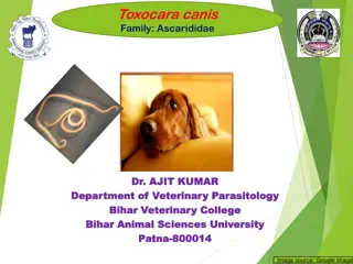
Ascarids, also known as roundworms, are large parasitic worms that specifically infect different animal hosts. Toxocara canis affects dogs, while Toxocara cati affects cats. These worms have distinct characteristics and can cause health issues like pot-belly in young animals. Understanding their life cycle, route of infection, and impact can help in prevention and treatment.
Download Presentation

Please find below an Image/Link to download the presentation.
The content on the website is provided AS IS for your information and personal use only. It may not be sold, licensed, or shared on other websites without obtaining consent from the author.If you encounter any issues during the download, it is possible that the publisher has removed the file from their server.
You are allowed to download the files provided on this website for personal or commercial use, subject to the condition that they are used lawfully. All files are the property of their respective owners.
The content on the website is provided AS IS for your information and personal use only. It may not be sold, licensed, or shared on other websites without obtaining consent from the author.
E N D
Presentation Transcript
VMP 930 lecture 20a Order Ascaridia Toxocara canis and Toxocara cati
Order ASCARIDIDA (Ascarids) Adult worms in small intestine are large! mouth surrounded by 3 fleshy lips Host-specific, adult stage Toxocara canis in dogs Toxocara cati in cats Ascaris suum in pigs Parascaris in horses Baylisascaris in raccoons
Order ASCARIDIDA (Ascarids) Eggs are thick-walled (highly resistant), distinctive, contain a single cell. Can persist in soil for years! Toxocara egg Ascaris suum egg thick rough shell thick rough shell
Pot-belly typical of large worm- burden in young
Toxocara canis very common parasitic problem in dogs posterior end of female thick, white, large 50-180 mm adult worms anterior end: cervical alae are expanded i.e. arrowhead worms cervical alae at anterior end
Toxocara canis Life Cycle Adult worms live in the small intestine Female worms produce a large number of eggs 1 cell develops into an infective larva within the eggshell in ~4 weeks Egg is ingested egg from fresh feces L3 hatching (in lab experiment)
Toxocara canis: Route of Infection Direct: Ingestion of infective egg containing larva. Ascarid L3 are infective when they hatch from egg in the small intestine. Indirect: Ingestion of paratenic host which contains larva
Toxocara canis: Route of Infection Larva from ingested eggs can take one of 2 routes depending on host age. 1. tracheal migration results in development of adult worms in the small intestine mostly in puppies under 3 months of age. 2. somatic migration results in infective larvae becoming arrested in tissue of mature dogs.
Toxocara canis: Route of Infection Larvae from ingestion of a paratenic host containing infective larvae in its tissues. Usually results in adult worm development without larval migration outside of the intestinal mucosa in a dog of any age.
Toxocara canis: Route of Infection Reactivation of arrested larvae in tissues of the mother dog during pregnancy Leads to transuterine infection of the puppies. Puppies have patent infections as soon as 3 weeks after birth. Transuterine infection does NOT occur in cats.
Toxocara canis: Prepatent Period Prepatent period 3 - 5 weeks ~5 weeks if infection starts with egg stage ~3 weeks if in utero infection ~3 weeks if ingestion of paratenic host
Pathogenesis & Clinical Signs Gastroenteritis Inflammation hypersensitivity Abdominal pain, pot-bellied, poor coat Fetid, mucoid diarrhea Respiratory signs are rare
Diagnosis Observation of Clinical signs Fecal can be negative unless > 3-5 weeks old Adult worms in vomit or in feces
Treatment & Control Adults and larvae in intestines - many drugs effective Arrested larvae - drugs less effective Deworm dam (timing of monthly prophylaxis) Deworm newborn puppies start at 2-3 weeks till monthly heartworm preventative started Environment Clean surfaces, dispose of feces wash hands thoroughly after handling
Toxocara cati small intestine of cats similar to T. canis but Cervical alae More prominent cervical alae Adult cats often get patent infections by ingestion of paratenic (transport) hosts Transmission to kittens: transmammary transmission is important but queen must have been infected during pregnancy no transuterine transmission PPP ~ 8weeks from ingested egg
Toxocara cati Treatment of kittens from 6-8 weeks of age Pyrantel, fenbendazole, ivermectin Visceral larva migrans in humans Avoid contaminated sand boxes and gardens
Zoonosis: Visceral larva migrans Migration of larvae in tissues of aberrant host (ex. Humans) ingestion of infective egg Beware cat frequented sand boxes and gardens Ocular larvae migrans children with granulomatous reaction to larvae in eye 14% of people have antibodies to Toxocara
Toxascaris leonina <1% prevalence - dogs, cats eggs oval, smooth shell Infection ingestion of eggs or infected paratenic host only PPP 8-10 weeks mild clinical signs no visceral larva migrans
Baylisascaris procyonis Baylisascaris egg Raccoons May infect dogs exposed to raccoon latrines Granular shell surface Very aggressive visceral larva migrans, neurological signs
VMP 930 lecture 20b Parascaris equorum in horses
Parascaris equorum The Ascarid of horses Common on breeding farms Target of routine deworming starting at 2-3 months of age. Why at this age? small intestine of young horses < 2 yrs This figure shows a ruptured small intestine that occurred shortly after a first-time deworming of a 4-month-old foal. adult worms are large, thick-bodied
Parascaris equorum Transmission Ingestion of an infective egg is the ONLY route of infection Fresh passed eggs require 10 14 days to develop infective larva within eggs.
Parascaris equorum Life Cycle Egg containing infective larva (takes ~10-14 days) Larvae migrate to liver, lungs, coughed up and swallowed, returning to the small intestine in 2-4 weeks after ingestion Prepatent period ~ 80 days
Parascaris equorum Pathogenesis Respiratory problems congestion due to parasite antigens/allergy migration of larvae Intestinal problems enteritis, obstruction, perforation
Parascaris equorum Clinical signs: diarrhea odorous potbellied appearance rough hair coat respiratory signs Suboptimal Growth
Parascaris equorum Treatment & Control: clean environment adult worms are very fecund eggs are very resistant and sticky! mare: clean teats & udder deworm foal at 2-3 months, q 2 months till ~1 year of age. Drug resistance to avermectins is common.
Parascaris equorum If you suspect a heavy infection, do NOT use a potent drug at full dosage (e.g. benzimidazole). WHY?
Parascaris equorum If you suspect a heavy infection, do NOT use a potent drug at full dosage (e.g. benzimidazole). WHY? LARGE worms causing impaction, anaphylaxis. So, use a lower dose or mild drug + mineral oil (For a clinical presentation of the consequences see: What is your diagnosis? in Supplemental Course Materials at https://parasitology.cvm.ncsu.edu/vmp930/supplement/par ascaris_impaction.pdf )
VMP 930 lecture 20c Ascaris suum in pigs Ascaridia in birds
Ascaris suum Ascarid of Pigs Eggs: thick shelled, rough, brownish, oval 1 female can produce 200,000 eggs/day Potential for large Ascaris suum infections Ascaris suum egg egg from fresh pig feces Impaction of pig jejunem with Ascaris suum
Ascaris suum Life Cycle Only 1 route of infection: INGESTION of infective egg requires 10-14 days for egg to be infective Larvae migrate, coughed up and swallowed back into the small intestine in 7-8 days p.i. Prepatent period ~ 60 days
Ascaris suum PATHOGENESIS Especially with repeated infection Lungs hemorrhage, edema, eosinophils/cells Liver focal fibrosis = milk spots Focal fibrosis $ loss, even though edible Intestine hypertrophy of muscle layer (causes distended abdomen and poor nutrient absorption)
Ascaris suum CLINICAL SIGNS coughing = thumps , rapid, shallow expiration stunted growth diarrhea
Ascaris suum Treatment & Control clean environment - adult worms are very fecund, eggs are very resistant and sticky! deworm sows 2 weeks before farrowing & wash thoroughly to get rid of those sticky eggs most drugs work PYRANTEL kills newly hatched larvae in the intestine when it is used as a feed additive Avermectin and benzimidazole class of drugs should kill migrating and adult stages
Ascaridia sp. One ascarid of birds Adults inhabit the Small intestine Relatively large worm, 5 10cm Males have distinctive pre-anal sucker Only route of infection is by ingestion of egg prepatent time = 4-8 weeks Pathology is in young birds (< 3 months) hemorrhagic enteritis, anemia and diarrhea blockage with heavy burdens
Ascaridia galli Posterior of male Ascaridia Pre-cloacal sucker Oval egg with smooth thick shell Ascaridia in small intestine of a bird
Heterakis gallinarum Another ascarid of birds AKA Cecal Worm cecum of chicken, turkeys, etc. bird is infected either by ingestion of egg containing infective larva OR infected transport host - earthworm Heterakis is relatively NON-pathogenic but it can carry a protozoa (Histomonas) that is deadly for turkeys.
Heterakis gallinarum in turkeys Blackhead disease in turkeys Pathology inflammation/necrosis of cecum & liver High mortality Doesn t affect chickens Epidemiology Etiological agent = Histomonas meleagridis (protozoan) Heterakis eggs and larvae are carrieirs of Histomonas. Turkeys become infected when they ingest Heterakis eggs or larvae, which carry Histomonas.
Heterakis gallinarum in turkeys To control blackhead disease Must control Heterakis nematode infections: Deworm Clean up the environment Don t house turkeys with chicken, or use areas that previously housed chickens
