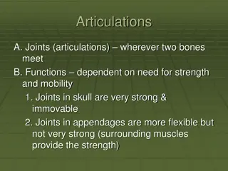
Articulations
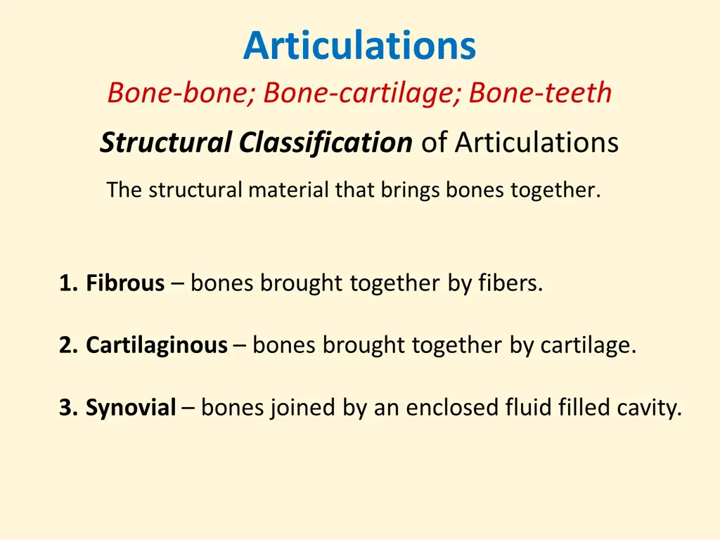


Articulations

Bone-bone; Bone-cartilage; Bone-teeth

Structural
Classification
of Articulations

1.
Fibrous
– bones brought together by fibers.
2.
Cartilaginous
– bones brought together by cartilage.
3.
Synovial
– bones joined by an enclosed fluid filled cavity.

The structural material that brings bones together.

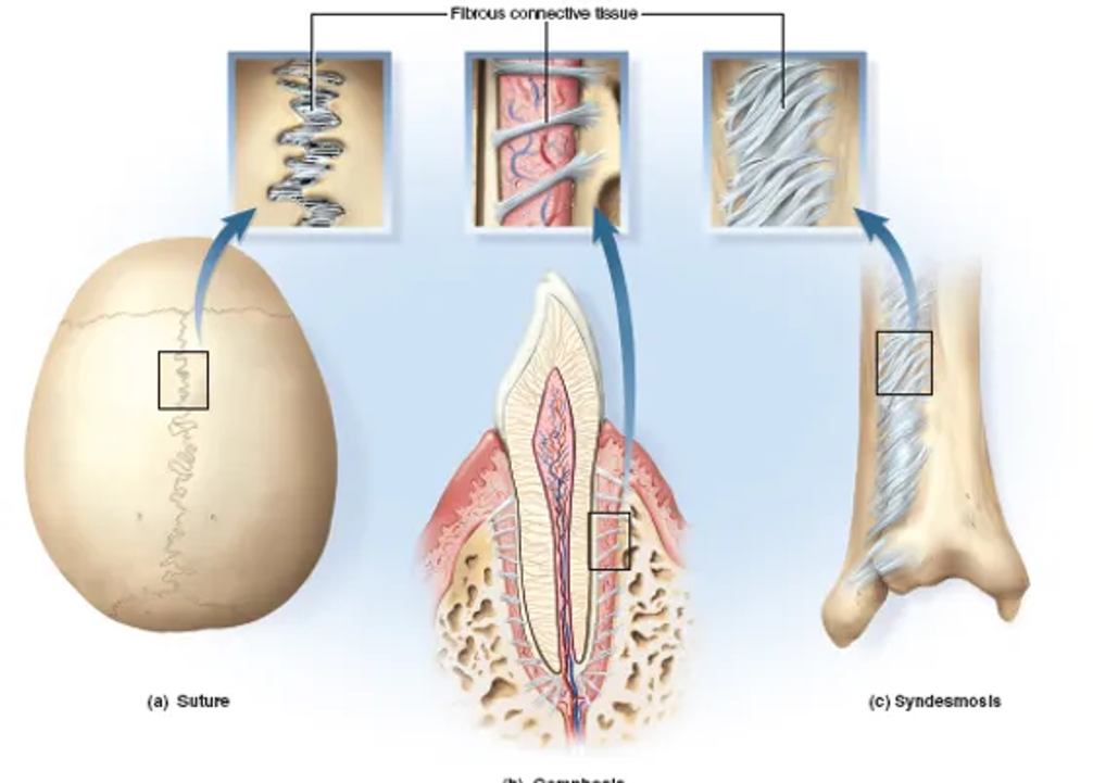


Fibrous Joints


Suture

Gomphosis

Syndesmosis

Synostosis
Synchondrosis
– joined by hyaline cartilage.
1
st
sternocostal joint
Pubic symphysis
intervertebral joints
Epiphyseal plate
Cartilaginous Joints
Symphysis
– joined by Fibrocartilage
Epiphysis
Diaphysis
Generalized Synovial Joint
A
r
t
i
c
u
l
a
r
(
j
o
i
n
t
)
C
a
p
s
u
l
e
-
I
n
n
e
r
‘
c
e
l
l
u
l
a
r
’
l
a
y
e
r
(synovial membrane)
-
O
u
t
e
r
‘
f
i
b
r
o
u
s
’
l
a
y
e
r
(dense irregular c.t.)
Synovial Joints
Functional
Classification
of Articulations
1.
Synarthrosis
– no movement possible at these joints.
2.
Amphiarthrosis
– slight movement possible at joint.
3.
Diarthrosis
– highly moveable at these joints.
Degree of movement permitted at articulation:
Ranges from: None, to a little bit, to highly movable.
Gomphoses
– tooth in socket
Synarthroses
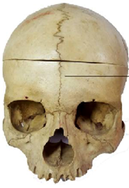
Sutures
– immovable joints of the skull
Symphysis
Slight movement with fibrocartilage
Amphiarthroses
Syndesmosis
Slightly movable fibrous joint.
Pubic symphysis of pelvic girdle
Cartilaginous joint uniting bodies
of adjacent vertebrae
Interosseous
membrane
①
②
①
②
Joints characterized by their high mobility and having a joint cavity within a synovial
membrane encased in the joint capsule.
Diarthroses
Diarthrotic
joints are also known as
Synovial joint
•
Gliding (Planar)
•
Hinge
•
Pivot
•
Ellipsoidal/Condyloid
•
Saddle
•
Ball and Socket
6 Types of
Synovial
Joints
Gliding Joint
These joints occur as short boxy bone surfaces move past each
other. See at wrist, ankles, and spine.
Carpals of the hand
Intercarpal joints
Tarsal joints
(navicular two cuneiforms)
Intertarsal joints
Vertebral Column
Facets
of intervertebral joints
Pivot Joint
1) Atlantoaxial joint
Formed between the
atlas
(C
1
) and the
axis
(C
2
) cervical vertebrae of neck.
For the “no” gesture,
Shaking head left to right.
2) Proximal radioulnar joint
Consists of the
radial head
articulating with the
radial
notch
of the
ulna
. The disc-like head of radius tightly
bound by the
annular ligament
securing it in place.
Allows the pivot action of pronation/supination.
Hinge Joint
Humeroulnar
joint of the elbow.
Interphalangeal
joints of the hand.
Tibiofemoral
join of the knee.
Condyloid/Ellipsoid Joint
Metacarpophalangeal
joints II through V.
Distal radiocarpal
joint of the wrist
Saddle Joint
1
st
Carpometacarpal
joint – the thumb joint (trapeziometacarpal)
Sternoclavicular
joint of the thorax
Calcaneocuboid
joint of the heel
Ball and Socket Joint
Hip
joint of the pelvic girdle
Shoulder
joint of pectoral girdle
The 6 Types of Synovial Joints
a)
Ball and Socket;
b)
Hinge;
c)
Saddle;
d)
Pivot;
e)
Ellipsoidal/Condyloid;
f)
Gliding (Planar).
a)
b)
c)
d)
e)
f)
Sternoclavicular
joint
Intercarpal and
Intertarsal
joints
Elbow joint
Knee joint
Ankle joint
Interphalangeal
joints
Atlantoaxial
joint
Proximal
Radioulnar
Joint
(
pronate/supinate
)
Radiocarpal
Joint
Metacarpophalangeal
Joints (2-5)
Trapeziometacarpal
Joint
(1
st
carpometacarpal)
Shoulder
joint
Hip
joint
Movements (
Actions
) at Articulations
Structures that Stabilize
Synovial Joints
Ligaments
Menisci
Bursae
Fat pads
Tendons
Diagrammatic Knee Joint
The Knee Joint
The Knee Joint
Iliofemoral
ligaments (n=2)
Pubofemoral
ligament
Ischiofemoral
ligament
The
Hip
Joint
Pattern
:
1
st
part = name of bone ligament is coming from on os coxa (3 choices).
2
nd
part = femoral.
Plus the don’t forget the
ligamentum teres
connecting the fovea
capitis to the acetabulum
Hip Joint
Hip Replacement Procedure
A ‘Hip Fracture’ is the breaking of which structure? ______________
Shoulder Joint
Acromioclavicular ligament
Coracoacromial ligament
Coracoclavicular ligaments
(n =2)
Segments of articular
(joint) capsule
Tendon for long head of biceps brachii
The Ligaments of
the
Shoulder
Joint
Pattern
: The coracoid is a big shot
and always gets named 1
st
; then
the acromion; the poor clavicle is
always last.
•
Osteoarthritis
: “Wear and tear” age-related arthritis,
due to use of the joint.
•
Arthritis
: Inflammation of a synovial joint, that is
often painful and restricts movement at that joint.
•
Rheumatoid Arthritis
: Autoimmune disease, in which
the body’s defense cells attack synovial joints.
Disorders of Articulations
•
Ankylosis
: Abnormal fusion of a joint, restricting
normal movement.
•
Gout (Gouty Arthritis)
: Uric acid deposits in synovial
joints and crystallizes there, causing pain and
restricting movement.
•
Bursitis
: Inflammation of a bursa, causing pain when
ever the associated ligament or tendon moves.
Disorders of Articulations
•
Sprain
: When a ligament is stretched beyond normal,
to where some collagen fibers are torn.
•
Luxation
: Dislocation, when articulating surfaces are
forced out of anatomical position.
The 6 Types of Synovial Joints
The different types of articulations, including fibrous, cartilaginous, and synovial joints, and learn about their structural classifications.

Download Presentation

Please find below an Image/Link to download the presentation.
The content on the website is provided AS IS for your information and personal use only. It may not be sold, licensed, or shared on other websites without obtaining consent from the author. Download presentation by click this link. If you encounter any issues during the download, it is possible that the publisher has removed the file from their server.
E N D
Presentation Transcript
Articulations Bone-bone; Bone-cartilage; Bone-teeth StructuralClassification of Articulations The structural material that brings bones together. 1. Fibrous bones brought together by fibers. 2. Cartilaginous bones brought together by cartilage. 3. Synovial bones joined by an enclosed fluid filled cavity.
Fibrous Joints Synostosis Suture Syndesmosis Gomphosis
Cartilaginous Joints Symphysis joined by Fibrocartilage Synchondrosis joined by hyaline cartilage. Epiphyseal plate intervertebral joints 1st sternocostal joint Pubic symphysis
Synovial Joints Generalized Synovial Joint Articular (joint) Capsule - Outer fibrous layer (dense irregular c.t.) - Inner cellular layer (synovial membrane)
Functional Classification of Articulations Degree of movement permitted at articulation: Ranges from: None, to a little bit, to highly movable. 1. Synarthrosis no movement possible at these joints. 2. Amphiarthrosis slight movement possible at joint. 3. Diarthrosis highly moveable at these joints.
Synarthroses Sutures immovable joints of the skull Gomphoses tooth in socket
Amphiarthroses Syndesmosis Slightly movable fibrous joint. Symphysis Slight movement with fibrocartilage Interosseous membrane Cartilaginous joint uniting bodies of adjacent vertebrae Pubic symphysis of pelvic girdle
Diarthroses Joints characterized by their high mobility and having a joint cavity within a synovial membrane encased in the joint capsule. Diarthroticjoints are also known as Synovial joint Gliding (Planar) Hinge 6 Types of Synovial Joints Pivot Ellipsoidal/Condyloid Saddle Ball and Socket
Gliding Joint These joints occur as short boxy bone surfaces move past each other. See at wrist, ankles, and spine. Carpals of the hand Intercarpal joints Tarsal joints (navicular two cuneiforms) Intertarsal joints Vertebral Column Facets of intervertebral joints
Pivot Joint 1) Atlantoaxial joint Formed between the atlas (C1) and the axis (C2) cervical vertebrae of neck. For the no gesture, Shaking head left to right. 2) Proximal radioulnar joint Consists of the radial head articulating with the radial notch of the ulna. The disc-like head of radius tightly bound by the annular ligament securing it in place. Allows the pivot action of pronation/supination.
Hinge Joint Humeroulnar joint of the elbow. Interphalangeal joints of the hand. Tibiofemoral join of the knee.
Condyloid/Ellipsoid Joint Metacarpophalangeal joints II through V. Distal radiocarpal joint of the wrist
Saddle Joint 1st Carpometacarpal joint the thumb joint (trapeziometacarpal) Sternoclavicular joint of the thorax Calcaneocuboid joint of the heel
Ball and Socket Joint Hip joint of the pelvic girdle Shoulder joint of pectoral girdle
The 6 Types of Synovial Joints a) b) c) d) e) f) a) Ball and Socket; b) Hinge; c) Saddle; d) Pivot; e) Ellipsoidal/Condyloid; f) Gliding (Planar).
Sternoclavicular joint Intercarpal and Intertarsal joints Elbow joint Knee joint Ankle joint Interphalangeal joints Atlantoaxial joint Proximal Radioulnar Joint (pronate/supinate)
Radiocarpal Joint Metacarpophalangeal Joints (2-5) Trapeziometacarpal Joint (1st carpometacarpal) Hip joint Shoulder joint
Structures that Stabilize Synovial Joints Ligaments Menisci Bursae Fat pads Tendons
Plus the dont forget the ligamentum teres connecting the fovea capitis to the acetabulum The Hip Joint Iliofemoral ligaments (n=2) Ischiofemoral ligament Pubofemoral ligament Pattern: 1st part = name of bone ligament is coming from on os coxa (3 choices). 2nd part = femoral.
A Hip Fracture is the breaking of which structure? ______________ Hip Replacement Procedure
Acromioclavicular ligament The Ligaments of the Shoulder Joint Coracoclavicular ligaments (n =2) Coracoacromial ligament Segments of articular (joint) capsule Pattern: The coracoid is a big shot and always gets named 1st ; then the acromion; the poor clavicle is always last. Tendon for long head of biceps brachii
Disorders of Articulations Arthritis: Inflammation of a synovial joint, that is often painful and restricts movement at that joint. Osteoarthritis: Wear and tear age-related arthritis, due to use of the joint. Rheumatoid Arthritis: Autoimmune disease, in which the body s defense cells attack synovial joints. Ankylosis: Abnormal fusion of a joint, restricting normal movement.
Disorders of Articulations Bursitis: Inflammation of a bursa, causing pain when ever the associated ligament or tendon moves. Gout (Gouty Arthritis): Uric acid deposits in synovial joints and crystallizes there, causing pain and restricting movement. Sprain: When a ligament is stretched beyond normal, to where some collagen fibers are torn. Luxation: Dislocation, when articulating surfaces are forced out of anatomical position.


 at Articulations")
 at Articulations")
 at Articulations")
 at Articulations")