Approach to a Thyroid Nodule: Causes, Diagnosis, and Management
When encountering a thyroid nodule, it is essential to consider both benign and malignant causes. Clinical features may vary, and investigations such as ultrasonography, FNAC, and thyroid scanning play crucial roles in evaluation. Understanding the findings suggestive of malignancy is key for appropriate management.

Download Presentation

Please find below an Image/Link to download the presentation.
The content on the website is provided AS IS for your information and personal use only. It may not be sold, licensed, or shared on other websites without obtaining consent from the author.If you encounter any issues during the download, it is possible that the publisher has removed the file from their server.
You are allowed to download the files provided on this website for personal or commercial use, subject to the condition that they are used lawfully. All files are the property of their respective owners.
The content on the website is provided AS IS for your information and personal use only. It may not be sold, licensed, or shared on other websites without obtaining consent from the author.
E N D
Presentation Transcript
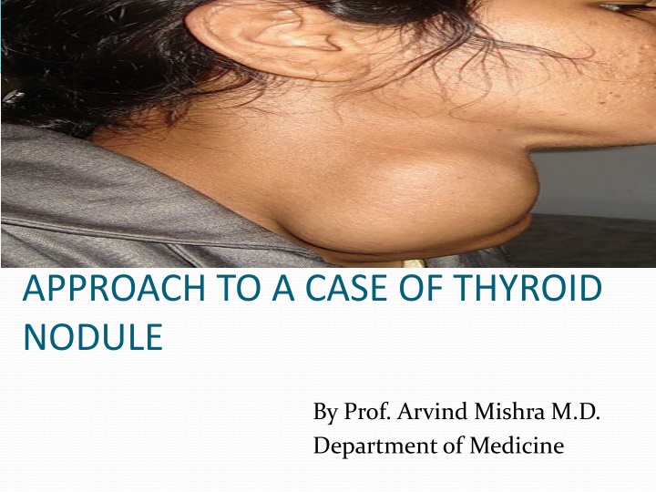
APPROACH TO A CASE OF THYROID NODULE By Prof. Arvind Mishra M.D. Department of Medicine
Causes of Thyroid Nodularity Benign Follicular Adenomas Multinodular goiter Hashimoto s thyroiditis Cysts (colloid, simple, hemorrhagic)
Malignant Papillary Carcinoma Follicular Carcinoma Medullary Carcinoma Anaplastic and poorly differentiated carcinoma Primary lymphoma of the thyroid Metastatic carcinoma
Clinical features Most thyroid nodules are asymptomatic Nodules that produce thyroid hormones in excess -palpitation -anxiety -clammy skin -increased appetite -weight loss -heat intolerance Nodules can press adjacent structures in neck causing -hoarseness of voice(recurrent laryngeal N compression) -dysphagia -dyspnoea -pain in neck
Nodules sometimes found in hashimotosthyroiditis, which may cause symptoms of an underactive thyroid gland -dry skin -face swelling -intolerance to cold -weight gain -decreased appetite -hair loss
INVESTIGATIONS ULTRASONOGRAPHY FNAC THYROID SCAN
Ultrasonography Most sensitive test to detect lesions in the thyroid Indicated in all patients who have a nodular thyroid, with a palpable solitary nodule or a multinodular goiter,beevaluated by US Not as screening test in general population 7
USG findings Number Size Extracapsular growth Cystic lesions Cervical LN 8
Findings suggestive of malignancy: Presence of halo Irregular border Presence of cystic components Presence of calcifications Heterogeneous echo pattern Extrathyroidal extension
Radionuclide Scanning Used to identify whether a nodule is functioning or not. Functioning nodules are nearly always benign Approximately 90 percent of nodules are nonfunctioning 5 percent of nonfunctioning nodules are malignant However even with suppressed level of serum TSH patient can have both functioning and non functioning nodules.Thus, even suppressed level of serum TSH may obviate the need for biopsy.
Usually either Technetium(Tc99) or Radioiodine(I123) used. Normal follicular cells will trap both but only radioiodine is added to tyrosine and stored in the colloid space Both benign and almost all malignant neoplastic tissue concentrate both radioisotopes less than normal thyroid tissue
Cold Nodules Cyst Non-functioning Adenoma Malignancy
Hot Nodules Functioning Adenoma Thyroiditis Multinodular goiter
Limitations of Thyroid scan Two dimensional scanning technique Inability to measure the size of a nodule accurately Missed malignant thyroid nodules
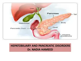
Other imaging tech CT and MRI not as routine. Can asses size, retrosternal extension, position and relation to the surrounding structure. 17
Images of a large, asymmetric multinodular goiter. (A) Chest radiography shows marked tracheal deviation to the right (arrow). (B) Chest CT confirmed the presence of a large substernal goiter on the left to the level of tracheal bifurcation. 18
USG guided FNAC Indicated if: Palpation-guided FNAC non-diagnostic Complex (solid/cystic) nodule Palpable small nodule (<1.5 cm) Impalpable nodule Abnormal cervical nodes Nodule with suspicious US features FNAC results are:70% Benign, 10% Malignant or suspicious of malignancy, and 20% Unsatisfactory 19
Malignant (+ve) cytology Commonest is PTC(Papillary thyroid carcinoma): Characteristics cytological feature- psammoma bodies, orphan annie eye nuclei (cleaved nuclei) Others include: Medullary thyroid carcinoma(amyloid deposits,intracytoplasmic calcitonin), anaplastic carcinoma(cellular anaplasia) and high-grade metastatic cancers 21
Suspicious cytology in FNAC Diagnosis cannot be made Inculdes: Follicular neoplasms, Atypical PTC, or Lymphoma 22
FNAC Limitations The absence of malignant cells in an acellular or hypocellular specimen does not exclude malignancy Inability to reliably distinguish a benign follicular adenoma from a follicular malignant tumour
TSH To detect early or subtle thyroid dysfunction. If TSH levels abnormal, free T3 & T4 should be measured to confirm the diagnosis TPOAb Thyroid peroxidase antibody Though characteristically observed in hypothyroidism, can also be seen in patients of hyperthyroidism and subacute thyroiditis
Serum Tg Correlates with iodine intake and the size of the thyroid gland rather than with the nature or function of the nodule Seldom used in nodule diagnosis Extremely elevated levels of Tg may suggest thyroid metastasis. Serum Calcitonin Good marker for medullary carcinoma and correlates well with tumor burden
1)Thyroid carcinoma associated with hypocalcemia is a)Follicular CA b)Medullary CA c)Anaplastic CA d)Papillary CA
2)Lab investigation of patient shows low T3, low T4 and highTSH.Diagnosis a)Primary hypothyroidism b)Grave s disease c)Hypothalamic failure d)Pituitary failure
3)Excess iodine intake causes hypothyroidism.It is known as a)Wolff chaikoff effect b)Jod basedow effect c)Thyrotoxicosis factitia d)De quervain s thyroiditis
4)FNAC can not differentiate between follicular adenoma and carcinoma because it can not clearly shows a)Vascular invasion b)Extracapsular extension c)a+b d)Nuclear pleomorphism
5)Subclinical hypothyroidism stands for biochemical evidence of hypothyroidism without any clinical features.Cut off TSH values are a)<5mU/L b)<8mU/L c)Normal d)<10mU/L


 or Radioiodine(I123)")


 Chest")


 cytology")


Thyroid carcinoma associated with")
Lab investigation of patient shows low T3, low T4 and")
Excess iodine intake causes hypothyroidism.It is")
FNAC can not differentiate between follicular")
Subclinical hypothyroidism stands for biochemical")