Anemia: Causes, Symptoms, and Diagnosis


History
Aetiology
Presentation
Investigations
Management


Anaemia Categorised
SBA 1
Dan is a 50-year-old man who reports 6kg of weight loss over the last 3
months alongside ‘stomach issues’. On examination, he has angular
cheilitis, koilonychia and appear pale.
A blood film shows hypochromic red blood cells with evident
anisopoikilocytosis and multiple pencil cells.
What is the diagnosis?
a)
Anaemia of Chronic Disease
b)
Thalassaemia major
c)
Iron Deficiency Anaemia
d)
Chronic Lymphocytic Leukaemia
e)
Hyperthyroidism
SBA 1
Dan is a 50-year-old man who reports
6kg of weight loss
over the last 3
months alongside ‘
stomach issues
’. On examination, he has
angular
cheilitis
,
koilonychia
and appear
pale
.
A blood film shows
hypochromic
red blood cells with evident
anisopoikilocytosis and multiple
pencil cells
.
What is the diagnosis?
a)
Anaemia of Chronic Disease
b)
Thalassaemia major
c)
Iron Deficiency Anaemia
d)
Chronic Lymphocytic Leukaemia
e)
Hyperthyroidism
Basic Features of Anaemia
Anaemia
= low blood
Hb
Pale
Conjunctivae
& Skin
Fatigue
RR↑ HR↑
(severe)
MICROCYTIC
Iron Deficiency Anaemia
Hypochromic &
microcytic
Pencil Cells
‘Aniso-
Poikilocytosis’
Blood Film
Causes of IDA
Reduced uptake
•
Malnutrition
•
Coeliac
•
IBD
Increased loss
•
GI Malignancy
•
Peptic ulcer
•
IBD
•
Menstruation
Increased requirement
Pregnancy
Breastfeeding
Colon
Cancer
PR
Bleeding
Unexplained
IDA
Change in
bowel
Habit
(
+
F
L
A
W
S
)
Anaemia of Chronic Disease
e.g. IBD
Hepcidin
uptake
↓
S
torage
↑
transport
↓
Ferroportin
Ferritin
Transferrin
IDA vs ACD on Blood Test
*Acute phase protein = ↑ during infections
A
C
D
i
s
o
f
t
e
n
N
O
R
M
O
C
Y
T
I
C
Thalassaemia
Investigations:
Microcytic anaemia +
Film
Normal Iron studies
Gel Electrophoresis
Management:
Regular red cell
transfusions every 2-4
weeks with iron
chelation regime
Thalassaemia
A
l
p
h
a
f
r
o
m
b
e
f
o
r
e
b
i
r
t
h
B
e
t
a
f
r
o
m
e
a
r
l
y
i
n
f
a
n
c
y
HbA1 = 2
α
+ 2
β
HbA2 = 2
α
+ 2
δ
HbF
P
= 2
α
+ 2
γ
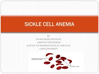
Sickle Cell Disease
Autosomal Recessive
Point mutation
β
Globin Gene
Chr 11
Sickle cell Trait
Asymptomatic*
Resistance to
Falciparum
Malaria
Sickling is predisposed by:
Hypoxia
Dehydration
Acidosis
Infection
2
0
%
o
f
t
r
o
p
i
c
a
l
A
f
r
i
c
a
p
o
p
u
l
a
t
i
o
n
h
a
v
e
S
i
c
k
l
e
c
e
l
l
t
r
a
i
t
Sickle Cell Disease
Howell-Jolly
Bodies
Sickled
Cells
Sickle Cell Crises
Sickle Cell Anaemia
Dactylitis
Haemolytic
anaemia
Acute Chest
syndrome
Conservative:
Trigger avoidance
Vaccination
Medical:
Vaccinations
Hydroxyurea
Hydroxycarbamide
Prophylactic ABx
Surgical:
Bone Marrow Transplant
(curative)
D
i
a
g
n
o
s
i
s
:
H
b
e
l
e
c
t
r
o
p
h
o
r
e
s
i
s
+
B
l
o
o
d
f
i
l
m
MACROCYTIC
SBA 2
Susan is a 12 year old girl presenting with tiredness and a tingling sensations in her
hands. She has recently modified her diet, taking part in Veganuary this year and is
following this through. Her mother has been diagnosed with Hashimoto’s Thyroiditis.
On examination she appears pale. The corners of her lips are notably sore.
What is the diagnosis?
a)
Dietary induced B12 deficiency
b)
Pernicious Anaemia
c)
Coeliac Disease
d)
Folate Deficiency
e)
Iron Deficiency
SBA 2
Susan is a 12 year old girl presenting with
tiredness
and a
tingling sensations in her
hands
. She has
recently
modified her diet, taking part in Veganuary this year and is
following this through. Her
mother
has been diagnosed with
Hashimoto’s
Thyroiditis.
On examination she appears pale. The corners of her lips are notably sore.
What is the diagnosis?
a)
Dietary induced B12 deficiency
b)
Pernicious Anaemia
c)
Coeliac Disease
d)
Folate Deficiency
e)
Iron Deficiency
Megaloblastic Anaemia
Folate
or
B12
Deficiency:
Hypersegmented
neutrophils
Macrocytic cells
Megaloblastic Anaemia Causes
V
i
t
a
m
i
n
B
1
2
F
o
l
a
t
e
Pernicious
Anaemia
Anti-folate
drugs
Pregnancy
IBD &
Coeliac
Malnutrition
Alcohol
IBD &
Coeliac
Alcohol
I
n
t
e
r
e
s
t
i
n
g
B
o
r
i
n
g
B12 Deficiency
B
1
2
i
s
f
u
n
d
a
m
e
n
t
a
l
f
o
r
R
B
C
m
a
t
u
r
a
t
i
o
n
:
Megaloblastic
+
Neuro signs
B
1
2
r
e
s
e
r
v
e
s
l
a
s
t
3
-
4
y
e
a
r
s
Glove & stocking parasthesiae
Hyporeflexia
Romberg’s +ve
“Subacute combined
degeneration of the cord”
A
n
t
i
-
P
a
r
i
e
t
a
l
c
e
l
l
s
A
n
t
i
-
I
n
t
r
i
n
s
i
c
f
a
c
t
o
r
+
P
e
r
n
i
c
i
o
u
s
A
n
a
e
m
i
a
Non-Megaloblastic Macrocytic
Myelodysplasia
Be
aware
of the following other causes:
Hypothyroidism
Liver disease
Alcohol
A
m
i
r
S
a
m
:
“
A
l
c
o
h
o
l
i
c
s
M
a
y
H
a
v
e
L
i
v
e
r
F
a
i
l
u
r
e
”
NORMOCYTIC
SBA 3
Which of these must be avoided in individuals with known G-6-PD
deficiency?
What is the diagnosis?
a)
Gluten products
b)
Cow’s Milk protein
c)
Broad beans
d)
ACE inhibitors
e)
Smoking
SBA 3
Which of these must be avoided in individuals with known G-6-PD
deficiency?
What is the diagnosis?
a)
Gluten products
b)
Cow’s Milk protein
c)
Broad beans
d)
ACE inhibitors
e)
Smoking
Features of Haemolytic Anaemia
Blood tests:
Hb low
Haptoglobin low*
Unconjugated bilirubin
raised
LDH raised
Hereditary Haemolytic Anaemias
D
i
s
o
r
d
e
r
s
o
f
t
h
e
R
B
C
i
t
s
e
l
f
:
Membrane
Enzymes
Haemoglobin
H
e
r
e
d
i
t
a
r
y
S
p
h
e
r
o
c
y
t
o
s
i
s
G
-
6
-
P
D
D
e
f
i
c
i
e
n
c
y
S
i
c
k
l
e
C
e
l
l
T
h
a
l
a
s
s
a
e
m
i
a
Glucose-6-Phosphate Deficiency
B
E
A
N
S
M
E
A
N
S
H
E
I
N
Z
Fava Beans
X-linked Recessive
Vulnerable to
Oxidative Stress
G-6-PD Deficiency Blood Film
H
e
i
n
z
B
o
d
i
e
s
B
i
t
e
C
e
l
l
s
(Active Haemolysis)
(Previous Haemolysis)
Hereditary Spherocytosis
Autosomal Dominant (75%)
S
p
h
e
r
o
c
y
t
e
s
Hereditary Spherocytosis
Beta Spectrin or Ankyrin deficiency
Pathogenesis
Investigations/Complications
Hereditary Spherocytosis
A
p
l
a
s
t
i
c
C
r
i
s
i
s
P
a
r
v
o
v
i
r
u
s
B
1
9
Hypotonic Solution
Lysis
O
s
m
o
t
i
c
F
r
a
g
i
l
i
t
y
T
e
s
t
+
C
o
o
m
b
s
T
e
s
t
N
e
g
a
t
i
v
e
SBA 4
Sophie is a 14 month old infant. She has a recent 5 day history of abdominal pain which her parents believe is
related to food she ate at their recent trip to the state fair. The paediatric registrar on call believes she has now
developed an AKI. Her blood tests reveal a normal PT, normocytic anaemia with leucocytosis and
thrombocytopaenia. She also has a high serum bilirubin. A section of her blood film is shown:
What is the most likely cause?
a)
ALL
b)
DIC
c)
HUS
d)
Pyelonephritis
e)
Sepsis
SBA 4
Sophie is a
14 month old infant
. She has a recent 5 day history of
abdominal pain
which her parents believe is
related to food she ate at their recent trip to the state fair. The paediatric registrar on call believes she has now
developed an
AKI
. Her blood tests reveal a normal PT, normocytic
anaemia with leucocytosis
and
thrombocytopaenia
. She also has a
high serum bilirubin
. A section of her blood film is shown:
What is the most likely cause?
a)
ALL
b)
DIC
c)
HUS
d)
Pyelonephritis
e)
Sepsis
Microangiopathic Haemolytic Anaemia
Sheared RBCs
Microthrombi formation
Haemolytic Uraemic Syndrome
DISEASE OF EARLY CHILDHOOD
+GI features
Abdominal Pain
Bloody Diarrhoea
AKI
MAHA
Jaundice
Conjunctival
pallor
Thrombo
cytope
nia
H
U
S
EHEC
O157:H7
Disseminated Intravascular Coagulation
Sepsis
Pancreatitis
ABO Reaction
Cancers
D
I
C
i
s
s
e
v
e
r
e
c
o
n
c
u
r
r
e
n
t
C
l
o
t
t
i
n
g
a
n
d
B
l
e
e
d
i
n
g
Causes include:
Trauma
Obstetric Complications
Disseminated Intravascular Coagulation
Clotting features
Prolonged APTT
Prolonged PT
Haemolytic features
Jaundice
Conjunctival pallor
Bleeding features
Petechiae
Ecchymoses
Haematuria
+ Features of
underlying
pathology
P
l
a
t
e
l
e
t
s
↓
F
i
b
r
i
n
o
g
e
n
↓
F
D
P
s
↑
D
-
d
i
m
e
r
↑
Pathophysiology of TTP
T
T
P
N
o
r
m
a
l
ADAMTS-13
functional
ADAMTS-13
dysfunctional
Thrombotic Thrombocytopaenic Purpura
•
A
ntiglobulin
negative
•
D
ecreased platelets
•
A
KI
•
M
AHA
•
T
emperature
•
S
winging CNS signs
D
e
f
u
n
c
t
A
D
A
M
T
S
-
1
3
e
n
z
y
m
e
:
A (rarely complete)
clinical pentad
Thrombotic Thrombocytopaenic Purpura
•
A
ntiglobulin
negative
•
Decreased platelets
•
AKI
•
MAHA
•
T
emperature
•
S
winging CNS signs
D
e
f
u
n
c
t
A
D
A
M
T
S
-
1
3
e
n
z
y
m
e
:
A (rarely complete)
clinical pentad
H
U
S
Plasmapharesis removes antibodies against ADAMTS-13
and also replaces the enzyme
Summary of MAHA
DAT/Coombs Negative
DAT/COOMBS Test
Auto-immune
or Not?
Other Acquired Haemolytic Anaemias
D
i
s
o
r
d
e
r
s
o
f
t
h
e
R
B
C
’
s
E
n
v
i
r
o
n
m
e
n
t
:
A
u
t
o
-
I
m
m
u
n
e
Anti-RBC antigen
antibodies
D
A
T
/
C
o
o
m
b
s
+
v
e
D
r
u
g
s
e.g. Dapsone
(Anti-leprosy ABx)
I
n
f
e
c
t
i
o
n
P
l
a
s
m
o
d
i
u
m
f
a
l
c
i
p
a
r
u
m
m
a
l
a
r
i
a
(
B
l
a
c
k
w
a
t
e
r
f
e
v
e
r
)
Agglutination:
Warm ≥ 37°C
IgG antibodies
Idiopathic
SLE
CLL
Cold ≤ 37°C
Ig
M
Antibodies
Idiopathic
M
ycoplasma
M
ononucleosis
Feedback
P
l
e
a
s
e
f
i
l
l
i
n
t
h
e
f
e
e
d
b
a
c
k
f
o
r
m
!
h
t
t
p
s
:
/
/
i
m
p
e
r
i
a
l
.
e
u
.
q
u
a
l
t
r
i
c
s
.
c
o
m
/
j
f
e
/
f
o
r
m
/
S
V
_
c
U
Q
S
3
X
N
c
J
L
O
w
J
o
i
SBA 5
Donald is an 85 year old owner of a nuclear powerplant. He is hypertensive and managed with
Amlodipine. He is referred to his doctor after his dentist notices he appears cachectic and his
dentures no longer fit. On examination, Mr Burns has massive splenomegaly. A blood test and
subsequent bone marrow biopsy are carried out:
What is the most likely cause of these findings?
a)
Multiple myeloma
b)
Acute myeloid leukaemia
c)
Essential thrombocytosis
d)
Myelofibrosis
e)
Myedlodysplasia
RBC – Low
Hb – Low
WCC – Low
Plts – High
Film: poikilocytosis
Biopsy: ‘dry tap’
SBA 5
Donald is an
85 year old owner
of a
nuclear powerplant
. He is hypertensive and managed with
Amlodipine. He is referred to his doctor after his dentist notices he appears
cachectic
and his
dentures no longer fit. On examination, Mr Burns has
massive splenomegaly
. A blood test and
subsequent bone marrow biopsy are carried out:
What is the most likely cause of these findings?
a)
Multiple myeloma
b)
Acute myeloid leukaemia
c)
Essential thrombocytosis
d)
Myelofibrosis
e)
Myedlodysplasia
RBC – Low
Hb – Low
WCC – Low
Plts – High
Film:
poikilocytosis
Biopsy: ‘
dry tap
’
Primary Myelofibrosis
Fibrosis
in response to a
BM
malignancy
Radiation
>65 years old
B
M
A
s
p
i
r
a
t
e
:
‘dry tap’
fibrosis
B
l
o
o
d
f
i
l
m
:
Tear drop
cells
Anaemia Categorised
Additional Conditions
•
Polycythaemia
•
Anti-phospholipid syndrome
•
Aplast
ic anaemia
Polycythaemia
Rubra Vera
EPO-secreting tumour
Hypoxic response
Polycythaemia Rubra Vera
Clinical features:
Older (~60)
Asymptomatic
Aquagenic pruritis
Hyper-viscosity syndrome
Elevated Hb
& haematocrit
+/- Thrombocytosis
A Philadelphia chromosome
negative
myeloproliferative
disorder
.
Almost all PCV is JAK2 V617F +ve
Anti-Phospholipid Syndrome
Auto-immune
mediated
thrombosis
– often manifesting during
pregnancy.
A
n
t
i
-
c
a
r
d
o
l
i
p
i
n
+
v
e
Lupus anti-coagulant test
+ve
Clinical features:
Recurrent miscarriages (3+)
VTE
Stroke/MIs, HTN
(arterial problems)
Livedo reticularis (mottled)
Aplastic Anaemia
Bone Marrow
Failure
causing
Pan
cytopaenia
Infections
Radiation
B
M
A
s
p
i
r
a
t
e
:
H
y
p
o
c
e
l
l
u
l
a
r
Anaemia features
:
+
EPO raised
Thrombocytopaenia
:
Bleeding
Petechiae
Leucopoenia
:
Sepsis
Recurrent infections
Fanconi’s
Anaemia
Auto-immune mediated
NOT
the same as an
Aplastic Crisis
Anemia is a condition characterized by low red blood cell count, leading to symptoms like fatigue, pale skin, and shortness of breath. Causes range from nutritional deficiencies to chronic diseases. Diagnosis involves blood tests and examination of red blood cells. This content delves into various types of anemia, their etiologies, clinical presentations, and management strategies.

Download Presentation

Please find below an Image/Link to download the presentation.
The content on the website is provided AS IS for your information and personal use only. It may not be sold, licensed, or shared on other websites without obtaining consent from the author.If you encounter any issues during the download, it is possible that the publisher has removed the file from their server.
You are allowed to download the files provided on this website for personal or commercial use, subject to the condition that they are used lawfully. All files are the property of their respective owners.
The content on the website is provided AS IS for your information and personal use only. It may not be sold, licensed, or shared on other websites without obtaining consent from the author.
E N D
Presentation Transcript
NICK SOON ns6118@ic.ac.uk MENTI CODE: 9373 6292
Aetiology History Presentation Investigations Management
Anaemia Micro Normo Macro Acute Blood Loss Sickle Cell ACD Myelofibrosis Alcohol Vit B12 Haemolytic Anaemias Thalassaemia IDA Aplastic Liver disease Folate Acquired Congenital Hypothyroid Myelodysplasia HUS Hereditary Spherocytosis Autoimmune Drugs DIC MAHA Infection G6PD TTP
Dan is a 50-year-old man who reports 6kg of weight loss over the last 3 months alongside stomach issues . On examination, he has angular cheilitis, koilonychia and appear pale. A blood film shows hypochromic red blood cells with evident anisopoikilocytosis and multiple pencil cells. What is the diagnosis? a) Anaemia of Chronic Disease b) Thalassaemia major c) Iron Deficiency Anaemia d) Chronic Lymphocytic Leukaemia e) Hyperthyroidism
Dan is a 50-year-old man who reports 6kg of weight loss over the last 3 months alongside stomach issues . On examination, he has angular cheilitis, koilonychia and appear pale. A blood film shows hypochromic red blood cells with evident anisopoikilocytosis and multiple pencil cells. What is the diagnosis? a) Anaemia of Chronic Disease b) Thalassaemia major c) Iron Deficiency Anaemia d) Chronic Lymphocytic Leukaemia e) Hyperthyroidism
Anaemia = low blood Hb Pale Conjunctivae & Skin Fatigue RR HR (severe)
Aniso- Poikilocytosis Pencil Cells Hypochromic & microcytic Blood Film
Reduced uptake Malnutrition Coeliac IBD Unexplained IDA Increased loss GI Malignancy Peptic ulcer IBD Menstruation Colon Cancer Change in bowel Habit PR Bleeding Increased requirement Pregnancy Breastfeeding (+ FLAWS)
Chronic Disease e.g. IBD Cytokines Hepcidin Hepcidin uptake transport Storage Fe Fe Fe Fe Fe FeFe Fe Fe Fe Ferroportin Transferrin Fe Fe Fe Fe Fe Fe Fe Fe Fe Fe Fe Fe Fe Fe Fe Fe Ferritin
Ferritin* TIBC IDA ACD /N *Acute phase protein = during infections ACD is often NORMOCYTIC
Investigations: Microcytic anaemia + Film Normal Iron studies Gel Electrophoresis Management: Regular red cell transfusions every 2-4 weeks with iron chelation regime Thalassaemia Minor Resistance to falciparum Malaria
Alpha from before birth Beta from early infancy HbA1 = 2 + 2 HbA2 = 2 + 2 HbFP = 2 + 2
Sickling is predisposed by: Point mutation Globin Gene Chr 11 Hypoxia Dehydration Acidosis Infection Autosomal Recessive Asymptomatic* Resistance to Falciparum Malaria 20% of tropical Africa population have Sickle cell trait Sickle cell Trait
Howell-Jolly Bodies Sickled Cells
Crisis Management Sickle Acute Painful Crisis Saturate (supportive Oxygen) Antibiotics (if needed) Pain relief Cannula (IV fluids) Crizanlizumab for prevention Acute Painful Crisis Stroke Exchange Blood Transfusion Sequestration Crisis Splenectomy Chronic Cholecystitis Cholecystectomy
Diagnosis: Hb electrophoresis + Blood film Conservative: Trigger avoidance Vaccination Medical: Vaccinations Hydroxyurea Hydroxycarbamide Prophylactic ABx Acute Chest syndrome Dactylitis Surgical: Bone Marrow Transplant (curative) Haemolytic anaemia Priapism Aplastic Crisis
Susan is a 12 year old girl presenting with tiredness and a tingling sensations in her hands. She has recently modified her diet, taking part in Veganuary this year and is following this through. Her mother has been diagnosed with Hashimoto s Thyroiditis. On examination she appears pale. The corners of her lips are notably sore. What is the diagnosis? a) Dietary induced B12 deficiency b)Pernicious Anaemia c) Coeliac Disease d)Folate Deficiency e) Iron Deficiency
Susan is a 12 year old girl presenting with tiredness and a tingling sensations in her hands. She has recently modified her diet, taking part in Veganuary this year and is following this through. Her mother has been diagnosed with Hashimoto s Thyroiditis. On examination she appears pale. The corners of her lips are notably sore. What is the diagnosis? a) Dietary induced B12 deficiency b)Pernicious Anaemia c) Coeliac Disease d)Folate Deficiency e) Iron Deficiency
Folate or B12 Deficiency: Hypersegmented neutrophils Macrocytic cells
Vitamin B12 Pernicious Anaemia IBD & Coeliac Alcohol Malnutrition Folate IBD & Coeliac Anti-folate drugs Pregnancy Alcohol
B12 is fundamental for RBC maturation: Megaloblastic + Neuro signs Glove & stocking parasthesiae Hyporeflexia Romberg s +ve Subacute combined degeneration of the cord +Pernicious Anaemia Anti-Parietal cells Anti-Intrinsic factor B12 reserves last 3-4 years
Be aware of the following other causes: Alcohol Hypothyroidism Liver disease Myelodysplasia Amir Sam: Alcoholics May Have Liver Failure
Which of these must be avoided in individuals with known G-6-PD deficiency? What is the diagnosis? a) Gluten products b)Cow s Milk protein c) Broad beans d)ACE inhibitors e) Smoking
Which of these must be avoided in individuals with known G-6-PD deficiency? What is the diagnosis? a) Gluten products b)Cow s Milk protein c) Broad beans d)ACE inhibitors e) Smoking
Scleral Icterus Pale Conjunctivae & Skin Blood tests: Hb low Haptoglobin low* Unconjugated bilirubin raised LDH raised
Disorders of the RBC itself: Hereditary Spherocytosis Membrane Enzymes G-6-PD Deficiency Haemoglobin Sickle Cell Thalassaemia
BEANS MEANS HEINZ Vulnerable to Oxidative Stress Fava Beans X-linked Recessive
Heinz Bodies Bite Cells (Active Haemolysis) (Previous Haemolysis)
Spherocytes Autosomal Dominant (75%)
Pathogenesis Beta Spectrin or Ankyrin deficiency
Hereditary Spherocytosis Osmotic Fragility Test Aplastic Crisis Lysis Hypotonic Solution Parvovirus B19 + Coombs Test Negative
Sophie is a 14 month old infant. She has a recent 5 day history of abdominal pain which her parents believe is related to food she ate at their recent trip to the state fair. The paediatric registrar on call believes she has now developed an AKI. Her blood tests reveal a normal PT, normocytic anaemia with leucocytosis and thrombocytopaenia. She also has a high serum bilirubin. A section of her blood film is shown: What is the most likely cause? a) ALL b)DIC c) HUS d)Pyelonephritis e) Sepsis
Sophie is a 14 month old infant. She has a recent 5 day history of abdominal pain which her parents believe is related to food she ate at their recent trip to the state fair. The paediatric registrar on call believes she has now developed an AKI. Her blood tests reveal a normal PT, normocytic anaemia with leucocytosis and thrombocytopaenia. She also has a high serum bilirubin. A section of her blood film is shown: What is the most likely cause? a) ALL b)DIC c) HUS d)Pyelonephritis e) Sepsis
Sheared RBCs Microthrombi formation
DISEASE OF EARLY CHILDHOOD EHEC O157:H7 Thrombocytopenia +GI features Abdominal Pain Bloody Diarrhoea HUS MAHA Jaundice Conjunctival pallor AKI
DIC is severe concurrent Clotting and Bleeding Causes include: Pancreatitis Obstetric Complications Sepsis Cancers Trauma ABO Reaction
Bleeding features Petechiae Ecchymoses Haematuria Clotting features Prolonged APTT Prolonged PT Haemolytic features Jaundice Conjunctival pallor Platelets Fibrinogen FDPs D-dimer + Features of underlying pathology
TTP Normal ADAMTS-13 functional ADAMTS-13 dysfunctional
Defunct ADAMTS-13 enzyme: Antiglobulin negative Decreased platelets AKI MAHA Temperature Swinging CNS signs A (rarely complete) clinical pentad
Defunct ADAMTS-13 enzyme: Antiglobulin negative Decreased platelets AKI MAHA Temperature Swinging CNS signs HUS A (rarely complete) clinical pentad Plasmapharesis removes antibodies against ADAMTS-13 and also replaces the enzyme
DIC HUS TTP AKI MAHA Thrombocytopaenia Child (<5) Clinical Pentad Swinging CNS Coombs ve Clotting + Bleeding DAT/Coombs Negative Pl E. coli Shiga Toxin ITU-related Enzyme dysfunction
Auto-immune or Not?
Disorders of the RBCs Environment: Agglutination: Auto-Immune Anti-RBC antigen antibodies DAT/Coombs +ve Warm 37 C IgG antibodies Idiopathic SLE CLL Drugs e.g. Dapsone (Anti-leprosy ABx) Cold 37 C IgM Antibodies Idiopathic Mycoplasma Mononucleosis Infection Plasmodium falciparum malaria (Blackwater fever)
Please fill in the feedback form! https://imperial.eu.qualtrics.com/jfe/form/ SV_cUQS3XNcJLOwJoi
Donald is an 85 year old owner of a nuclear powerplant. He is hypertensive and managed with Amlodipine. He is referred to his doctor after his dentist notices he appears cachectic and his dentures no longer fit. On examination, Mr Burns has massive splenomegaly. A blood test and subsequent bone marrow biopsy are carried out: RBC Low Hb Low WCC Low Plts High What is the most likely cause of these findings? a) Multiple myeloma b)Acute myeloid leukaemia c) Essential thrombocytosis d)Myelofibrosis e) Myedlodysplasia Film: poikilocytosis Biopsy: dry tap
Donald is an 85 year old owner of a nuclear powerplant. He is hypertensive and managed with Amlodipine. He is referred to his doctor after his dentist notices he appears cachectic and his dentures no longer fit. On examination, Mr Burns has massive splenomegaly. A blood test and subsequent bone marrow biopsy are carried out: RBC Low Hb Low WCC Low Plts High What is the most likely cause of these findings? a) Multiple myeloma b)Acute myeloid leukaemia c) Essential thrombocytosis d)Myelofibrosis e) Myedlodysplasia Film: poikilocytosis Biopsy: dry tap
Fibrosis in response to a BM malignancy >65 years old BM Aspirate: dry tap fibrosis Blood film: Tear drop cells Radiation