Hemoglobin: Functions, Medical Applications, and Conditions
Hemoglobin, a crucial protein in red blood cells, plays a vital role in carrying oxygen throughout the body. This article delves into its functions, detection methods, medical applications, and common conditions associated with hemoglobin, such as anemia and polycythemia. Learn about the various types of anemia, including iron deficiency anemia and megaloblastic anemia, and how they affect the body's overall health.

Download Presentation

Please find below an Image/Link to download the presentation.
The content on the website is provided AS IS for your information and personal use only. It may not be sold, licensed, or shared on other websites without obtaining consent from the author.If you encounter any issues during the download, it is possible that the publisher has removed the file from their server.
You are allowed to download the files provided on this website for personal or commercial use, subject to the condition that they are used lawfully. All files are the property of their respective owners.
The content on the website is provided AS IS for your information and personal use only. It may not be sold, licensed, or shared on other websites without obtaining consent from the author.
E N D
Presentation Transcript
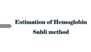
ESTIMATION OF ESTIMATION OF HEMOGLOBIN HEMOGLOBIN Physiology lab
HEMOGLOBIN found in the RBC. responsible for carrying oxygen to all cells in the body. also binds to carbon dioxide and carries it to the lungs from the cells to be released. buffer against change in [h+] hemoglobin detection can also gives the health care worker an idea about:- patient s oxygen carrying capacity current blood loss and recovery from blood loss treatment of rbc disorders, like anemia.
HEMOGLOBIN HEMOGLOBIN makes up 98% of the protein found in the rbc and gives blood its red color. composed of two parts . heme , 4 iron atoms in the ferrous state (fe2+) and porphyrin ring globin , 4 protein chains the most common globin forms are alpha and beta chains.
Variant forms of hemoglobin Oxyhemoglobin: hemoglobin combined with oxygen. Carbaminohemoglobin: hemoglobin combined with CO2. Methemoglobin: Ferrous iron in converted into ferric iron.
MEDICAL APPLICATION MEDICAL APPLICATION the body will respond for the decrease in haemoglobin level (for slightly lower than normal haemoglobin levels) as a compensatory mechanism:- the heart will beat faster and more forcefully. the lungs breath rate will increase. when the level drops too low for us, we start to feel tired, breathless and may start to run into problems with too little oxygen getting to important organs like the heart and brain. this can cause palpitations, angina (chest pains), headache or dizzy spells.
MEDICAL CONDITIONS MEDICAL CONDITIONS anemia :- is a decrease of hemoglobin concentration. polycythemia :- is an increase of hb concentration.
Types of anemia: Types of anemia: Iron deficiency anemia: commonest cause of anemia in most parts of the world cause either due to loss of iron due to bleeding or an inadequate diet or mal-absorption. Megaloblastic anemia: caused by deficiency of vitamin B12 or folate deficiency or both of them. Pernicious anemia :- an autoimmune destruction of gastric parietal cells leading to a lack of intrinsic factor which important for vitamin B12 absorption in the gut.
Hemolytic anemia includes 1- Hereditary spherocytosis:- 2- Glucose 6 Phosphate dehydrogenase deficiency Favism (hemolytic anemia from the ingestion of the broad beans) 3- Hemoglobinopathies : Sickle cell anemia: Thalassaemia: 4-Acquired hemolytic anemia Autoimmune hemolytic anemia Hemolytic disease of newborn ABO incompatibility Rhesus (Rh) incompatibility 5-Anemia of chronic disease.
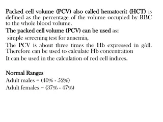
Materials and Instruments SAHLI S METHOD All Hb is converted into acid hematine and the intensity of the color is measured by comparing it with the standered Sahli Haemometer consists of A color standard Pipette marked to contain 20 microliter of blood. Graduated tube Distilled water (D.W.) 0.1 normal HCl Anticoagulated whole blood or capillary blood can be used.
Procedure : 1. Place into the Hb tube the HCL (0.1 N) by pipette till 20 marks. 2. Add blood from EDTA tube to Hb tube till the 20 m ml, and transfer the blood into the mixing tube. Mix it well by glass stirrer. 1. Add distal water drop by drop, mixing well by a glass rod and compare the color of this tube with the standard tube. 2. After 5 minutes, read the hemoglobin content from the scale on the graduated mixing tube.
NORMAL VALUE male 13.5-17.5 gm/dl female 11.5-15.5 gm/dl newborn 21 gm/dl
Date---------- Names of the students: Group-------- Name of the experiment: Aim of the experiment: Materials Procedure: 1.Fill the graduated tube to mark (2 or 10) with 0.1 normal HCl. 2.Draw blood by hemoglobin pipette to mark 20Ml. 3.Dip the tip of the pipette in the graduated tube to blow the blood into the tube, mix content with stirrer. 4.Place the tube in the hemoglobinometer for 10 minutes for complete reaction. 5.Add drop by drop D.W. until the color in the graduated tube is identical to the color of the standard. 6.Read the result in gm/dl. Result: Hb = .. Discussion: