Anatomy of Permanent Maxillary Central Incisor Teeth


L
e
c
.
4
a
n
d
5
I
n
d
i
v
i
d
u
a
l
A
n
a
t
o
m
y
o
f
P
e
r
m
a
n
e
n
t
T
e
e
t
h

M
a
x
i
l
l
a
r
y
c
e
n
t
r
a
l
i
n
c
i
s
o
r
T
h
e
m
a
x
i
l
l
a
r
y
c
e
n
t
r
a
l
i
n
c
i
s
o
r
i
s
a
h
u
m
a
n
i
n
t
h
e
f
r
o
n
t
u
p
p
e
r
j
a
w
,
o
r
,
a
n
d
i
s
u
s
u
a
l
l
y
t
h
e
m
o
s
t
v
i
s
i
b
l
e
o
f
a
l
l
t
e
e
t
h
i
n
t
h
e
m
o
u
t
h
.
I
t
i
s
l
o
c
a
t
e
d
(
c
l
o
s
e
r
t
o
t
h
e
m
i
d
l
i
n
e
o
f
t
h
e
f
a
c
e
)
t
o
t
h
e
.
A
s
w
i
t
h
a
l
l
,
t
h
e
i
r
f
u
n
c
t
i
o
n
i
s
f
o
r
o
r
c
u
t
t
i
n
g
f
o
o
d
d
u
r
i
n
g
(
c
h
e
w
i
n
g
)
.
T
h
e
r
e
a
r
e
n
o
o
n
t
h
e
t
e
e
t
h
.
I
n
s
t
e
a
d
,
t
h
e
s
u
r
f
a
c
e
a
r
e
a
o
f
t
h
e
t
o
o
t
h
u
s
e
d
i
n
e
a
t
i
n
g
i
s
c
a
l
l
e
d
a
n
r
i
d
g
e
o
r
i
n
c
i
s
a
l
e
d
g
e
.
F
o
r
m
a
t
i
o
n
o
f
t
h
e
s
e
t
e
e
t
h
b
e
g
i
n
a
t
1
4
w
e
e
k
s
i
n
u
t
e
r
o
f
o
r
t
h
e
(
b
a
b
y
)
s
e
t
a
n
d
3
–
4
m
o
n
t
h
s
o
f
a
g
e
f
o
r
t
h
e
s
e
t
.tnenamrepsuoudicedlasicnispsucnoitacitsamgniraehssrosicnirosicni laretalyrallixamlaisemallixamhtoot
T
h
e
r
e
a
r
e
s
o
m
e
m
i
n
o
r
d
i
f
f
e
r
e
n
c
e
s
b
e
t
w
e
e
n
t
h
e
d
e
c
i
d
u
o
u
s
m
a
x
i
l
l
a
r
y
c
e
n
t
r
a
l
i
n
c
i
s
o
r
a
n
d
t
h
a
t
o
f
t
h
e
p
e
r
m
a
n
e
n
t
m
a
x
i
l
l
a
r
y
c
e
n
t
r
a
l
i
n
c
i
s
o
r
.
T
h
e
d
e
c
i
d
u
o
u
s
t
o
o
t
h
a
p
p
e
a
r
s
i
n
t
h
e
m
o
u
t
h
a
t
3
–
1
8
m
o
n
t
h
s
o
f
a
g
e
,
w
i
t
h
6
m
o
n
t
h
s
b
e
i
n
g
t
h
e
a
v
e
r
a
g
e
a
n
d
i
s
r
e
p
l
a
c
e
d
b
y
t
h
e
p
e
r
m
a
n
e
n
t
t
o
o
t
h
a
r
o
u
n
d
7
–
8
y
e
a
r
s
o
f
a
g
e
.
T
h
e
p
e
r
m
a
n
e
n
t
t
o
o
t
h
i
s
l
a
r
g
e
r
a
n
d
i
s
t
h
a
n
i
t
i
s
w
i
d
e
.
T
h
e
m
a
x
i
l
l
a
r
y
c
e
n
t
r
a
l
i
n
c
i
s
o
r
s
c
o
n
t
a
c
t
e
a
c
h
o
t
h
e
r
a
t
t
h
e
m
i
d
l
i
n
e
o
f
t
h
e
f
a
c
e
.
T
h
e
a
r
e
t
h
e
o
n
l
y
o
t
h
e
r
t
y
p
e
o
f
t
e
e
t
h
t
o
d
o
s
o
.
T
h
e
p
o
s
i
t
i
o
n
o
f
t
h
e
s
e
t
e
e
t
h
m
a
y
d
e
t
e
r
m
i
n
e
t
h
e
e
x
i
s
t
e
n
c
e
o
f
a
n
o
p
e
n
b
i
t
e
o
r
.
A
s
w
i
t
h
a
l
l
t
e
e
t
h
,
v
a
r
i
a
t
i
o
n
s
o
f
s
i
z
e
,
s
h
a
p
e
,
a
n
d
c
o
l
o
r
e
x
i
s
t
a
m
o
n
g
p
e
o
p
l
e
.
S
y
s
t
e
m
i
c
d
i
s
e
a
s
e
,
s
u
c
h
a
s
,
m
a
y
a
f
f
e
c
t
t
h
e
a
p
p
e
a
r
a
n
c
e
o
f
t
e
e
t
hsilihpysametsaidsrosicni lartnec ralubidnamregnol
•
T
h
e
p
e
r
m
a
n
e
n
t
m
a
x
i
l
l
a
r
y
c
e
n
t
r
a
l
i
n
c
i
s
o
r
i
s
t
h
e
w
i
d
e
s
t
t
o
o
t
h
m
e
s
i
o
d
i
s
t
a
l
l
y
i
n
c
o
m
p
a
r
i
s
o
n
t
o
a
n
y
o
t
h
e
r
a
n
t
e
r
i
o
r
t
o
o
t
h
.
I
t
i
s
l
a
r
g
e
r
t
h
a
n
t
h
e
n
e
i
g
h
b
o
r
i
n
g
l
a
t
e
r
a
l
i
n
c
i
s
o
r
a
n
d
i
s
u
s
u
a
l
l
y
n
o
t
a
s
c
o
n
v
e
x
o
n
i
t
s
l
a
b
i
a
l
s
u
r
f
a
c
e
.
A
s
a
r
e
s
u
l
t
,
t
h
e
c
e
n
t
r
a
l
i
n
c
i
s
o
r
a
p
p
e
a
r
s
t
o
b
e
m
o
r
e
r
e
c
t
a
n
g
u
l
a
r
o
r
s
q
u
a
r
e
i
n
s
h
a
p
e
.
T
h
e
m
e
s
i
a
l
i
n
c
i
s
a
l
a
n
g
l
e
i
s
s
h
a
r
p
e
r
t
h
a
n
t
h
e
d
i
s
t
a
l
i
n
c
i
s
a
l
a
n
g
l
e
.
W
h
e
n
t
h
i
s
t
o
o
t
h
i
s
n
e
w
l
y
e
r
u
p
t
e
d
i
n
t
o
t
h
e
m
o
u
t
h
,
t
h
e
i
n
c
i
s
a
l
e
d
g
e
s
h
a
v
e
t
h
r
e
e
r
o
u
n
d
e
d
f
e
a
t
u
r
e
s
c
a
l
l
e
d
m
a
m
m
e
l
o
n
s
.
M
a
m
m
e
l
o
n
s
d
i
s
a
p
p
e
a
r
w
i
t
h
t
i
m
e
a
s
t
h
e
e
n
a
m
e
l
w
e
a
r
s
a
w
a
y
b
y
f
r
i
c
t
i
o
n
.
•
G
e
n
e
r
a
l
l
y
,
t
h
e
r
e
a
r
e
g
e
n
d
e
r
d
i
f
f
e
r
e
n
c
e
s
i
n
t
h
e
a
p
p
e
a
r
a
n
c
e
o
f
t
h
i
s
t
o
o
t
h
.
I
n
m
a
l
e
s
,
t
h
e
s
i
z
e
o
f
t
h
e
m
a
x
i
l
l
a
r
y
c
e
n
t
r
a
l
i
n
c
i
s
o
r
i
s
l
a
r
g
e
r
u
s
u
a
l
l
y
t
h
a
n
i
n
f
e
m
a
l
e
s
.
G
e
n
d
e
r
d
i
f
f
e
r
e
n
c
e
s
i
n
e
n
a
m
e
l
t
h
i
c
k
n
e
s
s
a
n
d
d
e
n
t
i
n
w
i
d
t
h
a
r
e
l
o
w
.
A
g
e
d
i
f
f
e
r
e
n
c
e
s
i
n
t
h
e
g
i
n
g
i
v
a
l
-
i
n
c
i
s
a
l
l
e
n
g
t
h
o
f
m
a
x
i
l
l
a
r
y
c
e
n
t
r
a
l
i
n
c
i
s
o
r
s
a
r
e
s
e
e
n
a
n
d
a
r
e
a
t
t
r
i
b
u
t
e
d
t
o
n
o
r
m
a
l
a
t
t
r
i
t
i
o
n
o
c
c
u
r
r
i
n
g
t
h
r
o
u
g
h
o
u
t
l
i
f
e
.
T
h
u
s
,
y
o
u
n
g
e
r
i
n
d
i
v
i
d
u
a
l
s
h
a
v
e
a
g
r
e
a
t
e
r
g
i
n
g
i
v
a
l
i
n
c
i
s
a
l
l
e
n
g
t
h
o
f
t
h
e
t
e
e
t
h
t
h
a
n
o
l
d
e
r
i
n
d
i
v
i
d
u
a
l
s
L
a
b
i
a
l
v
i
e
w
•
T
h
e
l
a
b
i
a
l
v
i
e
w
o
f
t
h
i
s
t
o
o
t
h
c
o
n
s
i
d
e
r
s
t
h
e
p
o
r
t
i
o
n
o
f
t
h
e
t
o
o
t
h
v
i
s
i
b
l
e
f
r
o
m
t
h
e
s
i
d
e
w
h
e
r
e
t
h
e
l
i
p
s
w
o
u
l
d
b
e
.
•
1
-
T
h
e
m
e
s
i
a
l
o
u
t
l
i
n
e
o
f
t
h
e
t
o
o
t
h
i
s
s
t
r
a
i
g
h
t
o
r
s
l
i
g
h
t
l
y
c
o
n
v
e
x
,
w
h
e
r
e
a
s
t
h
e
d
i
s
t
a
l
o
u
t
l
i
n
e
i
s
m
u
c
h
m
o
r
e
c
o
n
v
e
x
.
•
2
-
T
h
e
h
e
i
g
h
t
o
f
c
u
r
v
a
t
u
r
e
(
t
h
e
p
o
i
n
t
f
u
r
t
h
e
s
t
a
w
a
y
f
r
o
m
t
h
e
c
e
n
t
r
a
l
a
x
i
s
o
f
t
h
e
t
o
o
t
h
)
i
s
c
l
o
s
e
r
t
o
t
h
e
m
e
s
i
o
i
n
c
i
s
a
l
a
n
g
l
e
o
n
t
h
e
m
e
s
i
a
l
s
i
d
e
w
h
i
l
e
m
o
r
e
a
p
i
c
a
l
o
n
t
h
e
d
i
s
t
a
l
s
i
d
e
.
•
3
-
t
h
e
d
i
s
t
a
l
o
u
t
l
i
n
e
o
f
t
h
e
c
r
o
w
n
i
s
m
o
r
e
c
o
n
v
e
x
t
h
a
n
t
h
e
m
e
s
i
a
l
o
u
t
l
i
n
e
•
4
-
t
h
e
d
i
s
t
o
i
n
c
i
s
a
l
a
n
g
l
e
i
s
n
o
t
a
s
s
h
a
r
p
a
s
t
h
e
m
e
s
o
i
n
c
i
s
a
l
a
n
g
l
e
.
•
5
-
A
f
t
e
r
t
h
e
m
a
m
m
e
l
o
n
s
a
r
e
w
o
r
n
a
w
a
y
,
t
h
e
i
n
c
i
s
a
l
e
d
g
e
o
f
t
h
e
m
a
x
i
l
l
a
r
y
c
e
n
t
r
a
l
i
n
c
i
s
o
r
i
s
s
t
r
a
i
g
h
t
m
e
s
i
o
d
i
s
t
a
l
l
y
.
•
6
-
T
h
e
c
e
r
v
i
c
a
l
l
i
n
e
,
w
h
i
c
h
i
s
s
e
e
n
a
s
t
h
e
b
o
r
d
e
r
b
e
t
w
e
e
n
t
h
e
c
r
o
w
n
a
n
d
t
h
e
r
o
o
t
o
f
t
h
e
t
o
o
t
h
,
i
s
c
l
o
s
e
r
t
o
t
h
e
a
p
e
x
o
f
t
h
e
r
o
o
t
i
n
t
h
e
c
e
n
t
e
r
o
f
t
h
e
t
o
o
t
h
.
T
h
i
s
m
a
k
e
s
t
h
e
c
e
r
v
i
c
a
l
l
i
n
e
a
p
p
e
a
r
a
s
a
s
e
m
i
c
i
r
c
l
e
i
n
s
h
a
p
e
.
•
7
-
t
h
e
r
o
o
t
i
s
b
l
u
n
t
a
n
d
c
o
n
e
-
s
h
a
p
e
d
.
A
l
t
h
o
u
g
h
t
h
e
r
e
i
s
a
l
a
r
g
e
a
m
o
u
n
t
o
f
v
a
r
i
a
t
i
o
n
b
e
t
w
e
e
n
p
e
o
p
l
e
,
t
h
e
l
e
n
g
t
h
o
f
t
h
e
r
o
o
t
i
s
u
s
u
a
l
l
y
2
–
3
m
m
l
o
n
g
e
r
t
h
a
n
t
h
e
l
e
n
g
t
h
o
f
t
h
e
c
r
o
w
n
.
L
a
r
g
e
c
u
r
v
a
t
u
r
e
s
o
f
t
h
e
r
o
o
t
a
r
e
u
s
u
a
l
l
y
n
o
t
s
e
e
n
i
n
t
h
i
s
t
o
o
t
h
•
T
h
e
l
i
n
g
u
a
l
v
i
e
w
o
f
t
h
i
s
t
o
o
t
h
c
o
n
s
i
d
e
r
s
t
h
e
p
o
r
t
i
o
n
o
f
t
h
e
t
o
o
t
h
v
i
s
i
b
l
e
f
r
o
m
t
h
e
s
i
d
e
w
h
e
r
e
t
h
e
t
o
n
g
u
e
w
o
u
l
d
b
e
.
•
1
-
T
h
e
l
i
n
g
u
a
l
s
i
d
e
o
f
t
h
e
m
a
x
i
l
l
a
r
y
c
e
n
t
r
a
l
i
n
c
i
s
o
r
h
a
s
a
s
m
a
l
l
c
o
n
v
e
x
i
t
y
,
c
a
l
l
e
d
a
c
i
n
g
u
l
u
m
n
e
a
r
t
h
e
c
e
r
v
i
c
a
l
l
i
n
e
a
n
d
•
2
-
T
h
e
r
e
i
s
a
l
a
r
g
e
c
o
n
c
a
v
i
t
y
,
c
a
l
l
e
d
t
h
e
l
i
n
g
u
a
l
f
o
s
s
a
.
•
3
-
A
l
o
n
g
t
h
e
m
e
s
i
a
l
a
n
d
d
i
s
t
a
l
s
i
d
e
s
a
r
e
s
l
i
g
h
t
l
y
r
a
i
s
e
d
p
o
r
t
i
o
n
s
c
a
l
l
e
d
m
a
r
g
i
n
a
l
r
i
d
g
e
s
.
T
h
e
l
i
n
g
u
a
l
i
n
c
i
s
a
l
e
d
g
e
i
s
a
l
s
o
r
a
i
s
e
d
s
l
i
g
h
t
l
y
t
o
t
h
e
l
e
v
e
l
o
f
t
h
e
m
a
r
g
i
n
a
l
r
i
d
g
e
s
.
•
4
-
T
h
e
l
i
n
g
u
a
l
f
o
s
s
a
i
s
b
o
r
d
e
r
e
d
i
n
c
i
s
a
l
l
y
b
y
t
h
e
l
i
n
g
u
a
l
i
n
c
i
s
a
l
e
d
g
e
,
m
e
s
i
a
l
l
y
b
y
t
h
e
m
e
s
i
a
l
m
a
r
g
i
n
a
l
r
i
d
g
e
,
d
i
s
t
a
l
l
y
b
y
t
h
e
d
i
s
t
a
l
m
a
r
g
i
n
a
l
r
i
d
g
e
,
a
n
d
c
e
r
v
i
c
a
l
l
y
b
y
t
h
e
c
i
n
g
u
l
u
m
.
•
5
-
D
e
v
e
l
o
p
m
e
n
t
a
l
g
r
o
o
v
e
s
a
r
e
f
o
u
n
d
o
n
t
h
e
c
i
n
g
u
l
u
m
a
n
d
l
y
i
n
g
i
n
t
o
t
h
e
l
i
n
g
u
a
l
f
o
s
s
a
.
L
i
n
g
u
a
l
v
i
e
w
•
T
h
e
m
e
s
i
a
l
v
i
e
w
o
f
t
h
i
s
t
o
o
t
h
c
o
n
s
i
d
e
r
s
t
h
e
p
o
r
t
i
o
n
o
f
t
h
e
t
o
o
t
h
v
i
s
i
b
l
e
f
r
o
m
t
h
e
s
i
d
e
c
l
o
s
e
s
t
t
o
w
h
e
r
e
t
h
e
m
i
d
d
l
e
l
i
n
e
o
f
t
h
e
f
a
c
e
w
o
u
l
d
b
e
.
•
1
-
T
h
e
m
e
s
i
a
l
s
i
d
e
o
f
t
h
e
m
a
x
i
l
l
a
r
y
c
e
n
t
r
a
l
i
n
c
i
s
o
r
s
h
o
w
s
t
h
e
c
r
o
w
n
o
f
t
h
e
t
o
o
t
h
a
s
a
t
r
i
a
n
g
l
e
w
i
t
h
t
h
e
p
o
i
n
t
a
t
t
h
e
i
n
c
i
s
a
l
e
d
g
e
a
n
d
t
h
e
b
a
s
e
a
t
t
h
e
c
e
r
v
i
x
.
•
2
-
T
h
e
r
o
o
t
a
p
p
e
a
r
s
c
o
n
e
s
h
a
p
e
d
w
i
t
h
a
b
l
u
n
t
a
p
e
x
.
•
3
-
U
n
l
i
k
e
m
o
s
t
o
t
h
e
r
t
e
e
t
h
,
a
l
i
n
e
d
r
a
w
n
t
h
r
o
u
g
h
t
h
e
c
e
n
t
e
r
o
f
t
h
e
i
n
c
i
s
a
l
e
d
g
e
w
i
l
l
a
l
s
o
c
r
o
s
s
t
h
r
o
u
g
h
t
h
e
c
e
n
t
e
r
o
f
t
h
e
r
o
o
t
a
p
e
x
.
T
h
i
s
a
l
s
o
o
c
c
u
r
s
i
n
m
a
x
i
l
l
a
r
y
l
a
t
e
r
a
l
i
n
c
i
s
o
r
s
.
•
4
-
T
h
e
c
r
e
s
t
o
f
c
u
r
v
a
t
u
r
e
f
o
r
t
h
e
p
a
l
a
t
a
l
a
n
d
l
a
b
i
a
l
s
u
r
f
a
c
e
s
i
s
l
o
c
a
t
e
d
i
n
t
h
e
c
e
r
v
i
c
a
l
t
h
i
r
d
.
•
5
-
T
h
e
l
a
b
i
a
l
s
u
r
f
a
c
e
o
f
t
h
e
c
r
o
w
n
i
s
c
o
n
v
e
x
f
r
o
m
t
h
e
c
r
e
s
t
o
f
c
u
r
v
a
t
u
r
e
t
o
t
h
e
i
n
c
i
s
a
l
e
d
g
e
.
T
h
e
l
i
n
g
u
a
l
s
u
r
f
a
c
e
o
f
t
h
e
c
r
o
w
n
i
s
c
o
n
v
e
x
n
e
a
r
t
h
e
c
i
n
g
u
l
u
m
a
n
d
n
e
a
r
t
h
e
i
n
c
i
s
a
l
e
d
g
e
,
b
u
t
f
o
r
t
h
e
m
o
s
t
p
a
r
t
i
s
c
o
n
c
a
v
e
a
l
o
n
g
t
h
e
s
u
r
f
a
c
e
b
e
t
w
e
e
n
t
h
o
s
e
t
w
o
a
r
e
a
s
.
•
6
-
M
o
r
e
t
h
a
n
a
n
y
o
t
h
e
r
t
o
o
t
h
i
n
t
h
e
m
o
u
t
h
,
t
h
e
c
e
r
v
i
c
a
l
l
i
n
e
f
r
o
m
t
h
i
s
v
i
e
w
c
u
r
v
e
s
t
o
w
a
r
d
t
h
e
i
n
c
i
s
a
l
e
d
g
e
.
I
n
a
n
a
v
e
r
a
g
e
c
r
o
w
n
l
e
n
g
t
h
o
f
1
0
.
5
t
o
1
1
m
m
,
t
h
e
c
u
r
v
a
t
u
r
e
o
f
t
h
e
c
e
r
v
i
c
a
l
l
i
n
e
i
n
a
m
a
x
i
l
l
a
r
y
c
e
n
t
r
a
l
i
n
c
i
s
o
r
i
s
3
t
o
3
.
5
m
m
.
M
e
s
i
a
l
v
i
e
w
D
i
s
t
a
l
v
i
e
w
•
T
h
e
d
i
s
t
a
l
v
i
e
w
o
f
t
h
i
s
t
o
o
t
h
c
o
n
s
i
d
e
r
s
t
h
e
p
o
r
t
i
o
n
o
f
t
h
e
t
o
o
t
h
v
i
s
i
b
l
e
f
r
o
m
t
h
e
s
i
d
e
f
u
r
t
h
e
s
t
f
r
o
m
w
h
e
r
e
t
h
e
m
i
d
d
l
e
l
i
n
e
o
f
t
h
e
f
a
c
e
w
o
u
l
d
b
e
.
•
1
-
T
h
i
s
s
i
d
e
o
f
t
h
e
t
o
o
t
h
i
s
v
e
r
y
s
i
m
i
l
a
r
t
o
t
h
e
m
e
s
i
a
l
s
i
d
e
.
•
2
-
A
g
r
e
a
t
e
r
p
o
r
t
i
o
n
o
f
t
h
e
t
o
o
t
h
l
a
b
i
a
l
s
u
r
f
a
c
e
f
r
o
m
t
h
i
s
v
i
e
w
c
o
m
p
a
r
e
d
t
o
t
h
e
m
e
s
i
a
l
v
i
e
w
b
e
c
a
u
s
e
t
h
e
l
a
b
i
a
l
s
u
r
f
a
c
e
t
i
l
t
s
d
i
s
t
a
l
l
y
a
n
d
l
i
n
g
u
a
l
l
y
.
•
3
-
t
h
e
c
e
r
v
i
c
a
l
l
i
n
e
c
u
r
v
e
s
l
e
s
s
i
n
c
o
m
p
a
r
i
s
o
n
t
o
t
h
e
m
e
s
i
a
l
v
i
e
w
.
Incisal view
The incisal view of this tooth considers the portion of the
tooth visible from the side where the incisal edge is located.
From this angle, only the crown of the tooth is visible, and
overall the tooth looks bilateral. The labial surface appears
broad and flat. The lingual surface tapers toward the
cingulum. The distance between the mesioincisal angle to
the cingulum is slightly longer than the distance between
the distoincisal angle to the cingulum.
•
I
n
t
e
r
p
r
o
x
i
m
a
l
c
o
n
t
a
c
t
s
•
C
o
n
t
a
c
t
w
i
t
h
a
d
j
a
c
e
n
t
t
e
e
t
h
i
n
t
h
e
s
a
m
e
a
r
c
h
i
s
r
e
f
e
r
r
e
d
t
o
a
s
i
n
t
e
r
p
r
o
x
i
m
a
l
c
o
n
t
a
c
t
s
.
T
h
e
m
a
x
i
l
l
a
r
y
c
e
n
t
r
a
l
i
n
c
i
s
o
r
s
a
r
e
o
n
e
o
f
o
n
l
y
t
w
o
t
y
p
e
s
o
f
t
e
e
t
h
w
h
i
c
h
h
a
s
a
n
i
n
t
e
r
p
r
o
x
i
m
a
l
c
o
n
t
a
c
t
w
i
t
h
i
t
s
e
l
f
.
T
h
e
o
t
h
e
r
t
y
p
e
o
f
t
e
e
t
h
i
s
t
h
e
m
a
n
d
i
b
u
l
a
r
c
e
n
t
r
a
l
i
n
c
i
s
o
r
s
.
I
n
u
s
u
a
l
l
y
p
r
e
f
e
r
r
e
d
a
n
d
h
e
a
l
t
h
y
s
t
a
t
u
s
,
t
h
e
c
e
n
t
r
a
l
i
n
c
i
s
o
r
s
t
o
u
c
h
i
n
t
h
e
i
n
c
i
s
a
l
t
h
i
r
d
o
f
t
h
e
t
e
e
t
h
.
O
n
t
h
e
o
t
h
e
r
h
a
n
d
,
t
h
e
c
o
n
t
a
c
t
b
e
t
w
e
e
n
t
h
e
c
e
n
t
r
a
l
i
n
c
i
s
o
r
a
n
d
t
h
e
l
a
t
e
r
a
l
i
n
c
i
s
o
r
i
s
n
e
a
r
e
r
t
h
e
g
i
n
g
i
v
a
a
t
t
h
e
l
o
c
a
t
i
o
n
b
e
t
w
e
e
n
t
h
e
i
n
c
i
s
a
l
a
n
d
m
i
d
d
l
e
t
h
i
r
d
s
o
f
t
h
e
t
o
o
t
h
'
s
c
r
o
w
n
.
P
r
i
n
c
i
p
l
e
i
d
e
n
t
i
f
y
i
n
g
f
e
a
t
u
r
e
s
o
f
m
a
x
i
l
l
a
r
y
c
e
n
t
r
a
l
i
n
c
i
s
o
r
1
.
I
t
i
s
t
h
e
w
i
d
e
s
t
a
n
t
e
r
i
o
r
t
o
o
t
h
m
e
s
i
o
-
d
i
s
t
a
l
y
.
2
.
I
t
h
a
s
a
s
q
u
a
r
e
o
r
r
e
c
t
a
n
g
u
l
a
r
a
p
p
e
a
r
a
n
c
e
.
3
.
S
t
r
a
i
g
h
t
m
e
s
i
a
l
o
u
t
l
i
n
e
a
n
d
r
o
u
n
d
e
d
d
i
s
t
a
l
o
u
t
l
i
n
e
.
4
.
S
h
a
r
p
m
e
s
i
o
-
i
n
c
i
s
a
l
a
n
g
l
e
a
n
d
r
o
u
n
d
e
d
d
i
s
t
o
-
i
n
c
i
s
a
l
a
n
g
l
e
.
5
.
M
a
m
m
e
l
o
n
e
s
o
n
t
h
e
i
n
c
i
s
a
l
r
i
d
g
e
(
i
n
n
e
w
l
y
e
r
u
p
t
e
d
t
e
e
t
h
)
.
6
.
W
e
l
l
m
a
r
k
e
d
m
a
r
g
i
n
a
l
r
i
d
g
e
s
,
l
i
n
g
u
a
l
f
o
s
s
a
a
n
d
w
e
l
l
d
e
v
e
l
o
p
e
d
c
i
n
g
u
l
u
m
.
7
.
S
i
n
g
l
e
t
a
p
e
r
e
d
r
o
o
t
.
M
a
x
i
l
l
a
r
y
l
a
t
e
r
a
l
i
n
c
i
s
o
r
T
h
e
m
a
x
i
l
l
a
r
y
l
a
t
e
r
a
l
i
n
c
i
s
o
r
r
e
s
e
m
b
l
e
s
t
h
e
m
a
x
i
l
l
a
r
y
c
e
n
t
r
a
l
i
n
c
i
s
o
r
i
n
f
u
n
c
t
i
o
n
,
f
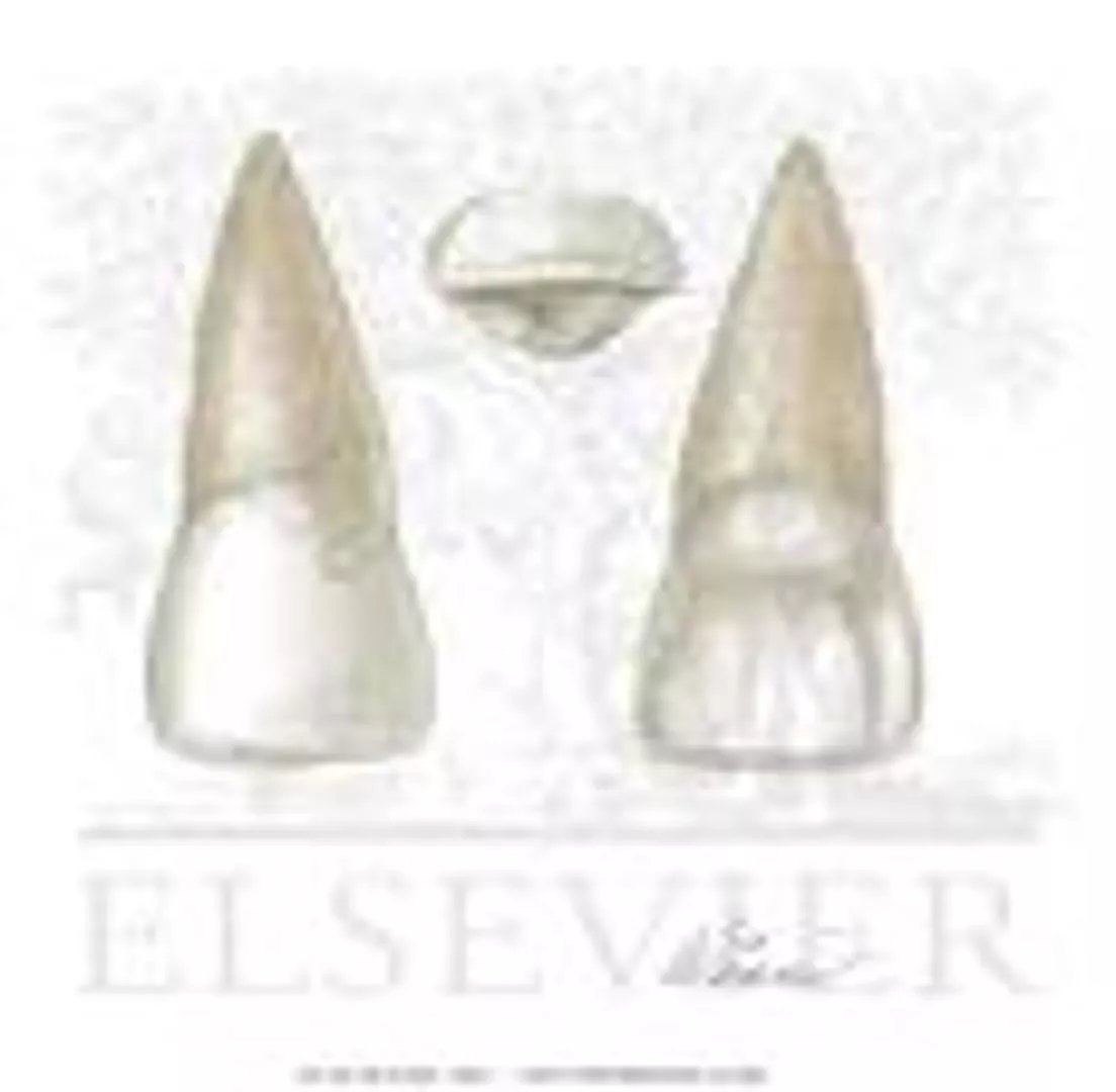
o
r
m
a
n
d
a
n
a
t
o
m
y
,
b
u
t
i
s
g
e
n
e
r
a
l
y
s
m
a
l
l
e
r
i
n
a
l
l
d
i
m
e
n
s
i
o
n
s
e
x
c
e
p
t
l
e
n
g
t
h
o
f
t
h
e
r
o
o
t
.
P
r
i
n
c
i
p
l
e
i
d
e
n
t
i
f
y
i
n
g
f
e
a
t
u
r
e
s
1
.
T
h
e
c
r
o
w
n
i
s
m
o
r
e
r
o
u
n
d
e
d
,
s
h
o
r
t
e
r
a
n
d
n
a
r
r
o
w
e
r
m
e
s
i
o
-
d
i
s
t
a
l
l
y
t
h
a
n
t
h
e
m
a
x
i
l
l
a
r
y
c
e
n
t
r
a
l
i
n
c
i
s
o
r
.
2
.
T
h
e
m
e
s
i
o
-
i
n
c
i
s
a
l
a
n
g
l
e
i
s
a
c
u
t
e
a
n
d
t
h
e
d
i
s
t
o
-
i
n
c
i
s
a
l
a
n
g
l
e
i
s
m
o
r
e
r
o
u
n
d
e
d
.
3
.
I
t
h
a
s
a
s
i
n
g
l
e
r
o
o
t
w
i
t
h
a
t
a
p
e
r
e
d
,
d
i
s
t
a
l
y
c
u
r
v
e
d
,
p
o
i
n
t
e
d
a
p
e
x
.
4
.
T
h
e
l
i
n
g
u
a
l
f
o
s
s
a
i
s
m
o
r
e
c
o
n
c
a
v
e
t
h
a
n
t
h
a
t
o
f
t
h
e
m
a
x
i
l
l
a
r
y
c
e
n
t
r
a
l
i
n
c
i
s
o
r
.
3
.
T
h
e
m
e
s
i
a
l
o
u
t
l
i
n
e
r
e
s
e
m
b
l
e
s
t
h
a
t
o
f
t
h
e
m
a
x
i
l
l
a
r
y
c
e
n
t
r
a
l
i
n
c
i
s
o
r
,
w
i
t
h
m
o
r
e
r
o
u
n
d
e
d
m
e
s
i
o
-
i
n
c
i
s
a
l
a
n
g
l
e
,
w
i
t
h
t
h
e
c
r
e
s
t
o
f
c
u
r
v
a
t
u
r
e
(
c
o
n
t
a
c
t
p
o
i
n
t
)
l
o
c
a
t
e
d
b
e
t
w
e
e
n
t
h
e
i
n
c
i
s
a
l
a
n
d
t
h
e
m
i
d
d
l
e
t
h
i
r
d
s
.
4
.
T
h
e
d
i
s
t
a
l
o
u
t
l
i
n
e
i
s
m
o
r
e
r
o
u
n
d
e
d
,
w
i
t
h
t
h
e
c
o
n
t
a
c
t
a
r
e
a
i
s
a
t
t
h
e
c
e
n
t
r
e
o
f
t
h
e
m
i
d
d
l
e
t
h
i
r
d
.
5
.
T
h
e
r
o
o
t
t
a
p
e
r
s
e
v
e
n
l
y
a
n
d
c
u
r
e
v
e
s
d
i
s
t
a
l
y
a
t
t
h
e
a
p
e
x
.
L
a
b
i
a
l
a
s
p
e
c
t
1
.
t
h
e
c
r
o
w
n
i
s
s
h
o
r
t
e
r
a
n
d
n
a
r
r
o
w
e
r
b
u
t
t
h
e
r
o
o
t
i
s
a
s
l
o
n
g
a
s
t
h
a
t
o
f
t
h
e
m
a
x
i
l
l
a
r
y
c
e
n
t
r
a
l
i
n
c
i
s
o
r
o
r
l
o
n
g
e
r
.
2
.
T
h
e
l
a
b
i
a
l
s
u
r
f
a
c
e
o
f
t
h
e
c
r
o
w
n
i
s
m
o
r
e
c
o
n
v
e
x
t
h
a
n
t
h
a
t
o
f
t
h
e
m
a
x
i
l
l
a
r
y
c
e
n
t
r
a
l
i
n
c
i
s
o
r
.
1
.
T
h
e
c
r
o
w
n
n
a
r
r
o
w
e
r
l
a
b
i
o
-
l
i
n
g
u
a
l
y
t
h
a
n
m
a
x
i
l
l
a
r
y
c
e
n
t
r
a
l
i
n
c
i
s
o
r
.
2
.
T
h
e
c
u
r
v
a
t
u
r
e
o
f
t
h
e
c
e
r
v
i
c
a
l
l
i
n
e
i
s
l
e
s
s
t
h
a
n
t
h
a
t
o
f
t
h
e
m
a
x
i
l
l
a
r
y
c
e
n
t
r
a
l
i
n
s
i
s
o
r
.
3
.
T
h
w
r
o
o
t
a
p
e
a
r
e
s
a
s
a
t
a
p
e
r
e
d
c
o
n
e
,
a
n
d
a
l
i
n
e
b
i
s
e
c
t
i
n
g
t
h
e
r
o
o
t
b
i
s
e
c
t
s
t
h
e
i
n
c
i
s
a
l
r
i
d
g
e
.
D
i
s
t
a
l
a
s
p
e
c
t
1
.
T
h
e
c
u
r
v
a
t
u
r
e
o
f
t
h
e
c
e
r
v
i
c
a
l
l
i
n
e
d
i
s
t
a
l
y
i
s
l
e
s
s
t
h
a
n
t
h
a
t
m
e
s
i
a
l
y
.
2
.
I
t
i
s
n
o
t
u
n
c
o
m
m
o
n
t
o
f
i
n
d
a
d
e
v
e
l
o
p
m
e
n
t
a
l
g
r
o
o
v
e
e
x
t
e
n
d
i
n
g
t
o
t
h
e
r
o
o
t
.
M
e
s
i
a
l
a
s
p
e
c
t
L
i
n
g
u
a
l
a
s
p
e
c
t
1
.
T
h
e
m
e
s
i
a
l
a
n
d
d
i
s
t
a
l
m
a
r
g
i
n
a
l
r
i
d
g
e
s
a
n
d
t
h
e
l
i
n
g
u
a
l
p
o
r
t
i
o
n
o
f
t
h
e
i
n
s
i
s
a
l
r
i
d
g
e
a
r
e
w
e
l
l
m
a
r
k
e
d
w
i
t
h
a
m
o
r
e
c
o
n
c
a
v
e
d
l
i
n
g
u
a
l
f
o
s
s
a
.
2
.
T
h
e
c
i
n
g
u
l
u
m
i
s
p
r
o
m
i
n
e
n
t
w
i
t
h
a
t
e
n
d
e
n
c
y
t
o
w
a
r
d
s
e
e
i
n
g
a
d
e
e
p
d
e
v
e
l
o
p
m
e
n
t
a
l
g
r
o
o
v
e
w
i
t
h
i
n
t
h
e
l
i
n
g
u
a
l
f
o
s
s
a
.
T
h
e
i
n
c
i
s
a
l
a
s
p
e
c
t
I
t
r
e
s
e
m
b
l
e
s
t
h
a
t
o
f
t
h
e
m
a
x
i
l
l
a
r
y
c
e
n
t
r
a
l
i
n
c
i
s
o
r
,
b
u
t
;
(a)
T
h
e
c
i
n
g
u
l
u
m
a
n
d
i
n
c
i
s
a
l
r
i
d
g
e
m
a
y
b
e
l
a
r
g
e
.
(b)
T
h
e
l
a
b
i
o
-
l
i
n
g
u
a
l
t
o
m
e
s
i
o
-
d
i
s
t
a
l
m
e
s
u
r
e
m
e
n
t
i
s
l
a
r
g
e
r
,
t
h
e
r
e
f
o
r
e
,
i
t
r
e
s
e
m
b
l
e
s
a
s
m
a
l
l
c
a
n
i
n
e
.
2
.
f
r
o
m
t
h
e
i
n
c
i
s
a
l
a
s
p
e
c
t
,
a
l
l
m
a
x
i
l
l
a
r
y
l
a
t
e
r
a
l
i
n
c
i
s
o
r
e
x
h
i
b
i
t
s
m
o
r
e
c
o
n
v
e
x
i
t
y
l
a
b
i
a
l
y
a
n
d
l
i
n
g
u
a
l
y
.
V
a
r
i
a
t
i
o
n
s
f
r
o
m
t
h
e
t
y
p
i
c
a
l
f
o
r
m
(
a
n
o
m
a
l
i
e
s
)
1
.
P
e
g
-
s
h
a
p
e
d
l
a
t
e
r
a
l
i
n
c
i
s
o
r
,
w
i
t
h
a
t
h
i
n
r
o
o
t
a
n
d
a
s
m
a
l
l
c
o
n
i
c
a
l
c
r
o
w
n
.
2
.
T
h
e
m
a
x
i
l
l
a
r
y
l
a
t
e
r
a
l
i
n
c
i
s
o
r
m
a
y
b
e
c
o
n
g
e
n
i
t
a
l
y
m
i
s
s
i
n
g
.
3
.
L
a
r
g
e
d
e
v
e
l
o
p
m
e
n
t
a
l
g
r
o
o
v
e
o
n
t
h
e
d
i
s
t
a
l
a
s
p
e
c
t
e
x
t
e
n
d
i
n
g
t
o
t
h
e
r
o
o
t
.
T
h
e
m
a
i
n
d
i
f
f
e
r
e
n
c
e
s
b
e
t
w
e
e
n
t
h
e
m
a
x
i
l
l
a
r
y
c
e
n
t
r
a
l
a
n
d
l
a
t
e
r
a
l
i
n
c
i
s
o
r
The maxillary central incisor is a prominent tooth in the upper front jaw, vital for cutting and shearing food. Differences between deciduous and permanent teeth, eruption timeline, and variations in shape and size are highlighted. Gender and age-related variances in appearance are discussed, along with details on the labial view and root structure.

Download Presentation

Please find below an Image/Link to download the presentation.
The content on the website is provided AS IS for your information and personal use only. It may not be sold, licensed, or shared on other websites without obtaining consent from the author. Download presentation by click this link. If you encounter any issues during the download, it is possible that the publisher has removed the file from their server.
E N D
Presentation Transcript
Lec. 4 and 5 Individual Anatomy of Permanent Teeth
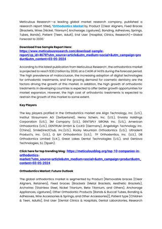
Maxillary central incisor Maxillary central incisor The maxillary central incisor is a human tooth in the front upper jaw, or maxilla, and is usually the most visible of all teeth in the mouth. It is located mesial (closer to the midline of the face) to the maxillary lateral incisor. As with all incisors, their function is for shearing or cutting food during mastication (chewing). There are no cusps on the teeth. Instead, the surface area of the tooth used in eating is called an incisal ridge or incisal edge. Formation of these teeth begin at 14 weeks in utero for the deciduous (baby) set and 3 4 months of age for the permanent set. There are some minor differences between the deciduous maxillary central incisor and that of the permanent maxillary central incisor. The deciduous tooth appears in the mouth at 3 18 months of age, with 6 months being the average and is replaced by the permanent tooth around 7 8 years of age. The permanent tooth is larger and is longer than it is wide. The maxillary central incisors contact each other at the midline of the face. The mandibular central incisors are the only other type of teeth to do so. The position of these teeth may determine the existence of an open bite or diastema. As with all teeth, variations of size, shape, and color exist among people. Systemic disease, such as syphilis, may affect the appearance of teeth
The permanent maxillary central incisor is the widest tooth mesiodistally in comparison to any other anterior tooth. It is larger than the neighboring lateral incisor and is usually not as convex on its labial surface. As a result, the central incisor appears to be more rectangular or square in shape. The mesial incisal angle is sharper than the distal incisal angle. When this tooth is newly erupted into the mouth, the incisal edges have three rounded features called mammelons.Mammelons disappear with time as the enamel wears away by friction. Generally, there are gender differences in the appearance of this tooth. In males, the size of the maxillary central incisor is larger usually than in females. Gender differences in enamel thickness and dentin width are low. Age differences in the gingival-incisal length of maxillary central incisors are seen and are attributed to normal attrition occurring throughout life. Thus, younger individuals have a greater gingival incisal length of the teeth than older individuals
Labial view Labial view The labial view of this tooth considers the portion of the tooth visible from the side where the lips would be. 1-The mesial outline of the tooth is straight or slightly convex, whereas the distal outline is much more convex. 2-The height of curvature (the point furthest away from the central axis of the tooth) is closer to the mesioincisal angle on the mesial side while more apical on the distal side. 3-the distal outline of the crown is more convex than the mesial outline 4-the distoincisal angle is not as sharp as the mesoincisal angle. 5- After the mammelons are worn away, the incisal edge of the maxillary central incisor is straight mesiodistally. 6-The cervical line, which is seen as the border between the crown and the root of the tooth, is closer to the apex of the root in the center of the tooth. This makes the cervical line appear as a semicircle in shape. 7- the root is blunt and cone-shaped. Although there is a large amount of variation between people, the length of the root is usually 2 3 mm longer than the length of the crown.Large curvatures of the root are usually not seen in this tooth
Lingual view Lingual view The lingual view of this tooth considers the portion of the tooth visible from the side where the tongue would be. 1-The lingual side of the maxillary central incisor has a small convexity, called a cingulum near the cervical line and 2-There is a large concavity, called the lingual fossa. 3-Along the mesial and distal sides are slightly raised portions called marginal ridges. The lingual incisal edge is also raised slightly to the level of the marginal ridges. 4-The lingual fossa is bordered incisally by the lingual incisal edge, mesially by the mesial marginal ridge, distally by the distal marginal ridge, and cervically by the cingulum. 5-Developmental grooves are found on the cingulum and lying into the lingual fossa.
Mesial Mesial view view The mesial view of this tooth considers the portion of the tooth visible from the side closest to where the middle line of the face would be. 1- The mesial side of the maxillary central incisor shows the crown of the tooth as a triangle with the point at the incisal edge and the base at the cervix. 2-The root appears cone shaped with a blunt apex. 3-Unlike most other teeth, a line drawn through the center of the incisal edge will also cross through the center of the root apex.This also occurs in maxillary lateral incisors. 4-The crest of curvature for the palatal and labial surfaces is located in the cervical third. 5-The labial surface of the crown is convex from the crest of curvature to the incisal edge. The lingual surface of the crown is convex near the cingulum and near the incisal edge, but for the most part is concave along the surface between those two areas. 6- More than any other tooth in the mouth, the cervical line from this view curves toward the incisal edge. In an average crown length of 10.5 to 11 mm, the curvature of the cervical line in a maxillary central incisor is 3 to 3.5 mm.
Distal view The distal view of this tooth considers the portion of the tooth visible from the side furthest from where the middle line of the face would be. 1-This side of the tooth is very similar to the mesial side. 2- A greater portion of the tooth labial surface from this view compared to the mesial view because the labial surface tilts distally and lingually. 3- the cervical line curves less in comparison to the mesial view. Incisal view The incisal view of this tooth considers the portion of the tooth visible from the side where the incisal edge is located. From this angle, only the crown of the tooth is visible, and overall the tooth looks bilateral. The labial surface appears broad and flat. The lingual surface tapers toward the cingulum. The distance between the mesioincisal angle to the cingulum is slightly longer than the distance between the distoincisal angle to the cingulum.
Interproximal contacts Contact with adjacent teeth in the same arch is referred to as interproximal contacts. The maxillary central incisors are one of only two types of teeth which has an interproximal contact with itself. The other type of teeth is the mandibular central incisors. In usually preferred and healthy status, the central incisors touch in the incisal third of the teeth.On the other hand, the contact between the central incisor and the lateral incisor is nearer the gingiva at the location between the incisal and middle thirds of the tooth's crown. Principle identifying features of maxillary central incisor 1.It is the widest anterior tooth mesio-distaly. 2.It has a square or rectangular appearance. 3.Straight mesial outline and rounded distal outline. 4.Sharp mesio-incisal angle and rounded disto-incisal angle. 5.Mammelones on the incisal ridge (in newly erupted teeth). 6.Well marked marginal ridges, lingual fossa and well developed cingulum. 7.Single tapered root.
Maxillary lateral incisor The maxillary lateral incisor resembles the maxillary central incisor in function,form and anatomy, but is generaly smaller in all dimensions except length of the root. Principle identifying features 1.The crown is more rounded,shorter and narrower mesio-distally than the maxillary central incisor. 2.The mesio-incisal angle is acute and the disto-incisal angle is more rounded. 3.It has a single root with a tapered, distaly curved, pointed apex. 4.The lingual fossa is more concave than that of the maxillary central incisor.
Labial aspect 1.the crown is shorter and narrower but the root is as long as that of the maxillary central incisor or longer. 2.The labial surface of the crown is more convex than that of the maxillary central incisor. 3.The mesial outline resembles that of the maxillary central incisor, with more rounded mesio-incisal angle, with the crest of curvature (contact point) located between the incisal and the middle thirds. 4.The distal outline is more rounded, with the contact area is at the centre of the middle third. 5.The root tapers evenly and cureves distaly at the apex.
Mesial aspect Mesial aspect 1. The crown narrower labio-lingualy than maxillary central incisor. 2.The curvature of the cervical line is less than that of the maxillary central insisor. 3.Thw root apeares as a tapered cone, and a line bisecting the root bisects the incisal ridge. Distal aspect 1.The curvature of the cervical line distaly is less than that mesialy. 2.It is not uncommon to find a developmental groove extending to the root.
Lingual aspect Lingual aspect 1. The mesial and distal marginal ridges and the lingual portion of the insisal ridge are well marked with a more concaved lingual fossa. 2.The cingulum is prominent with a tendency toward seeing a deep developmental groove within the lingual fossa. The incisal aspect It resembles that of the maxillary central incisor, but; The cingulum and incisal ridge may be large. (a) The labio-lingual to mesio-distal mesurement is larger, therefore, it resembles a small canine. (b) 2.from the incisal aspect, all maxillary lateral incisor exhibits more convexity labialy and lingualy.
Variations from the typical form( Variations from the typical form(anomalies) anomalies) 1.Peg- shaped lateral incisor, with a thin root and a small conical crown. 2.The maxillary lateral incisor may be congenitaly missing. 3.Large developmental groove on the distal aspect extending to the root.
The main differences between the maxillary central and lateral The main differences between the maxillary central and lateral incisor incisor Maxillary central incisor Maxillary lateral incisor Labial aspect 1.The crown longer and wider mesio-distally. 2.Mesial outline is slightly convex with the contact area near the mesio-incisal angle. 1.The crown is shorter and narrower. 2.Mesial outline is similar to the maxillary central incisor with acute and more rounded mesio-incisal angle with the crest of curvature at the junction between middle and incisal thirds. 3.Distal outline is more convex than mesial outline with the crest of curvature closer to the 3.Distal outline is more rounded and the crest of curvature is at the centre of the middle middle third. third. 4.The root is shorter with a cone shape and 4.The root is longer than maxillary central incisor,tapers evenly and curves distally in a blunt apex. pointed apex. Mesial aspect 5.The crown is wider labio-lingualy with a pronounced curvature of the cervical line. 5.The crown is narrower labio-lingualy with the curvature of the cervical line less pronounced. Distal aspect 6.No developmental groove extending to the 6.A developmental groove extend from the side of the cingulum to the root may be root. found. Lingual aspect 7.Well defined lingual anatomical features . 7.More prominent cingulum with more concave lingual fossa.