Comprehensive Overview of Maxillary Sinus Anatomy and Function
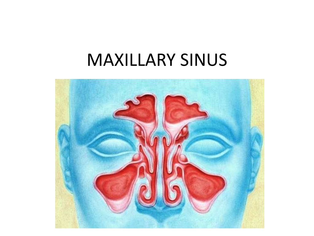

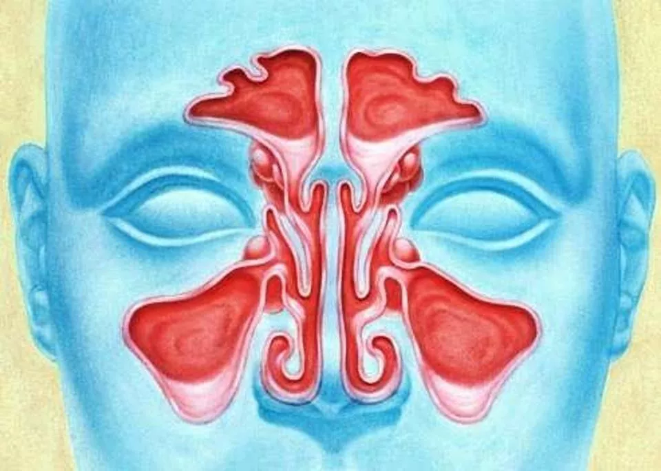
MAXILLARY SINUS
Anatomy
•
The maxillary sinus was first described in
1651, by Nathaniel Highmore. Therefore,
it is also known as antrum of Highmore
•
Maxillary sinuses are two in number,
one on either side of the maxilla, and
they are the largest of the paranasal air
sinuses
•
Dimensions –
3.5 × 3.2 × 2.5 cm (Turner 1902)
•
Volume- 15 to 30 ml
•
The ostium (3 to 6 mm diameter) opens
into middle meatus
•
It can be described as pyramidal in shape
•
Maxillary Sinus consist of
The base -lateral wall of the nose.
The apex -zygomatic process of maxilla;
The four walls are formed by:
(i)
the roof of antrum or the floor of orbit,
(ii)
the anterior, and
(iii)
infratemporal surfaces of body of maxilla, and
(iv)
floor of sinus- the alveolar process of maxilla which is the
Teeth
in
proximity
•
2
nd,
1
st
,
molar>3
rd
molar>2
nd
pm>1
st
pm>canine
Embryology (Growth of Maxillary
Sinus)
•
In early stages, maxillary sinus is high in maxilla. Later
gradually grows downward, by a process of pneumatization
•
Birth Tubular: 2 cm × 1 cm × 1 cm 3 mm per year -Tubular
9 years 60% of adult size-Ovoid
18 years Adult size-Pyramidal
Physiology
Pseudostratified columnar
ciliated
epithelium
Also Known as ‘Schneiderian membrane’
Mucociliary mechanism is useful means for removal of particulate
matter, bacteria etc.
The cilia move the mucus and other debris towards the ostium and
subsequently discharged in the middle meatus.
Functions
of
sinus
1.
Impart resonance to the voice.
2.
Increase the surface area and lighten the skull.
3.
Moisten and warm the inspired air.
4.
Filter the debris from the inspired air.
5.
Sinuses are located in front of the forebrain, olfactory region,
etc. They create “air padding” to provide thermal insulation to
the important tissues mentioned above.
Vascularization
&
innervation
Nerve Supply
•
superior dental nerves (anterior, middle and
posterior), and the greater palatine nerve.
Applied Surgical Anatomy
•
Relation of the Root Apices with the Floor of the Sinus
In adults, there is a distance of approximately, 1-1.25 cm between the floor of
the sinus and the root apices of maxillary posterior teeth
•
Low Incidence of Oroantral Fistula in Children under Fifteen Years
The maxillary sinus reaches its normal adult size by the age of 15 years
•
Lining of Maxillary Sinus
The wall of the sinus is very thin in this area. This area is used for following: (i)
diagnostic aspiration, and (ii) the site for Caldwell-Luc operation; that is,
the antral exploration with or without an intraoral antrostotomy
•
Fractures of the Middle Third of Face
LeForte I, II, and III, show disturbance in the walls of maxillary sinus.
•
Paraesthesia in Maxillary Teeth Following Surgical Procedures
Radiology of Maxillary Sinus
•
The various radiographs useful are as follows:
•
1. Extraoral views: (i) Occipitomental (OM), (ii) Lateral skull, (iii)
Submentovertex (SMV), (iv) Linear tomography, (v) Orthopantomography,
and (vi) Computed axial tomography (CAT).
•
2. Intraoral: (i) Occlusal, (ii) Lateral occlusal, and (iii) Periapical.
Maxillary
sinusitis
Group
of
diseases
mainly
inflammation
&
infection which affect
the nasal
mucosa
and
PNS
Acute Maxillary Sinusitis
•
Duration- 7 days-4 week
•
May be suppurative or non
suppurative
•
Symptoms
•
Cold 3-4 days prior to acute attack
•
Postnasal discharge with
pharyngitis
•
Constant throbbing pain
•
Generalized constitutional
symptoms
•
Signs
•
Tenderness & mild swelling
over cheek
•
Paresthesia over cheek
•
Fetor oris, discharge of pus in
to mouth through OAF
Chronic Maxillary Sinusitis
•
Causes
•
It may be due to-
•
Persistent dental focus
•
Chronic rhinitis
•
Allergic condition
•
Chronic infection of frontal &
ethamoidal sinuses
•
Pathophysiology-
•
Hyperplasia or atrophy of Mucus
membrane
•
Cilia are lost, multiple polyp
formation
•
Ostium shows oedematous leads to
complete blockage
•
Clinical features-
•
Asymptomatic
•
Sometimes pain & tenderness
in antrum area
•
Unilateral foul discharge from
posterior nares
•
Fetid odour with bad taste
•
Classical antral regimen includes:
Bed rest, plenty of fluids, maintenance of oral hygiene. The
regimen should be carried out for at least five to seven days.
Antimicrobials
The most suitable against common antral
pathogens are: (1) Macrolides: Erythromycin 250-500 mg six
hourly for 5 days. (2) Broad spectrum group: Amoxicillin 250
to 500 mg eight hourly for five days.
•
Decongestants
e.g. Ephedrine sulphate— 0.5 to 1 per cent in normal saline, six hourly.
ii. Xylometazolin hydrochloride 0.1 per cent.
•
Mucolytic agents
•
Drugs used—volatile oil preparations are used, Tinc.benzoin, camphor,
menthol, chlorbutol, etc. or simple steam inhalation every four hourly can
be used.
•
Non-steroidal anti-inflammatory analgesic agents
(i) Aspirin (ii)
Paracetamol (iii) Ibuprofen
Oroantral
fistula
•
It is an epitheliazed, pathological, unnatural
communication between the oral cavity and maxillary
sinus
Duration
and width of lumen
contributes
to
infection
of
sinus.
OAC
OAF(incidence:
0.3-3.8
%)
Oroantral
fistula
OAC
OAF
Defect
> 5mm
diameter
No
approximation
of
gingival
tissues
Post
op
regime
not
followed
Loss
of
clot
or
wound
dehiscence
Cyst
enucleation
Smoking,
drinking
Oroantral
fistula
Etiology
•
Iatrogenic
(50%)
•
Presence
of periapical
lesions
•
Injudicious
use
of
instruments
•
During
attempted
extraction
•
Trauma(7.5%)
•
Chronic
infections(11%)
•
Malignant
diseases(18.5%)
•
Infected
maxillary
dentures(3.7%)
•
h/o sinus
surgery(7.5%)
Symptoms of fresh
oroantral
communication (5E’s)
•
Escape of
fluids
•
Epistaxis
•
Escape of
air
•
Enhanced column
of
air.
•
Excruciating
pain
•
Symptoms of
established oroantral
fistula (5P’s)
•
Pain.
•
Persistent
purulent
unilateral
nasal
discharge.
•
Post
nasal drip.
•
Popping out of antral
polyp.
•
Possible sequalae of generalized systemic condition
Oroantral
fistula
Diagnosis
h/o
previous
extraction
Nose blowing test
Mouth mirror
test
Cotton
wisp
test
Inspection
Radiological
IOPA
OPG
O
ccipitomental view
Management
•
3mm-5mm
heals
spontaneously(HANAZANE)
•
Ideal
treatment
:immediate
surgery
followed
by
Ab
prophylaxis
•
Treatment of OAF within 24hrs of accident (Early)
- If edges of wounds are clean & not complicated by
displacement of tooth or root in to antrum then
closed by buccal flap and sutured under LA
- If complicated then treated by immediate surgery
under GA
•
Treatment of delayed cases-
•
Cases seen after 24hrs after accident-
•
- Defer the surgical closure until gingival edges show
sound healing i.e. 3weeks.
•
Cases seen 1 month after OAF- Surgical closure required.
Oroantral
fistula
1)
antibiotics
:
P
enicilli
n
&
derivatives
2)
nasal
decongestants:
Ephedrine
drops
Inhalations
(steam,benzoin
,menthol)
3)
Analgesics:
Aspirin
500mg
Paracetamol
500mg
Ibuprofen
400
mg
4)
Antral
lavage
Oroantral
fistula
Temporary Therapeutic measures
•
Whitehead’s
varnish
pack
•
Composition-
Benzoin 44g
Storax 33g
Balsum of tolu 22g
Iodoform 44g
Solvent ether 100 parts
Oroantral
fistula
•
Acrylic
plates
Surgical
closure
•
T
e
mp
oralis
flap
•
Forehead
flap
Overview
of
the
treatment
modalities
of
Oro-Antral
Communications
C
l
o
s
u
r
e
o
f
O
r
o
a
n
t
r
a
l
C
o
m
m
u
n
i
c
a
t
i
o
n
s
:
A
R
e
v
i
e
w
o
f
t
h
e
L
i
t
e
r
a
t
u
r
e
,
S
u
s
a
n
H
.
V
i
s
s
c
h
e
r
e
t
a
l
,
J
O
r
a
l
M
a
x
i
l
l
o
f
a
c
S
u
r
g
6
8
:
1
3
8
4
-
1
3
9
1
,
2
0
1
0
Surgical
closure
Factors
determining
flap
selection
Size
of
communication
Timeline of
diagnosing
Presence
of
infection
O
p
e
r
a
t
i
v
e
t
e
c
h
n
i
q
u
e
Buccal flap
advancement
O
peration
(
V
on Reher
m
an)
7
2
oroantral fistula closure by buccal advancement flap. Modified Rehrmann’s procedure:(1) OAF,
(2 and 3)
Outline of buccal flap, (4 and 5) Reflection of buccal mucoperiosteal flap. Relieving incision high up through
theperiosteum, (6) Sagittal section-Rehrmann buccal flap, (7) Modified Rehrmann flap with de-
epithelialization of the margin of the buccal flap, which is tucked under the palatal flap over the periosteum.
This ensures double layer closure. Buccal and some palatal alveolar bones are reduced with rongeurs, (8)
Initial mattress suturing to pull the margin of the flap and then interrupted suturing is carried out
ww
w
.
ind
i
andentalacadem
y
.
c
om
33
PALATAL
FLAPS
Rotational
advancement.(Ashley
1939)
34
SUBMUCOUS CONNECTIVE
TISSUE
FLAP(
Ito
et
al 1980)
35
BUCCAL
FAT
PAD(Hanazawa
et
al
1995
)
36
Combined
flap
Tongue
flap
Introduced by
lexer,1909
Technique
Advantages
Disadvantages
G
r
a
fts
Caldwell
luc
Operation
By
George Caldwell in 1893 from New York described a method of gaining
entry into the maxillary sinus via canine fossa with nasal antrostomy
Indications
Open procedure for removal of
root fragments, teeth or foreign
body or an antrolith
(stone) from the maxillary sinus.
2. To treat chronic
maxillary sinusitis
with hyperplastic lining and
polypoid degeneration of the mucosa.
3. Removal
of cysts or benign growths
from the maxillary sinus.
4. Management
of haematoma
in the maxillary sinus and to control
post-traumatic haemorrhage in the sinus.
5.
Zygomaticomaxillary complex fractures
involving floor of the
orbit and anterior wall of the maxillary sinus.
6. Removal of
impacted canine
or impacted third molar.
7. Along with closure of
chronic oroantral fistula
, associated with
chronic maxillary sinusitis.
Caldwell
luc
sinusotomy
Antibiotics, analgesics, antiinflammatory drugs for 5 days
The maxillary sinus, also known as the antrum of Highmore, is a key structure in the paranasal air sinuses. Described first in 1651, it plays a vital role in skull lightening, voice resonance, and air filtration. This pyramidal-shaped sinus is lined with Schneiderian membrane and has complex vascularization and innervation. Understanding its embryology, physiology, and proximity to teeth is crucial for dental and ENT professionals.

Download Presentation

Please find below an Image/Link to download the presentation.
The content on the website is provided AS IS for your information and personal use only. It may not be sold, licensed, or shared on other websites without obtaining consent from the author. Download presentation by click this link. If you encounter any issues during the download, it is possible that the publisher has removed the file from their server.
E N D
Presentation Transcript
Anatomy The maxillary sinus was first described in 1651, by Nathaniel Highmore. Therefore, it is also known as antrum of Highmore Maxillary sinuses are two in number, one on either side of the maxilla, and they are the largest of the paranasal air sinuses Dimensions 3.5 3.2 2.5 cm (Turner 1902) Volume- 15 to 30 ml The ostium (3 to 6 mm diameter) opens into middle meatus
It can be described as pyramidal in shape Maxillary Sinus consist of The base -lateral wall of the nose. The apex -zygomatic process of maxilla; The four walls are formed by: (i) the roof of antrum or the floor of orbit, (ii) the anterior, and (iii) infratemporal surfaces of body of maxilla, and (iv) floor of sinus- the alveolar process of maxilla which is the
Teeth inproximity 2nd, 1st , molar>3rd molar>2nd pm>1stpm>canine
Embryology (Growth of Maxillary Sinus) In early stages, maxillary sinus is high in maxilla. Later gradually grows downward, by a process of pneumatization Birth Tubular: 2 cm 1 cm 1 cm 3 mm per year -Tubular 9 years 60% of adult size-Ovoid 18 years Adult size-Pyramidal
Physiology Pseudostratified columnar ciliatedepithelium Also Known as Schneiderian membrane Mucociliary mechanism is useful means for removal of particulate matter, bacteria etc. The cilia move the mucus and other debris towards the ostium and subsequently discharged in the middle meatus.
Functions ofsinus Impart resonance to the voice. 1. Increase the surface area and lighten the skull. 2. Moisten and warm the inspired air. 3. Filter the debris from the inspired air. 4. Sinuses are located in front of the forebrain, olfactory region, 5. etc. They create air padding to provide thermal insulation to the important tissues mentioned above.
Vascularization & innervation a) Nasal MucosalVasculature SP,Ethmoid Arterial Supply b) OsseousVasculature IO, PSA, ASA, GP, Facial a) Medial wall - SP VenousDrainage b) Other walls PterygomaxillaryPlexus LymphaticDrainage Collecting vessels in middlemeatus NerveInnervation ION, GP, PSA, MSA, ASA
Nerve Supply superior dental nerves (anterior, middle and posterior), and the greater palatine nerve.
Applied Surgical Anatomy Relation of the Root Apices with the Floor of the Sinus In adults, there is a distance of approximately, 1-1.25 cm between the floor of the sinus and the root apices of maxillary posterior teeth The maxillary sinus reaches its normal adult size by the age of 15 years Low Incidence of Oroantral Fistula in Children under Fifteen Years The wall of the sinus is very thin in this area. This area is used for following: (i) diagnostic aspiration, and (ii) the site for Caldwell-Luc operation; that is, the antral exploration with or without an intraoral antrostotomy Lining of Maxillary Sinus
LeForte I, II, and III, show disturbance in the walls of maxillary sinus. Fractures of the Middle Third of Face Paraesthesia in Maxillary Teeth Following Surgical Procedures
Radiology of Maxillary Sinus The various radiographs useful are as follows: 1. Extraoral views: (i) Occipitomental (OM), (ii) Lateral skull, (iii) Submentovertex (SMV), (iv) Linear tomography, (v) Orthopantomography, and (vi) Computed axial tomography (CAT). 2. Intraoral: (i) Occlusal, (ii) Lateral occlusal, and (iii) Periapical.
Maxillarysinusitis Group of diseases mainly inflammation& infection which affect the nasal mucosa and PNS
Acute Maxillary Sinusitis Duration- 7 days-4 week Signs May be suppurative or non suppurative Symptoms Tenderness & mild swelling over cheek Cold 3-4 days prior to acute attack Paresthesia over cheek Postnasal discharge with pharyngitis Fetor oris, discharge of pus in to mouth through OAF Constant throbbing pain Generalized constitutional symptoms
Chronic Maxillary Sinusitis Pathophysiology- Causes It may be due to- Persistent dental focus Hyperplasia or atrophy of Mucus membrane Cilia are lost, multiple polyp formation Ostium shows oedematous leads to complete blockage Chronic rhinitis Allergic condition Chronic infection of frontal & ethamoidal sinuses
Clinical features- Asymptomatic Sometimes pain & tenderness in antrum area Unilateral foul discharge from posterior nares Fetid odour with bad taste
Classical antral regimen includes: Bed rest, plenty of fluids, maintenance of oral hygiene. The regimen should be carried out for at least five to seven days. Antimicrobials The most suitable against common antral pathogens are: (1) Macrolides: Erythromycin 250-500 mg six hourly for 5 days. (2) Broad spectrum group: Amoxicillin 250 to 500 mg eight hourly for five days.
Decongestants e.g. Ephedrine sulphate 0.5 to 1 per cent in normal saline, six hourly. ii. Xylometazolin hydrochloride 0.1 per cent. Mucolytic agents Drugs used volatile oil preparations are used, Tinc.benzoin, camphor, menthol, chlorbutol, etc. or simple steam inhalation every four hourly can be used. Non-steroidal anti-inflammatory analgesic agents (i) Aspirin (ii) Paracetamol (iii) Ibuprofen
Oroantral fistula It is an epitheliazed, pathological, unnatural communication between the oral cavity and maxillary sinus Duration and width of lumen contributesto infection of sinus. OAC OAF(incidence: 0.3-3.8 %)
Oroantral fistula OAC OAF Defect > 5mmdiameter No approximation of gingivaltissues Post op regime notfollowed Lossof clotorwounddehiscence Cystenucleation Smoking,drinking
Oroantral fistula Etiology Iatrogenic(50%) Presence of periapicallesions Injudicious use ofinstruments During attemptedextraction Trauma(7.5%) Chronic infections(11%) Malignantdiseases(18.5%) Infected maxillarydentures(3.7%) h/o sinussurgery(7.5%)
Symptoms of fresh oroantral communication (5Es) Escape of fluids Epistaxis Escape of air Enhanced column of air. Excruciating pain Symptoms of established oroantral fistula (5P s) Pain. Persistent purulent unilateral nasal discharge. Post nasal drip. Popping out of antral polyp. Possible sequalae of generalized systemic condition
Oroantral fistula Diagnosis h/o previous extraction Nose blowing test Mouth mirrortest Cotton wisptest Inspection Radiological IOPA OPG Occipitomental view
Management 3mm-5mm healsspontaneously(HANAZANE) Ideal treatment :immediate surgery followed byAb prophylaxis Treatment of OAF within 24hrs of accident (Early) - If edges of wounds are clean & not complicated by displacement of tooth or root in to antrum then closed by buccal flap and sutured under LA - If complicated then treated by immediate surgery under GA
Treatment of delayed cases- Cases seen after 24hrs after accident- - Defer the surgical closure until gingival edges show sound healing i.e. 3weeks. Cases seen 1 month after OAF- Surgical closure required.
Oroantral fistula 1) antibiotics : Penicillin &derivatives 2) nasaldecongestants: Ephedrinedrops Inhalations (steam,benzoin,menthol) 3) Analgesics: Aspirin500mg Paracetamol 500mg Ibuprofen 400 mg Antral lavage 4)
Oroantral fistula Temporary Therapeutic measures Whitehead svarnish pack Composition- Benzoin 44g Storax 33g Balsum of tolu 22g Iodoform 44g Solvent ether 100 parts
Oroantral fistula Acrylicplates
Surgicalclosure Temporalis flap Forehead flap Overview of the treatment modalities of Oro-AntralCommunications Closure of Oroantral Communications:A Review of the Literature, Susan H. Visscher et al, J OralMaxillofac Surg68:1384-1391, 2010
Surgicalclosure Factors determining flapselection Size ofcommunication Timeline ofdiagnosing Presence of infection
Operative technique Buccal flap advancement Operation (Von Reherman) oroantral fistula closure by buccal advancement flap. Modified Rehrmann s procedure:(1) OAF, (2 and 3) Outline of buccal flap, (4 and 5) Reflection of buccal mucoperiosteal flap. Relieving incision high up through theperiosteum, (6) Sagittal section-Rehrmann buccal flap, (7) Modified Rehrmann flap with de- epithelialization of the margin of the buccal flap, which is tucked under the palatal flap over the periosteum. This ensures double layer closure. Buccal and some palatal alveolar bones are reduced with rongeurs, (8) Initial mattress suturing to pull the margin of the flap and then interrupted suturing is carried out 7
PALATALFLAPS Rotational advancement.(Ashley 1939) 34
SUBMUCOUS CONNECTIVE TISSUE FLAP( Ito et al 1980) 35
BUCCAL FAT PAD(Hanazawa et al 1995 ) 36
Tongueflap Introduced bylexer,1909 Technique Advantages Disadvantages
Caldwell luc Operation By George Caldwell in 1893 from New York described a method of gaining entry into the maxillary sinus via canine fossa with nasal antrostomy Indications Open procedure for removal of root fragments, teeth or foreign body or an antrolith (stone) from the maxillary sinus. 2. To treat chronic maxillary sinusitis with hyperplastic lining and polypoid degeneration of the mucosa. 3. Removal of cysts or benign growths from the maxillary sinus. 4. Management of haematoma in the maxillary sinus and to control post-traumatic haemorrhage in the sinus. 5. Zygomaticomaxillary complex fractures involving floor of the orbit and anterior wall of the maxillary sinus. 6. Removal of impacted canine or impacted third molar. 7. Along with closure of chronic oroantral fistula, associated with chronic maxillary sinusitis.
Caldwell luc sinusotomy Antibiotics, analgesics, antiinflammatory drugs for 5 days


")