Anatomy and Skeletal System Overview
Delve into the fundamental concepts of anatomy and the skeletal system in this comprehensive lecture by Dr. Essam Salama. Explore various anatomical fields, terminology, positions, movements, planes, and bone classifications, aiding in a deeper understanding of the human body's structural framework.

Download Presentation

Please find below an Image/Link to download the presentation.
The content on the website is provided AS IS for your information and personal use only. It may not be sold, licensed, or shared on other websites without obtaining consent from the author.If you encounter any issues during the download, it is possible that the publisher has removed the file from their server.
You are allowed to download the files provided on this website for personal or commercial use, subject to the condition that they are used lawfully. All files are the property of their respective owners.
The content on the website is provided AS IS for your information and personal use only. It may not be sold, licensed, or shared on other websites without obtaining consent from the author.
E N D
Presentation Transcript
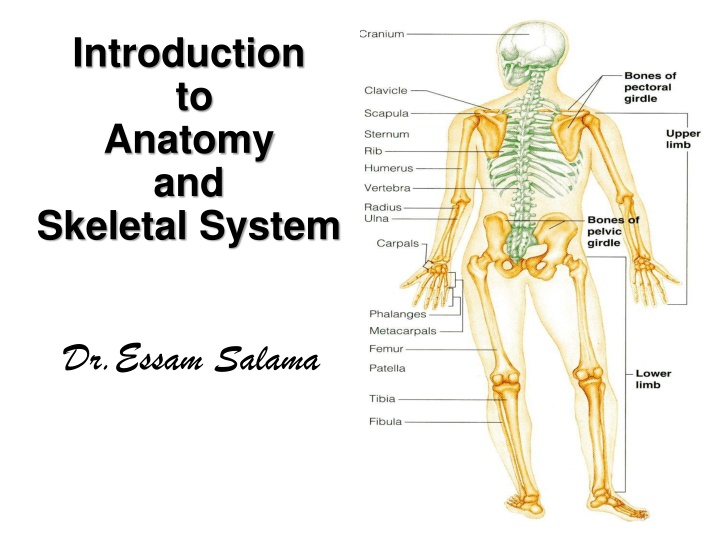
Introduction to Anatomy and Skeletal System Dr.Essam Salama
OBJECTIVES By the end of the lecture, you should be able to: Define the word Anatomy . Enumerate the different anatomical fields. Describe the anatomical position. Describe different anatomical terms of position & movements as well different anatomical planes. Classify bones according to shape, structure & development. Enumerate different bones of both axial & appendicular skeleton.
WHAT IS ANATOMY? The word anatome is of Greek origin meaning cutting up (ana= up; tome= cutting). Gross (macroscopic) anatomy: Study of human body with naked eye. Microscopic anatomy; (Histology): Study of fine structure (cells & tissues) of the human body with the help of microscope. Developmental anatomy; ( Embryology). Radiological anatomy. Applied anatomy. Surface anatomy. Surgical anatomy.
ANATOMICAL POSITION It is the standard position in which the body assume to describe its parts. Body is erect Arms hanging by the side Palm facing forward Feet are parallel
ANATOMICAL TERMINOLOGY TERMS OF POSITION Superior (cranial): near to head. X Inferior (caudal): away from head. Anterior (ventral): near to front. X Posterior (dorsal): near to back. Medial: near to median plane. X Lateral: away from median plane Proximal: near to trunk. X Distal: away from trunk. Superficial: near to skin (surface). X Deep: away from skin.
ANATOMICAL TERMINOLOGY TERMS OF MOVEMENT Flexion: approximation of 2 parts (decreasing the angle between 2 parts). X Extension: straightening (increasing the angle between 2 parts). Abduction: away from median plane. X Adduction: toward median plane. Lateral rotation: rotation away from median plane. X Medial rotation: rotation toward median plane. Circumduction: combined movements of flexion, extension, abduction & adduction.
ANATOMICAL PLANES & SECTIONS Sagittal (median): divides the body into 2 equal halves (right & left). Parasagittal (paramedian): divides the body into 2 unequal parts (right & left). Frontal (coronal): divides the body into anterior & posterior parts. Transverse (cross): divides the body into superior & inferior .parts
PLANES, TERMS OF POSITION & TERMS OF MOVEMENT
BODY CAVITIES Ventral body cavity: divided by diaphragm into: 1. Thoracic cavity: superior to diaphragm, contains heart & lungs. 2. Abdominal cavity: inferior to diaphragm, contains stomach, intestine, liver, urinary bladder, etc Dorsal body cavity: divided into 2 parts continuous with each other: 1. Cranial cavity: space inside skull, contains brain 2. Spinal cavity: space inside vertebral column, contains spinal cord
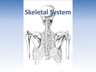
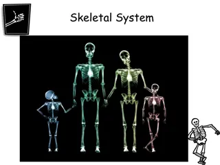
SKELETAL SYSTEM Includes: Bones Joints: articulations between bones
FUNCTIONS OF BONE 1. Support: of the body. 2. Storage: of fat and minerals e.g. calcium and phosphorus. 3. Protection: of soft body organs. 4. Attachment: of muscles. 5. Movement: of the body as a whole, or of the body parts. 6. Blood cell formation.
CLASSIFICATION OF BONE Bones are classified on the bases of their: Shape: Long, Short, Flat, Irregular. Structure: Compact, Spongy. Development: Membrane, Cartilage.
THE SKELETON Formed of 206 bones. Divided into: 1. Axial skeleton: Bones forming the trunk (longitudinal axis) of body. 2. Appendicular skeleton: Bones forming the girdles & limbs.
BONES OF AXIAL SKELETON SKULL Consists of: Cranium: bones enclosing brain: Frontal, Occipital, Parietal, Temporal. Facial bones: bones of face: Maxilla, Nasal, Zygomatic, Mandible.
BONES OF AXIAL SKELETON VERTEBRAL COLUMN Number: 33 vertebrae. Functions: protects spinal cord and supports the body. Formed of: 7 cervical vertebrae. 12 thoracic vertebrae. 5 lumbar vertebrae. 5 sacral vertebrae fused to form sacrum. 4 coccygeal vertebrae fused to form coccyx.
BONES OF AXIAL SKELETON STERNUM Has 3 parts: Manubrium, Body & Xiphoid process. RIBS 12 pairs: All ribs articulate with vertebrae. Only upper 7 pairs articulate with sternum, (true ribs). 8th ,9th & 10th ribs are false ribs. 11th & 12th ribs are floating ribs.
BONES OF APPENDICULAR SKELETON PECTORAL GIRDLE Connects upper limb with axial skeleton. Formed of:(two bones on each side) Clavicle & Scapula. PELVIC GIRDLE Connects lower limb with axial skeleton. Formed of:(one only on each side Hip bone.
BONES OF APPENDICULAR SKELETON UPPER LIMB Bone of arm: Humerus. Bones of forearm: Radius(lateral) & ulna (medial). Bones of the hand: 8 carpal bones. 5 metacarpal bones. 14 phalanges: 2 for thumb & 3 for each of medial 4 fingers.
BONES OF APPENDICULAR SKELETON LOWER LIMB Bone of thigh: Femur. Bones of leg: Fibula(lateral) & Tibia (medial). Patella. Bones of foot: 7 tarsal bones. 5 metatarsal bones. 14 phalanges: 2 for big toe & 3 for each of lateral 4 toes.
LONG BONES Formed of: A shaft (diaphysis): composed of compact bone. Two ends (epiphysis): composed of spongy bone. Metaphysis: This is the region of contact between epiphysis & diaphysis. The metaphysis contains epiphyseal plate of cartilage responsible for linear bone growth.
Reference book CLINICAL ANATOMY RICHARD S. SNELL 7TH EDITION LIPPINCOTT WILLIAMS & WILKINS
TEST YOURSELF! QUESTION 1 Which one of the following bones is a bone of the axial skeleton? 1. Femur. 2. Humerus. 3. Scapula. 4. Sternum.
QUESTION 2 Which one of the following bones is an example of an irregular bone? 1. Femur. 2. Vertebra. 3. Scapula. 4. Sternum.
QUESTION 3 Which one of the following planes divides the body into superior & inferior parts? 1. Frontal (coronal) plane. 2. Sagittal (median) plane. 3. Parasagittal (Paramedian) plane 4. Transverse plane.
THANK YOU THANK YOU AND AND GOOD LUCK GOOD LUCK