Acute Coronary Syndromes: Causes, Symptoms, and Risk Factors
Acute coronary syndromes (ACS) encompass a spectrum of conditions such as unstable angina, non-ST-segment elevated MI (NSTEMI), and ST-segment elevated MI (STEMI) that result from coronary artery occlusion. Risk factors for ACS include family history of heart disease, obesity, smoking, and hypertension. ACS typically occurs due to thrombus progression leading to myocardial oxygen supply-demand imbalance. Recognizing symptoms like chest pain radiating to the arm, neck, or jaw is crucial for prompt diagnosis and treatment.

Download Presentation

Please find below an Image/Link to download the presentation.
The content on the website is provided AS IS for your information and personal use only. It may not be sold, licensed, or shared on other websites without obtaining consent from the author.If you encounter any issues during the download, it is possible that the publisher has removed the file from their server.
You are allowed to download the files provided on this website for personal or commercial use, subject to the condition that they are used lawfully. All files are the property of their respective owners.
The content on the website is provided AS IS for your information and personal use only. It may not be sold, licensed, or shared on other websites without obtaining consent from the author.
E N D
Presentation Transcript
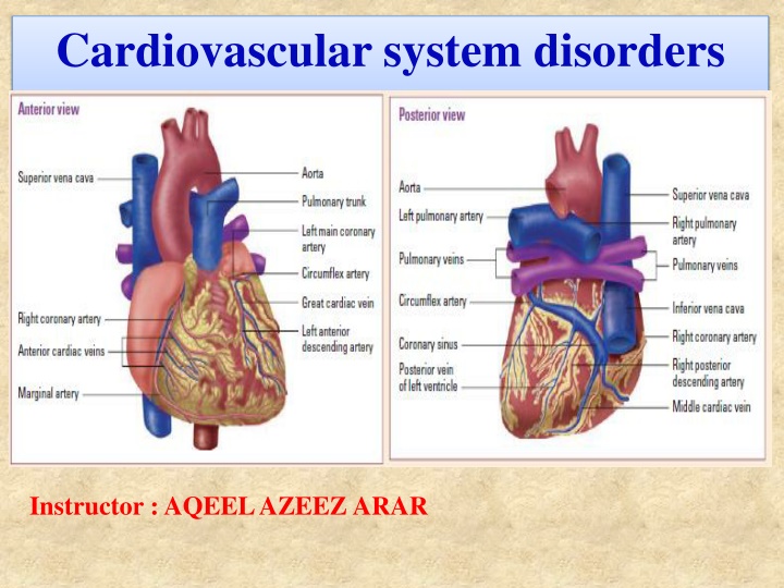
Cardiovascular system disorders Instructor : AQEEL AZEEZ ARAR
Common cardiovascular disorders include: Acute coronary syndromes Aneurysms Cardiac arrhythmias Cardiac tamponade Cardiogenic shock Cardiomyopathy Heart failure Hypertensive crisis Pericarditis valvular heart disease.
1. Acute coronary syndromes Patients with acute coronary syndromes have some degree of coronary artery occlusion. The degree of occlusion defines whether the ACS is: Unstable angina. Non ST-segment elevated MI (NSTEMI). ST segment elevated MI (STEMI). Plaque s place The development of any acute coronary syndrome begins with a rupture or erosion of plaque an unstable and lipid-rich substance. The rupture results in platelet adhesions, fibrin clot formation, and activation of thrombin.
What causes it? Risk factors appear to face a greater likelihood of developing an ACS: Family history of heart disease Menopause Obesity Stress Smoking Diabetes High-fat, high-carbohydrate diet Hypertension Sedentary lifestyle Hyperlipoproteinemia.
How it happens An acute coronary syndrome most commonly results when a thrombus progresses and occludes blood flow. (An early thrombus doesn t necessarily block blood flow.) The effect is an imbalance in myocardial oxygen supply and demand. Degree and duration The degree and duration of blockage dictate the type of infarct that occurs: If the patient has unstable angina, a thrombus partially occludes occluded vessel may have distal microthrombi that cause necrosis in some myocytes.
If smaller vessels infarct, the patient is at higher risk for MI, which may progress to NSTEMI. Usually, only the innermost layer of the heart is damaged. STEMI results when reduced blood flow through one of the coronary arteries causes myocardial ischemia, injury, and necrosis. The damage extends through all myocardial layers. What to look for A patient with angina typically experiences: burning squeezing crushing tightness in the substernal or precordial chest that may radiate to the left arm, neck, jaw, or shoulder blade.
atypical chest pain in women Women with coronary artery disease may experience typical chest pain, but commonly experience atypical chest pain, vague chest pain, or a lack of chest pain. They re more likely than men to experience a toothache or pain in the arm, shoulder, jaw, neck, throat, back, breast, or stomach. It hurts when I do this Angina most frequently follows physical exertion but may also follow emotional excitement, exposure to cold, or a large meal. Angina is commonly relieved by nitroglycerin and rest. It s less severe and shorter-lived than the pain of acute MI.
Four forms Angina has four major forms: 1- Stable predictable pain, in frequency and duration, which can be relieved with nitrates and rest 2- Unstable increased pain, which is easily induced 3- Prinzmetal s or a variant pain from unpredictable coronary artery spasm 4- Microvascular angina-like chest pain due to impairment of vasodilator reserve in a patient with normal coronary arteries.
MI pain A patient with MI experiences severe, persistent chest pain that isn t relieved by rest or nitroglycerin. He may describe pain as crushing or squeezing. The pain is usually substernal, but may radiate to the left arm, jaw, neck, or shoulder blades. Other signs and symptoms of MI include: a feeling of impending doom fatigue nausea and vomiting shortness of breath cool extremities perspiration anxiety hypotension or hypertension palpable precordial pulse muffled heart sounds.
What tests tell you These tests are used to diagnose CAD: ECG during an anginal episode shows ischemia. Serial 12-lead ECGs may be normal or inconclusive during the first few hours after an MI. Abnormalities include serial ST-segment depression in NSTEMI and ST-segment elevation and Q waves, representing scarring and necrosis, in STEMI. (See Locating myocardial damage, page 256.) Coronary angiography reveals coronary artery stenosis or obstruction and collateral circulation and shows the condition of the arteries beyond the narrowing. Myocardial perfusion imaging with thallium-201 during treadmill exercise discloses ischemic areas of the myocardium, visualized as cold spots.
With MI, serial serum cardiac marker measurements show elevated CK, especially the CK-MB isoenzyme (the cardiac muscle fraction of CK), troponin T and I, myoglobin, and ischemia modified albumin. C-reactive protein (CRP) levels help measure cardiac risk. Patients with chest pain and a higher CRP level have an increased risk of CAD. The PLAC test is a new test that also helps identify patients at a higher risk for CAD. With STEMI, echocardiography shows ventricular wall dyskinesia.
For patients with angina, the goal is to reduce oxygen demand or increase oxygen supply.
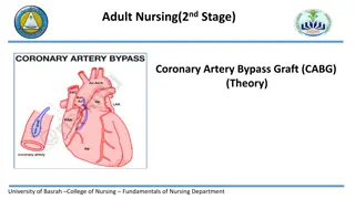
How its treated The goal treatment of angina is to reduce myocardial oxygen demand or increase oxygen supply. These treatments are used to manage angina: Nitrates reduce myocardial oxygen consumption. Beta-adrenergic blockers may be administered to reduce the workload and oxygen demands of the heart. If angina is caused by coronary artery spasm, calcium channel blockers may be given. Antiplatelet drugs minimize platelet aggregation and the danger of coronary occlusion. Antilipemic drugs can reduce elevated serum cholesterol or triglyceride levels. Obstructive lesions may necessitate CABG or PTCA. Other alternatives include laser angioplasty, minimally invasive surgery, atherectomy, or stent placement.
Myocardial Infarction Relief The goals of treatment for MI are to relieve pain, stabilize heart rhythm, revascularize the coronary artery, preserve myocardial tissue and reduce cardiac workload. some guidelines for treatment: Thrombolytic therapy should be started within 6 hours of the onset of symptoms (unless contraindications exist). Thrombolytic therapy involves administration of streptokinase (Streptase), alteplase (Activase), or reteplase (Retavase). PTCA or stent placement are options for opening blocked or narrowed arteries. Oxygen is administered to increase oxygenation of the blood. Nitroglycerin is administered sublingually to relieve chest pain, unless systolic blood pressure is less than 90 mm Hg or heart rate is less than 50 or greater than 100 beats/minute.
Heart ache Morphine is administered as analgesia because pain stimulates the sympathetic nervous system, leading to an increase in heart rate and vasoconstriction. Aspirin and anti-platelet drugs are administered to inhibit platelet aggregation. I.V. heparin is given to patients who have received tissue plasminogen activator to increase the chances of patency in the affected coronary artery. Lidocaine, transcutaneous pacing patches (or a transvenous pacemaker), defibrillation, or epinephrine may be used if arrhythmias are present. Physical activity is limited for the first 12 hours to reduce cardiac workload, thereby limiting the area of necrosis.
I.V. nitroglycerin is administered for 24 to 48 hours in patients without hypotension, bradycardia, or excessive tachycardia, to reduce afterload and preload and relieve chest pain. Glycoprotein IIb/IIIa inhibitors (such as abciximab [ReoPro]) are administered to patients with continued unstable angina or acute chest pain, or following invasive cardiac procedures, to reduce platelet aggregation. I.V. beta-adrenergic blocker is administered early to patients with evolving acute MI; it s followed by oral therapy to reduce heart rate and contractibility and reduce myocardial oxygen requirements.
ACE inhibitors are administered to those with evolving MI with ST- segment elevation or left bundle-branch block, to reduce afterload and preload and prevent remodeling. Laser angioplasty, atherectomy, or stent placement may be initiated. Lipid-lowering drugs are administered to patients with elevated LDL and cholesterol levels. What to do During anginal episodes, monitor blood pressure and heart rate. Take an ECG before administering nitroglycerin or other nitrates. Record duration of pain, amount of medication required to relieve it, and accompanying symptoms. During episodes of chest pain, monitor ECG, blood pressure, and PA catheter readings for changes.
CCU, ECG, and more! On admission to coronary care unit, monitor and record the patient s ECG, blood pressure, temperature, and heart and breath sounds. Also, assess and record the severity, location, type, and duration of pain. Obtain a 12-lead ECG and assess heart rate and blood pressure when the patient experiences acute chest pain. Monitor the patient s hemodynamic status closely. Be alert for indicators suggesting decreased cardiac output, such as decreased blood pressure, increased heart rate, increased PAP, increased PAWP, decreased cardiac output measurements, and decreased right atrial pressure. Assess urine output hourly. Monitor the patient s oxygen saturation levels and notify the practitioner if oxygen saturation falls below 90%. Check the patient s blood pressure after giving nitroglycerin, especially the first dose.
During episodes of chest pain, monitor ECG, blood pressure, and PA catheter readings (if applicable) to determine changes. Frequently monitor ECG rhythm strips to detect heart rate changes and arrhythmias. Obtain serial measurements of cardiac enzyme levels as ordered. Watch for crackles, cough, tachypnea, and edema, which may indicate impending left-sided heart failure. Carefully monitor dailyweight, intake and output, respiratory rate, serum enzyme levels, ECG waveforms, and blood pressure. Auscultate for S3 or S4 gallops. Prepare the patient for reperfusion therapy as indicated. Administer and titrate medications as ordered. Avoid giving I.M. injections; I.V. administration provides more rapid symptom relief.
I need a break Organize patient care and activities to allow rest periods. If the patient is immobilized, turn him often and use intermittent compression devices. Gradually increase the patient s activity level as tolerated. Provide a clear-liquid diet until nausea subsides. Anticipate a possible order for a low-cholesterol, low-sodium diet without caffeine. Provide a stool softener to prevent straining during defecation. Remember, I.V. administration of medications provides more rapid relief of the patient s MI symptoms.