Understanding Acute Coronary Syndrome Differentiation and Treatment
Learn to distinguish between STE and NSTE acute coronary syndromes in terms of diagnosis, prognosis, and treatment strategies. Understand the pathophysiological differences between ACS and chronic coronary syndromes. Gain insight into diagnostic approaches, treatment regimens, and prognosis for various types of ACS. Develop pharmacotherapy plans for patients undergoing primary PCI or fibrinolytic therapy for STEMI, as well as those with NSTE-ACS. Design therapeutic regimens pre-discharge and discover online resources for guideline implementation.

Uploaded on Sep 15, 2024 | 0 Views
Download Presentation

Please find below an Image/Link to download the presentation.
The content on the website is provided AS IS for your information and personal use only. It may not be sold, licensed, or shared on other websites without obtaining consent from the author. Download presentation by click this link. If you encounter any issues during the download, it is possible that the publisher has removed the file from their server.
E N D
Presentation Transcript
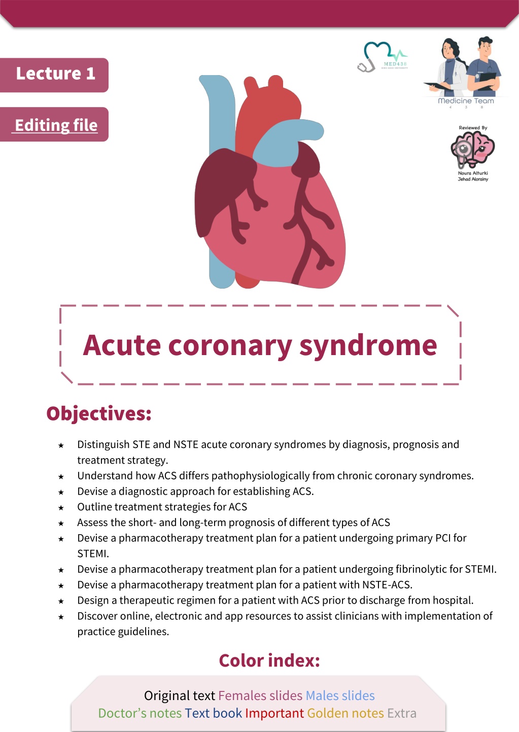
Lecture 1 Editing file Acute coronary syndrome Objectives: Distinguish STE and NSTE acute coronary syndromes by diagnosis, prognosis and treatment strategy. Understand how ACS differs pathophysiologically from chronic coronary syndromes. Devise a diagnostic approach for establishing ACS. Outline treatment strategies for ACS Assess the short-and long-term prognosis of different types of ACS Devise a pharmacotherapy treatment plan for a patient undergoing primary PCI for STEMI. Devise a pharmacotherapy treatment plan for a patient undergoing fibrinolytic for STEMI. Devise a pharmacotherapy treatment plan for a patient with NSTE-ACS. Design a therapeutic regimen for a patient with ACS prior to discharge from hospital. Discover online, electronic and app resources to assist clinicians with implementation of practice guidelines. Color index: Color index: Original text Females slides Males slides Doctor s notes Text bookImportant Golden notes Extra
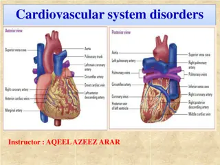
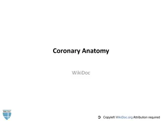
Introduction 2 Anatomy EXTRA The left main and right coronary arteries arise from the left and right sinuses of the aortic root, distal to the aortic valve: Left main coronary artery:1 Left Anterior Descending (LAD): Supply anterior 2/3 of interventricular septum, anterolateral papillary muscle, and anterior surface of LV. Most commonly occluded. Left Circumflex (LCX):Supply the lateral, posterior and inferior segments of the LV. Right main coronary artery:2supplies SA node3 Posterior Descending Artery (PDA): Supplies AV node (dependent on dominance), posterior 1/3 of interventricular septum, posterior 2/3 walls of ventricles, and posteromedial papillary muscle. Acute Marginal Artery (AMA): Supplies RV. ACS Introduction to ACS NSTE-ACS STE-ACS Acute coronary syndrome is a term that encompasses both unstable anginaand myocardial infarction (NSTEMI & STEMI). Acute coronary syndrome almost always occurs in patients who have atherosclerosis1. UA STEMI NSTEMI ACS: Acute coronary syndromes Unstable angina NSTE-ACS: Non-ST elevation ACS STE-ACS: ST elevation ACS elevation MI UA: NSTEMI: Non-ST elevation MI STEMI: ST Unstable angina No myocardial necrosis NSTEMI STEMI Subendocardial infarcts Transmural infarcts Thrombosis with incomplete coronary artery occlusion Normal or nonspecific (e.g. ST depression) on ECG but NOcardiac biomarker elevation Characterised by angina on minimal exertion or angina at rest in the absence of myocardial damage. Lasts less than 20 min. Subendocardium (inner 1/3) especially vulnerable to ischemia; if the blood doesn t return it progresses to STEMI. Normal or nonspecific (e.g. ST depression) and cardiac biomarker elevation. Full thickness of myocardial wall involved. Completer occlusion. ST elevation and cardiac biomarker elevation. Lasts more than 20 min. The pain does not usually respond to sublingual glyceryl trinitrate. Q: Clinically, how would you differentiate between STEMI, NSTEMI & UA? STEMI: Symptoms+ ST elevation+ Elevated Cardiac biomarkers(Troponin & CK-MB) NSTEMI: Symptoms+ (+/-) ST depression+ Elevated Cardiac biomarkers (Troponin & CK-MB) UA: Symptoms+ (+/-) ST depression+ Normal Cardiac biomarkers 1- The atherosclerosis process starts from birth, when we start eating and drinking and as we evolve and start eating lipid containing diet; the process of deposits of cholesterol in the walls of the vessels happens, Multiple factors (e.g. being a male, eating unhealthy diet, smoking, inflammatory diseases like rheumatoid arthritis and others mentioned in next page) determine the rapidity of forming atheroma that eventually lead to the occlusion of the coronaries. It s inevitable that humans will have atherosclerosis it s only the rate of progression that differs. And as you can see in KSA the population of MI are younger because atherosclerosis risk factors are prevalent ( of the population are diabetics). The pathophysiology of ACS will be discussed in the next page.
Acute coronary syndrome cont. 3 Pathogenesis of ACS The common mechanism to all ACS is: Rupture1or erosion of the fibrous cap of an atherosclerotic plaque, expose collagen and vWF Platelet aggregation and adhesion, Platelets will release: a. Thromboxane A2 (aspirin works here): Vasoconstriction & Activate nearby platelets b. ADP ( clopidogrel and Ticagrelor work here): Activate nearby platelets c. Serotonin: Vasoconstriction These signals will attract more platelets which will attach to each other by fibrinogen to form thrombus which will close the vessel; distal thrombus embolization is possible. During evolution of an atherosclerotic plaque, monocytes and other inflammatory cells bind to receptors expressed by endothelial cells. Subsequently, they migrate into the intima, and take up oxidised low-density lipoprotein (LDL) particles by phagocytosis to become lipid-laden macrophages or foam cells. Risk factors for ACS2 1. 2. 3. 4. Complete occlusion: STEMI Incomplete occlusion: UA & NSTEMI EXTRA but important CHOSED AGood Food Major risk factors Minor risk factors 1) Physical inactivity (Exercise) Poor diet Emotional stress Excess alcohol ingestion. Obesity Modifiable Non-Modifiable Age & Gender3 Family history & genetics: considered a risk factor if ACS happened in premature4first degree relatives5 1) 2) 3) 4) Diabete mellitus:The worst Smoking Hypertension: Most common Hyperlipidemia (Cholesterol): LDL & HDL (High HDL is protective) 1) 2) 2) 3) 4) 5) Smoking cessationamong other risk factors modification shows the most immediate effect. Giving up smoking is the single most effective contribution a patient can make to his or her future. The goal of LDL level is below 100 mg/dL. Statins ,among other lipid lowering agents, is the only one that reduces mortality rate. Overview on the approach to ACS 1 Clinical presentation Chest pain: Ischemic pain is described as dull or sore: Squeezing/ pressure like NOT sharp(knifelike) or pointlike. It s also NOT tender, positional or pleuritic. If the patient presents with chest pain ALWAYS begin with ECG to determine whether it s STE-ACS or NSTE-ACS, if the ECG showed NSTE then do cardiac enzymes to determine whether it s NSTEMI or UA. 2 ECG 3 Elevated CK-MB and troponin-I indicate STEMI or NSTEMI, while Normal CK-MB and troponin-I indicate unstable angina Cardiac Biomarkers 1- In a fixed stenosis (NO rupture), a 75% stenosis of the epicardial arteries is required to eliminate the flow in the coronaries by 90% which will eventually lead to stable angina with exertional symptoms (symptoms that come with activity or emotional stress). In contrast, plaques that only occlude 20% may be more aggressive if rupture occurs, once it ruptures a clot will be formed and it will lead to ACS. The presence of a rich lipid pool within the plaque and a thin fibrous cap are associated with an increased risk of rupture.Bottom line: Ischemia due to fixed stenosis (no rupture) Stable angina, Plaque rupture with superimposed thrombus ACS 2- If the cause of atherosclerosis is genetics (Non-modifiable) then you can t control it, you can only slow the progression with medications and treating the other reversible factors, whereas for the modifiable factors we can control them by counseling patients, advocating the community to reduce unhealthy diet, smoking and exert exercise. 3- Pre-menopausal women have lower rates of disease than men, although the gender difference disappears after the menopause. 4- Premature: Age<50 in men and <55 in women; family history doesn t convey a risk for the patient if the heart disease developed in elderly relatives! 5- First degree relatives: Siblings and parents
Clinical presentation ECG Cardiac biomarkers 1 1-ACS Clinical presentation 4 2 Approach 3 Signs & Symptoms of ACS Severe, persistent, typically Substernal1 Characteristic pain Dull, Squeezing, tightness, heaviness, pressure, aching. NOTsharp, knifelike, pins, stabbing. Quality Shoulders, arms, neck, lower jaw, teeth, epigastrium Radiation Sympathetic Diaphoresis, Cool and clammy skin Parasympathetic (Vagal effect)2 Weakness, Nausea & vomiting Associated symptoms Inflammatory Mild fever S4 (and S3 if systolic dysfunction present) gallop. Dyskinetic bulge (in anterior wall MI) Systolic murmur (if mitral regurgitation or VSD) Cardiac findings Pulmonary rales/crackles(If heart failure present) Jugular venous distention (IF HF or right ventricular MI) Other S&S that increase/ decrease the likelihood of ACS3 LR: Likelihood rate CI: Confidence interval The higher the number, the higher the likelihood of ACS. Levine's sign is a clenched fist held over the chest to describe ischemic chest pain. The Levine Sign has a poor sensitivityfor chest pain related to myocardial ischemia or infarction. A patient pointing to a specific point on the chest likely does not have discomfort due to cardiac ischemia or myocardial infarction. Larger areas of chest discomfort correlate with a greater likelihood of cardiac ischemia or myocardial infarction. High likelihood of ACS High likelihood of ACS Low likelihood of ACS Levine sign 1-If it s not substernal that doesn t mean it s not ACS, but it will decrease the likelihood of ACS. 2-Especially if it was related to the Right coronary artery (RCA) 3-It s important to know what symptoms increase/decrease the likelihood of ACS the most. The Q in the exam may list the symptoms for you and ask what symptom has the highest/lowest likelihood of ACS (Check the table and LR rate). If the pain changes with respiration (pleuritic), changes with body position or if there is tenderness of chest wall (indicates a nonurgent musculoskeletal type of pain), cardiac cause of pain is highly unlikely.
Clinical presentation ECG Cardiac biomarkers 1 1- ACS Clinical presentation (cont.) 5 2 Approach 3 Differential diagnosis of chest pain in ER You have to know how to differentiate between them Click here for a quick summary of these conditions Life threatening causes of CP: PPPPAA Cardiac Non-Cardiac 1-Acute coronary syndrome 4-Acute pulmonary embolism Sudden onset, pleuritic, dyspnea, tachycardia, tachypnea, hypoxia, evidence of lower extremity deep venous thrombosis Substernal, radiating to arm, dyspnea on exertion, diaphoresis, worse with exertion. 2-Aortic dissection1 5-Tension pneumothorax Sudden onset, severe, tearing, radiating to the back (associated with neurologic deficits, AR), unequal arm BP >20 mmHg, wide mediastinum Sudden onset, sharp, pleuritic, decreased breath sounds and chest excursion, hyperresonant percussion, hypoxia 3-Acute pericarditis & tamponade 6-Esophageal rupture/perforation Sudden onset, pleuritic, better with sitting forward, radiating to the back, pericardial rub, tamponade (distant heart sounds, hypotension, JVD) Severe, increase with swallowing, fever, abdominal pain, history of endoscopy, foreigns body ingestion, trauma, vomiting Stable angina VS Unstable angina Unstable angina Stable angina New onset2with normal activities. Crescendo, increase in severity. NOTrelieved by rest or nitroglycerin. Duration: Less than 20 min. Typical3 (Definite) Atypical (Probable) 1. Substernal chest pain or discomfort. Provoked by exertion or emotional stress. Relieved by rest or nitroglycerin Meets only 2 from the 3 typical anginal characteristics. 2. 3. Can ACS present without chest pain? Yes, 33% of all ACS cases. They have worse prognosis, bc they don t seek medical care early. More likely in elderly (>70y/o), diabetic individuals, women, or patients with prior HF, Cardiac transplant In patients with severe diabetes, chest pain may be completely absent (silent MI) due to polyneuropathy Autonomic symptoms (e.g. nausea, diaphoresis) are often the chief complaint 1-If the patient has aortic dissection every 1hr delay will increase the mortality by 3-5%, so by 10 hrs it will be ~50% (very high). 2-New onset means angina of less than 3 months duration. In general all angina will start as unstable then either progress to NSTEMI or stabilize and become stable angina. 3-Be Careful there s another type of CP which is Non-cardiac chest pain: in which it meets only 1 or none of the typical anginal characteristics
Clinical presentation ECG Cardiac biomarkers 1 2-ACS: ECG 6 2 Approach 3 ECG Changes Click here for a quick review of ECG basics These findings are seen in the absence of left ventricular hypertrophy and left bundle branch block1) ECG should be performed immediately once ACS is suspected (Best initial)2, followed by measurement of cardiac biomarkers. STEMI Within hours there is ST segment elevation at the J-point with the cut point: 1 mm in two or more contiguous leads (This cut point applies to all leads except V2-V3) The cut point for V2-V3: Men >40 years: 2mm Men <40 years: 2.5 mm Women regardless of age: 1.5mm What ECG changes are indicative of a prior Myocardial infarction? Any Q waves in leads V2-V3 >0,02s or QS complex in leads V2-V3. Q wave 0.03s and 1mm deep or QS complex in leads l, ll, aVL, aVF or V4-V6 in any 2 leads of a contiguous lead grouping (l, aVL; V1-V6; ll, lll, aVF). R wave >0.04s in V1-V2 and R/S >1 with a concordant positive T wave in absence of conduction defect. NSTEMI & Unstable angina Could be anything but NOTST-elevation:New horizontal or downsloping ST-depression3 0.5mm in 2 contiguous leads and/or T inversion>1mm in two contiguous leads with prominent R wave or R/S ratio >1 Do all NSTE-ACS present with ST depression? No, 50% of cases have normal ECG. You can t differentiate between NSTEMI and UA with ECG only, you have to do Cardiac enzymes. ECG lead V2-V5 V1-V3 V3-V4 V4-V6, aVL, l l,aVL ll,lll, aVF Infarct location Extensive anterior (Antero) septal (Antero) apical EXTRA (Antero) lateral Lateral Inferior Vessel involved Distal LAD Distal & Diagonal branch of LAD or LCX Proximal LCX Proximal LAD LAD RCA (PDA) 1-ST elevations can be masked by a LBBB. Therefore, a LBBB with typical MI symptoms is diagnosed as STEMI, and immediate reperfusion therapy with PCI is required. 2-What s the most accurate test for ACS? Coronary angiography, it should be considered with a view to revascularisation in all patients at moderate or high risk of a further events. 3-There are different types of ST-depression: 1-Downsloping 2-Upsloping 3-Horizontal. Downsloping and horizontal types are the worst because they indicate ischemia, whereas upsloping is a normal variant that occurs with stress.
Clinical presentation ECG Cardiac biomarkers 1 3-ACS: Cardiac biomarkers 7 2 Approach 3 Cardiac biomarkers Greater sensitivity and specificity than CK-MB for myocardial injury. Troponin I can be falsely elevated in patients with renal failure. Levels of troponins T and I increase within 3-6 hours, peak at about 12-24 hours and remain elevated for up to 14 days. Troponin1 Creatine kinase (CK), which is also produced by damaged skeletal muscle and brain, is less sensitive than troponin for myocardial damage. The myocardial-bound (MB) isoenzyme fraction of CK is cardio-specific Levels of CK-MB Increase within 4 to 8 hours and returns to normal in 48 to 72 hrs; reaches a peak in 24 hrs. CK-MB is used to detect reinfarction (because of its short half-life) Creatine kinase2 Levels of cardiac enzymes are: High in: STEMI & NSTEMI Normal in: Unstable angina Troponin is also high in other conditions, check the table. (and the mnemonic below) TIMI score Thrombolysis In MI (TIMI) score3 Predict 30d and 1yrs mortality in UA/NSTEMI: One point given for each of the following: Age 65 y/o Markers (Elevated cardiac biomarkers) ECG (ST-segment deviation ( 0.5mm) Risk factors ( 3 or more CAD risk factors) (risk factors are mentioned in page 3) Ischemic chest pain ( 2 or more angina events in < 24hrs) Coronary stenosis (Prior stenosis of 50% or more) Aspirin use in the past 7 days. Risk of major adverse cardiac events (all-cause mortality, new or recurrent MI or severe recurrent ischemia requiring urgent revascularization) at 14 days is based on number of points: 1. 2. 3. 4. 5. 6. 7. Calculate TIMI score with the app AMERICA 0-1: Low risk4 2-3: Moderate risk 4: High risk 0-1 points: 5% 2 points: 8% 3 points: 13% 4 points: 20% 5 points: 26% 6-7 points: 41% 1-In cases of STEMI, time is muscle so you shouldn t wait 3-6 hrs for the cardiac biomarkers, Chest pain + ECG suggestive of STEMI are enough to diagnose STEMI. Troponins can be used later to determine the extent of the myocardial injury. 2-CK-MB can be used in patients who have positive troponin chronically e.g. A renal dialysis patient who had surgery (e.g. CABG), post-op he became hemodynamically unstable, you can t use troponin to asses the heart in this case because it s already positive, so use CK-MB. 3-it s useful for assessing the type of treatment needed e.g. if the score is 0-1 points, the mortality rate is low in the next 14 days compared to 6-7 points, so you have time to investigate with echo and other noninvasive stuff, whereas patients with 6-7 point are at high risk and require immediate admission to CCU and may even need urgent cath. 4- Low-risk patients can be managed with oral aspirin, ADP-receptor antagonists, beta-blockers and nitrates. An exercise test should be performed; a negative result has a good prognosis but an early positive test should direct the patient to an invasive strategy. If the patient is unable to exercise satisfactorily, or if the baseline ECG is abnormal (e.g. left ventricular hypertrophy or left bundle branch block), then dobutamine stress echocardiography or myocardial perfusion scintigraphy is recommended.
Management of ACS 8 1) Concurrent treatment Always start withMONA if you suspect ACS Morphine Oxygen Nitrate Aspirin ACS Concurrent Treatments Anti-ischemic therapies Antithrombotic therapies Adjunctive therapies Antiplatelet agents (Use 2) Anticoagulants (use one) ?-blockers Nitrates +/- CCB Aspirin P2Y12 inhibitor +/- GP llb/llla inhibitor UFH LMWH Bivalirudin Statin ACE inhibitor 1- Beta-blockers (e.g. Metoprolol, Atenolol) 2- Statin Recommended within the first 24 hours of admission Avoid in patients with hypotension, features of heart failure, bradycardia, RCA occlusion, cardiogenic shock (e.g., large LV infarct, low ejection fraction) or asthma. BB reduces long-term mortality by 25% COMMIT-CCS trial Day 2-15. Reduces HR, decrease workload on the heart Reduced the endpoint of death/MI/cardiac arrest. 1 month up to 3 year for normal LVEF Early initiation of high-intensity statin (such as atorvastatin Lipitor 80mg) regardless of baseline cholesterol, LDL, and HDL levels. Stabilize plaques and lower cholesterol levels; should be part of acute and maintenance therapy Anti-ischemic & Adjunctive therapies 3- Nitroglycerin Others Sublingual or intravenous nitrate For symptomatic relief of chest pain All patients must be discharged with it Contraindications: inferior wall infarct (due to risk for hypotension), hypotension, and/or PDE 5 inhibitor (e.g., sildenafil) taken within last 24 hours 4) PPI: to prevent peptic ulcer caused by ASA 5) Morphine 6) Oxygen: in case of cyanosis, severe dyspnea or SpO2 <90%. 7) Regular activities1: 1wk if revascularized/ 1 month for sport. 8) ACEI2: ISIS- 46 weeks, PEACE no benefit 1- Aspirin (ASA) Reduces coronary reocclusion by inhibiting platelet aggregation (By inhibiting COX-1 enzyme) on top of the thrombus. Chewable 160 to 325 mg at presentation, then 75 to 325 mg daily ISIS-2: Within 24hr of STEMI, giving aspirin reduces CV mortality by 23% at 5 weeks f/u. Giving SK with aspirin decreases mortality by 42%. Antiplatelet agents3 2- P2Y12 inhibitor: Clopidogrel (Plavix), Ticagrelor4and Prasugrel More potent than ASA. CURE trial: Effects on clopidogrel in addition aspirin in patients with ACS without ST-elevation. 1- Resumption of activity can happen as soon as a week if the Pt can do activity without CP. However, you can also do a stress test to check their functional capacity especially if they were athletes. 2- Long-term treatment with ACE inhibitors such as enalapril (10 mg twice daily) or ramipril (2.5 5 mg twice daily) can counteract ventricular remodelling, prevent the onset of heart failure, improve survival, reduce recurrent MI and avoid rehospitalisation. 3- All patients with ACS should receive Dual Antiplatelet Therapy (DAPT) (Aspirin + P2Y12 inhibitors) immediately upon arrival to the ER. 4- Nowadays ticagrelor is prefered over clopidogrel to be given with aspirin, bc it has a rapid onset and in a subset of diabetic patients the benefit is extending and marginally better than plavix, but if the patient can t afford it you can give clopidogrel. Clopidogrel takes 6 hrs for the onset of action, whereas Ticagrelor 2hrs.
Management of ACS (cont.) 9 1) Concurrent treatment cont. Platelet activation: Activators: Collagen, vWF and Thrombin Consequences of activation: AA COX-1 TXA2 Release of granule content(ADP, Serotonin and Fibrinogen) Low molecular weight heparin (LMWH), Unfractionated heparin (UFH) and, Bivalirudin, Fondaparinux Initiate in all patients with MI; prevents progression of thrombus; however, has not been shown to decrease mortality LMWH, specifically enoxaparin, is preferred2over UFH Anticoagulants are typically stopped after the PCI. If PCI is not performed, anticoagulants are typically administered for at least 48 hours, and preferably longer, for the duration of hospitalization (up to 8 days.) Why is Low molecular weight heparin (LMWH) preferred over UFH? Greater anti-Xa activity (so greater thrombin inhibition). Greater release of tissue factor pathway inhibitor. Less thrombocytopenia. Higher bioavailability so s/c administration. Less binding to plasma protein so more consistenteffect and no monitoring required. Note: If the patient has a history of Heparin Induced Thrombocytopenia (HIT) give fondaparinux. Anticoagulants 1 2) Revascularization pathway Acute coronary syndrome Revascularization pathways STEMI UA & NSTEMI Risk Assessment (e.g. Troponin, ECG, TIMI Score)3 Emergent PCI available within 90 min? (120 min if transferring to a PCI-capable hospital) Yes Low High No Conservative Strategy (Proceed to cardiac cath if angina recurs or subsequent stress test shows substantial ischemia) Invasive strategy (Early cardiac cath with PCI or CABG as dictated by coronary anatomy) Primary PCI (First line) (It s both Diagnostic and therapeutic tool) Fibrinolytic therapy (If no contraindication) 1- A period of treatment with warfarin should be considered if there is persistent AF 2- If there were no contraindications to LMWH e.g. renal failure 3- Revascularization therapy is indicated (<24hrs) In high risk patients This includes those individuals with persistent or recurrent angina with ST changes 2mm or deep negative T-wave changes, clinical signs of heart failure or haemodynamic instability, or life-threatening arrhythmias (ventricular fibrillation, ventricular tachycardia).
Management of ACS (cont.) 10 2) Revascularization pathways cont. A) Invasive Angiography is indispensable in evaluating a patient for the possibility of revascularization, which is either Coronary artery Bypass Grafting (CABG) or Percutaneous Coronary Intervention (PCI). PCI1 CABG PCI is commonly referred to as angioplasty. PCI is unquestionably the best therapy in ACS. Ideally, door-to-PCItime should be < 90 minutes. It should not exceed 120 minutes. If PCI cannot be performed within 120min for any reason, and thrombolysis is contraindicated, the procedure should be performed as soon as practically possible. Not routinely recommended CABG lowers mortality only in a few circumstances: in case PCI was unsuccessful or if it was present with very severe disease such as: Three vessels disease Left main coronary artery occlusion. Two-vessel disease in a patient with diabetes B) Fibrinolytics (Thrombolytics) (tPA, reteplase or streptokinase) Only used with STEMI (NOT used in NSTEMI or UA) Used If primary PCI cannot be achieved in < 120 min after onset of STEMI Should be given within 12 hrs of admission. (Has no benefit after 12hrs) Even where thrombolysis successfully achieves reperfusion, PCI should be considered within 24hrs to prevent recurrent infarction and improve outcome. Uses 1) 2) Non-fibrin specific: Streptokinase Fibrin specific: Tenecteplase(TNK), Alteplase and reteplase (rPA) Types Major bleedinginto the bowel (melena) or brain (intracranial hemorrhage) Recent surgery (within the last 2 weeks) or major trauma Severe hypertension (>180/110 mmHg), pregnancy or peptic ulcer (within the last 21d) Nonhemorrhagic stroke within the last 3 months. Contraindications Drug Dosage Add on tPA Accelerated regimen 3 Doses 15mg IV bolus 0.75mg/kg (max 50) over 30min 0.5mg/kg (max 35) over 1hr Better than SK (GUSTO-1) 100mg over 90min rPA (2 Doses) 10U over 2min then 10U at 30min =tPA Single bolus over 10sec <60kg=30mg, 90 50mg 5mg increment/10kg =tPA (ASSENT-2) but less non-cerebral bleeding &Tx. TNK (1 Dose) Dual antiplatelet therapy (DAPT)should be continued for at least 12 months if PCIwas performed. After 1 year you can go into Single Antiplatelet Therapy (SAPT) if the Pt has high risk of bleeding or switch to clopidogrel if the Pt is intolerant to aspirin (e.g. develops dyspepsia) or has allergy. If the patient has low bleeding risk you can continue DAPT for 36 months. All patients get ASA load/UFH 60U/kg max 4000 then infusion 12U/kg max 1000U/hr PTT target 50-70 (UFH not beneficial with SK) 1- GP IIb/IIIa receptor antagonist (e.g., eptifibatide or tirofiban): should be considered in precatheterization setting.
MI types and complications 11 MI types1 MI type 1 MI type 2 Caused by CAD with atherothrombotic plaque rupture or erosion leading to either an occlusive or partially- occlusive thrombus. (The one discussed in this lecture) Due to an oxygen supply-demand imbalance, either alone or in combination with atherosclerosis without plaque rupture, vasospasm or coronary microvascular dysfunction, or non-atherosclerotic coronary dissection. MI Complications DARTHVADER 1) 2) 3) 4) 5) 6) 7) 8) 9) 10) Recurrence/Mitral regurgitation Death: Early death is usually due to ventricular arrhythmia and is independent of the extent of MI. Arrhythmia:AF and VF, especially if a scar was formed Rupture of papillary muscles: Usually present with pulmonary edema & shock due to MR. Tamponade Heart failure: of HF presentation to ER is due to ACS Valve disease Aneurysm of the ventricle Dressler s syndrome2 Embolism: due to ventricular thrombus3 ACS prognosis OBJ. In almost one-quarter of all cases of MI, death occurs within a few minutes without medical care. Half the deaths occur within 24 hours of the onset of symptoms and about 40% of all affected patients die within the first month. The prognosis of those who survive to reach hospital is much better, with a 28-day survival of more than 85%. Patients with unstable angina have a mortality of approximately half that of patients with MI. Early death is usually due to an arrhythmia and is independent of the extent of MI. However, late outcomes are determined by the extent of myocardial damage, and unfavourable features include poor left ventricular function, AV block and persistent ventricular arrhythmias. The prognosis is worse for anterior than for inferior infarcts. Bundle branch block and high cardiac marker levels both indicate extensive myocardial damage. Old age, depression and social isolation are also associated with a higher mortality. Of those who survive an acute attack, more than 80% live for a further year, about 75% for 5 years, 50% for 10 years and 25% for 20 years. 1- There are 5 types of MI but you only need to know these two. We can differentiate between them by the angiogram, if you see a clot it s most likely type 1 MI, If you don t it s most likely type 2. 2-characterised by persistent fever, pericarditis and pleurisy, and is probably due to autoimmunity. If the symptoms are prolonged or severe, treatment with high-dose aspirin, NSAIDs or even glucocorticoids may be required. 3-That s why you have to do echo before discharging the patient, especially if the Pt had an anterior MI, and in this case you will need to prolong anticoagulation therapy
Cases 12 Case study 1 A 70-year-old man presents to the emergency department complaining of central chest pain that has been getting worse in the past 3 days, although he reports he is not in pain currently. He reports a history of squeezing pain in his chest for the past 5 years that occurs occasionally with physical exertion and resolves with rest; this pain has been occurring at rest for the past 3 days, is occurring more frequently (up to 6 times per day), and lasts a few minutes longer than usual. His medical history is significant for hypertension, hypercholesterolemia, a 40-pack-year smoking history, and a family history of heart disease. Vital signs include a temperature of 37.0 C (98.6 F), blood pressure of 130/90 mm Hg, pulse rate of 80/min, respiratory rate of 12/min, and oxygen saturation of 99% on room air. His physical examination is remarkable for slightly decreased breath sounds bilaterally and a normal cardiac examination. X-ray of the chest reveals clear lungs with a normal-sized heart and mediastinum. An ECG showed ST-depression of > 1 mm in leads V4 V6. What is the diagnosis? This patient s clinical presentation and the ECG showing ST-segment depression of > 1 mm in leads V4 V6 is consistent with ischemia and should be considered acute coronary syndrome (ACS). ACS can be subdivided into ST-elevation myocardial infarction (STEMI), unstable angina and non ST-elevation myocardial infarction (NSTEMI); an ST-elevation MI cannot be diagnosed in this case given the absence of ST elevation on ECG. Unstable angina and NSTEMI can be distinguished by the presence of positive serum biomarkers for an NSTEMI (troponin I, CK, CK-MB); regardless, their treatment is similar. What are the immediate next steps in management? ABCs (Airway, Breathing, Circulation) assessed and secured, 12-lead ECG, Cardiac monitor, Oxygen, IV access, Resuscitation equipment at hand, Chewed oral aspirin (162 or 325 mg), Nitrates and morphine, Heparin, -blocker (if no hypotension, cardiogenic shock) What s the most appropriate next step in management?The next step, after immediate medical therapy as described above, is risk stratification using the Thrombolysis in Myocardial Infarction (TIMI) risk score to determine how aggressive a therapeutic approach is warranted. An early invasive strategy option is used for high-risk patients (i.e., patients with therapy-resistant chest pain, a TIMI score 3, troponin, or ST changes >1mm.) Case study 2 A 48-year-old man presents to the ED complaining of crushing substernal chest pain. He is diaphoretic, anxious, and dyspneic. His pulse is 110/min, blood pressure is 175/112 mm Hg, and respiratory rate is 30/min. His oxygen saturation is 94%. Aspirin, oxygen, sublingual nitroglycerin, and morphine are given, but they do not relieve his pain. ECG shows ST segment elevation in V2 to V4. The duration of symptoms is now approximately 30 minutes. What is the diagnosis? Acute STEMI What are the next steps in management? It depends on whether the hospital can provide PCI within 90 min or not, if they can then it s the treatment of choice. If not, then fibrinolytics can be given (Contraindications must be ruled out)
Summary Acute coronary syndrome Acute coronary syndrome (ACS) refers to acute myocardial ischemia and/or infarction due to partial or complete occlusion of a coronary artery. There are three clinical entities grouped under ACS: unstable angina pectoris, non-ST- segment elevation myocardial infarction (NSTEMI), and ST-segment elevation myocardial infarction (STEMI). Definition Typical cardiac chest pain is substernal in nature, often described as a feeling of pressure, and is relieved with rest and/or nitrate use. The pain may radiate to the lower jaw, neck, shoulders, epigastrium. Additionally, autonomic symptoms such as diaphoresis, nausea, and vomiting are common. Symptoms Best initial test: ECG Most accurate test: Coronary angiography STEMI: ST elevation + Elevated cardiac biomarkers NSTEMI: Normal or nonspecific (e.g. ST depression) NOST-elevation + Elevated cardiac biomarkers Unstable angina: Normal or nonspecific (e.g. ST depression) NOST-elevation + Normal cardiac biomarkers Diagnosis This figure is important, it will help you understand the proper order in which you should manage patients with ACS
Lecture Quiz Q1: A 57-year-old man presents to the ED with worsening substernal chest pain occurring over the past 20 minutes. He has a medical history significant for a 2-pack-per-day smoking his- tory, gout, obesity, hypercholesterolemia, hypertension, osteoarthritis of both knees, inflammatory bowel disease, and recently diagnosed type 2 diabetes that is well controlled on oral antiglycemics (hemoglobin A1c of 7.8%). On physical examination, he is in moderate distress, diaphoretic, and nauseous, and has a temperature of 37.5 C (99.5 F), a pulse of 112/min, blood pressure of 142/85 mm Hg, and a respiratory rate of 22/min. He tests positive for myocardial infarction (MI) by serial cardiac enzymes. He is started on the appropriate therapy and is ready for discharge the following evening. What is the number one preventive measurethe patient can take that will decreasehis immediate risk for asecondMI? A- Decrease the amount of cholesterol in his diet B- Exercise three times a week C- Lower his blood pressure to the 120/80 mm Hg range D-Quit smoking Q2: A middle-aged man is brought to the ED by ambulance after developing acute-onset hemiparesis and aphasia while at work. Brain imaging identifies a large ischemic area in the right anterior cerebral artery/middle cerebral artery distribution. Bedside carotid duplex shows complete occlusion of the right internal carotid artery just distal to the bifurcation. Lab- oratory tests show a platelet count of 250,000/mm3, blood glucose of 110 mg/dL, and normal coagulation times. His blood pressure is 140/85 mm Hg and he is afebrile. Due to confusion and aphasia, it is almost impossible to obtain a history from the patient. The physician is planning on administering thrombolytics, but needs certain key information beforehand. Suddenly, the patient s wife arrives in the ED. Which of the following items in the patient s recent medicalhistory isacontraindication to thrombolytic therapy? A- Bruise on the leg B- Documented carotid stenosis C- Myocardial infarction 10 years ago D-Stroke 2 months ago Q3: 63-year-old man is brought into the ED by his wife after he complains of acute-onset chest pain, like an elephant is sitting on my chest. After the patient is stabilized, his electrocardiogram is noted to have ST-segment elevations in leads II, III, and aVF. This patient is most likely suffering an acute myocardial infarction affecting which part of the heart? A- Anterior B- Inferior C- Posterior D-Lateral Q4: A 65-year-old man presents with central crushing chest pain for the first time. He is transferred immediately to the closest cardiac unit to undergo a primary percutaneous coronary intervention. There is thrombosis of the left circumflex artery only. Angioplasty is carried out and adrug-eluding stent is inserted. What are the most likely changes to have occurredon ECG during admission? A- ST elevation in leads V1 6 B- ST elevation in leads V5 6 C- ST elevation in leads II, III and AVF D-ST depression in leads V1 4 Q5: A 60-year-old man presents to accident and emergency with a 3-day history of increasingly severe chest pain. The patient describes the pain as a sharp, tearing pain starting in the centre of his chest and radiating straight through to his back between his shoulder blades. The patient looks in pain but there is no pallor, heart rate is 95, respiratory rate is20,temperature 37 C and blood pressureis155/95mmHg. The most likely diagnosis is:? A- STEMI B- NSTEMI C- Aortic dissection D-Pulmonary embolism Q6: A 61-year-old man presents with a 2-hour history of moderately severe retrosternal chest pain, which does not radiate and is not affected by respiration or posture. He complains of general malaise and nausea, but has not vomited. His ECG shows ST segment depression and T wave inversion in the inferior leads. Troponin levels are not elevated. He has already been given oxygen, aspirin and intravenous GTN; he is an occasional user of sublingual GTN and takes regular bisoprolol for stable angina. What would be the most appropriate next step in his management? A- IV LMWH B- Thrombolysis with alteplase C- Angiography with stenting D-Oral clopidogrel Answers Explanation File! Answers: Q1:D | Q2:D | Q3:B | Q4:B | Q5:C | Q6:A Answers Explanation File!
THANKS!! This lecture was done by: Mashal AbaAlkhail Females co-leaders: Raghad AlKhashan Amirah Aldakhilallah Males co-leaders: Mashal AbaAlkhail Ibrahim AlAsous Send us your feedback: We are all ears!


")
")