Understanding Bilirubin Metabolism and Different Types of Jaundice

Explore the detailed information about bilirubin metabolism and various types of jaundice, including their clinical presentations, etiologies, and management strategies. Learn to differentiate between different types of jaundice based on case scenarios and understand the functional anatomy and physiology of the liver to enhance your knowledge in hepatology.

Download Presentation

Please find below an Image/Link to download the presentation.
The content on the website is provided AS IS for your information and personal use only. It may not be sold, licensed, or shared on other websites without obtaining consent from the author. Download presentation by click this link. If you encounter any issues during the download, it is possible that the publisher has removed the file from their server.
E N D
Presentation Transcript
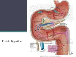
THE LIVER THE LIVER Bilirubin metabolism, types of jaundice Bilirubin metabolism, types of jaundice Dr. Muntadher Abdulkareem Abdullah M.B.Ch.B,CABM,FIBMS,FIBMS(GE.&HEP.)
At the end of this lecture , you must be able to answer these questions At the end of this lecture , you must be able to answer these questions Q1: 22 year old male patient , with history of recurrent self resolving jaundice since childhood, presented with jaundice at the end of heavy duty day , otherwise the patient is healthy with nonsignificant other finding, Investigations show : TSB=2.9mg/dl, direct = 0.4mg/dl , normal other Ix What is the type of jaundice in this patient? What is the most likely etiology? How can you prevent other episodes of jaundice?
Q2: 20 year old male patient , hx of UTI with use of trimethoprim- sulfamethoxazole ;during the last two days, today he notice that his sclera turn yellow Ix : TSB= 6 MG/;DL , direct=0.6 mgldl CBD SHOW Hb%=6 g/dl, corrected retic%= 6 What is the type of jaundice? What is the etiology of jaundice?
Q3 :18 year old male patient , presented with one day history of jaundice preceded by fever, nausea and repeated vomiting If the cause of jaundice is acute viral hepatitis A, What other sign and symptoms you may the patient has? What will be the LFTs most likely be in this patient How ;differ the signs, symptoms and LFTs will be , if this patient has cholestatic type of jaundice?
Functional anatomy and physiology Functional anatomy and physiology
The liver weighs 1.21.5 kg It is classically divided into left and right lobes by the falciform ligament useful functional division is into the right and left hemilivers, based on blood supply. These are further divided into eight segments, according to subdivisions of the hepatic and portal veins. Each segment has its own branch of the hepatic artery and biliary tree. A liver segment is made up of multiple smaller units known as lobules, comprised of a central vein, radiating sinusoids separated from each other by single liver cell (hepatocyte) plates, and peripheral portal tracts. The functional unit of the liver is the hepatic acinus.
Blood supply On entering the porta hepatis, the portal vein and the hepatic artery canals) the sinusoids the central vein hepatic veins interlobular branches(in the portal the inferior vena cava
Dual perfusion A majority of its supply via the portal vein: which drains blood from the gut via the splanchnic circulation and is the principal route for nutrient trafficking to the liver. A minority from the hepatic artery Venous drainage: hepatic veins
Functions of the liver Functions of the liver production of bile : this is the main digestive function of the liver, the bile is a complex substance required for the emulsifcation, hydrolysis, and uptake of fats in the duodenum. The liver is the major interface between the digestive system and the blood, as the organ in which nutrients absorbed in the small intestine are processed before distribution throughout the body. Synthesis and endocrine secretion into the blood of the major plasma proteins, including albumins, fibrinogen, apolipoproteins, transferrin, and many others. Conversion of amino acids into glucose (gluconeogenesis) Breakdown (detoxifcation) and conjugation of ingested toxins, including many drugs. Amino acid deamination, producing urea removed from blood in kidneys.
Storage of glucose in glycogen granules and triglycerides in small lipid droplets. Storage of vitamin A (in hepatic stellate cells) and other fat-soluble vitamins. Removal of effete erythrocytes (by specialized macrophages, or Kupffer cells). Storage of iron in complexes with the protein ferritin.
Bilirubin metabolism and bile Bilirubin metabolism and bile
Bilirubin is a breakdown product of heme (ferroprotoporphyrin IX). About 4 mg/kg body weight of bilirubin is produced each day, nearly 80% from the breakdown of hemoglobin in senescent red blood cells and prematurely destroyed erythroid cells in the bone marrow and the remainder from the turnover of hemoproteins such as myoglobin and cytochromes distributed throughout the body. The initial steps of bilirubin metabolism occur in reticuloendothelial cells, predominantly in the spleen. Heme is converted to biliverdin by the microsomal enzyme heme oxygenase. Biliverdin is then converted to bilirubin by the cytosolic enzyme biliverdin reductase
The liver plays a central role in the metabolism of bilirubin and is responsible for the production of bile. In the blood bilirubin is normally almost all unconjugated and, because it is not water-soluble, is bound to albumin and does not pass into the urine. Unconjugated bilirubin is taken up by hepatocytes at the sinusoidal membrane, where it is conjugated in the endoplasmic reticulum by UDP- glucuronyl transferase, producing bilirubin mono- and diglucuronide. Impaired conjugation by this enzyme is a cause of inherited hyperbilirubinaemias.
These bilirubin conjugates are water-soluble and are exported into the bile canaliculi by specific carriers on the hepatocyte membranes. The conjugated bilirubin is excreted in the bile and passes into the duodenal lumen. Once in the intestine, conjugated bilirubin is metabolized by colonic bacteria to form stercobilinogen, which may be further oxidized to stercobilin. Both stercobilinogen and stercobilin are then excreted in the stool, contributing to its brown colour. Biliary obstruction results in reduced stercobilinogen in the stool, and the stools become pale.
A small amount of stercobilinogen (4 mg/day) is absorbed from the bowel, passes through the liver and is excreted in the urine, where it is known as urobilinogen or, following further oxidization, urobilin. The liver secretes 1 2 L of bile daily. Bile contains bile acids (formed from cholesterol), phospholipids, bilirubin and cholesterol. Several biliary transporter proteins have been identified . Mutations in genes encoding these proteins have been identified in inherited intrahepatic biliary diseases presenting in childhood, and in adult-onset disease such as intrahepatic cholestasis of pregnancy and gallstone formation.
Investigations for liver diseases: Investigations for liver diseases: Serum bilirubin: Fractionation of serum bilirubin AMINOTRANSFERASES: the most sensitive markers of acute hepatocellular injury: ALT (formerly serum glutamic pyruvic transaminase, or SGPT) and AST (formerly serum glutamic oxaloacetic transaminase, or SGOT)
ALKALINE PHOSPHATASE: Hepatobiliary disease leads to increased serum ALP levels through induced synthesis of the enzyme and leakage into the serum, a process mediated by bile acids. Increase mainly in obstructive cholestatic liver diseases 5 -Nucleotidase and gamma GT: The primary use of these enzymes levels is to identify the source of an isolated elevation in the serum ALP level , as they are not elevated in bone disease
TESTS OF HEPATIC SYNTHETIC FUNCTION: Albumin: low in chronic liver disease , half life =2 weeks Prothrombin Time: clotting factors, all of which are produced in the liver except factor VIII, which is produced by vascular endothelial cells. TESTS TO DETECT HEPATIC FIBROSIS: single serum biochemical markers:(hyaluronan) multiparameter blood tests :FibroTest (marketed as FibroSure in the United States) FIBROSCAN Imaging of the liver: Uls , CT SCAN , MRI , MRI +MRCP LIVER BIOPESY
Jaundice Jaundice yellow discoloration of the skin, conjunctivae, and mucous membranes, resulting from widespread tissue deposition of the pigmented metabolite bilirubin. In adults, the normal bilirubin concentration is lower than 1 to 1.5 mg/dL. Jaundice is usually detectable clinically when the plasma bilirubin exceeds 40 mol/L (~2.5 mg/dL). Yellowish discoloration of skin also can result from excessive deposition of carotene in hypercarotenemia, how differ from hyperbilirubinemia clinically ? In a patient with jaundice it is useful to consider whether the cause might be pre-hepatic, hepatic or post-hepatic.
Pre-hepatic jaundice: This is caused either by haemolysis or by congenital hyperbilirubinaemia, and is characterised by an isolated raised bilirubin level. Usually mild , lemon colour , normal urine and stool . In haemolysis, destruction of red blood cells or their marrow precursors causes increased bilirubin production. Jaundice due to haemolysis is usually mild because a healthy liver can excrete a bilirubin load six times greater than normal before unconjugated bilirubin accumulates in the plasma. This does not apply to newborns, who have less capacity to metabolism bilirubin. The most common form of non-haemolytic hyperbilirubinaemia is Gilbert s syndrome
congenital unconjugated hyperbilirubinemia congenital unconjugated hyperbilirubinemia Gilbert s syndrome Crigler-Najjar syndrome types I and II
Gilberts syndrome has a prevalence of approximately 10% in white populations. Patients with Gilbert s syndrome typically present when isolated hyperbilirubinemia is detected as an incidental finding on routine multiphasic biochemical screening, and clinical jaundice is uncommon. Serum bilirubin levels may rise 2- to 3-fold with fasting or dehydration but are generally below 4 mg/dL. Reduction in transcription of the B-UGT gene UGT1A1 ( Glucuronyl transferase activity)
Although Gilberts syndrome has generally been thought to be an entirely benign condition, persons with this disorder may be at increased risk for gallstones and for toxicity of selected drugs like irinotecan that require glucuronidation for metabolic disposal. On the other hand, patients with Gilbert s syndrome may be at decreased risk for cardiovascular disease, because unconjugated bilirubin has antioxidant properties that are thought to retard atherosclerosis.
Crigler-Najjar syndrome : Mutations in the coding region of UGT1A1 appear to be responsible for Crigler- Najjar syndrome. In type I Crigler-Najjar syndrome, B-UGT activity is absent, and marked unconjugated hyperbilirubinemia is evident shortly after birth. Patients with type II Crigler-Najjar syndrome are not ill during the neonatal period and may not be diagnosed until early childhood. Most patients with type II Crigler-Najjar syndrome can be treated successfully with phenobarbital
Conjugated or Mixed Hyperbilirubinemia: Two autosomal inherited disorders, Dubin-Johnson syndrome: impaired canalicular export of conjugated bilirubin Coarse pigment in centrilobular hepatocytes on liver biopsy and Rotor s syndrome, are associated with conjugated or mixed hyperbilirubinemia (i.e., increase in serum concentrations of both conjugated and unconjugated bilirubin).
Rotors syndrome, are associated with conjugated or mixed hyperbilirubinemia (i.e., increase in serum concentrations of both conjugated and unconjugated bilirubin). Impaired canalicular export of conjugated bilirubin Normal liver biopsy
Hepatocellular jaundice Hepatocellular jaundice Hepatocellular jaundice results from an inability of the liver to transport bilirubin into the bile, occurring as a consequence of parenchymal disease. In hepatocellular jaundice, the concentrations of both unconjugated and conjugated bilirubin in the blood increase. There is tea colour urine with normal colour stool
Characteristically, jaundice due to parenchymal liver disease is associated with increases in transaminases (AST, ALT), but increases in other LFTs, including cholestatic enzymes (GGT, ALP), may occur and suggest specific aetiologies . Acute jaundice in the presence of an ALT of >1000 U/L is highly suggestive of an infectious cause (e.g. hepatitis A or B), drugs (e.g. paracetamol) or hepatic ischemia. Imaging is essential, in particular to identify features suggestive of cirrhosis, define the patency of the hepatic vasculature and obtain evidence of portal hypertension. Liver biopsy has an important role in defining the aetiology of hepatocellular jaundice and the extent of liver injury
Obstructive (cholestatic) jaundice Obstructive (cholestatic) jaundice Cholestatic jaundice may be caused by: failure of hepatocytes to initiate bile flow obstruction of the bile ducts or portal tracts obstruction of bile flow in the extrahepatic bile ducts between the porta hepatis and the papilla of Vater
Clinical features and complications of cholestatic jaundice Clinical features and complications of cholestatic jaundice Cholestasis Early features: Jaundice Dark urine Pale stools Pruritus Late features: Malabsorption (vitamins A, D, E and K): weight loss, Steatorrhoea, Osteomalacia, bleeding tendency Xanthelasma and xanthomas Cholangitis Fever Rigors
Key history points in patients with jaundice: Symptoms* Itching preceding jaundice Abdominal pain (suggests stones) Weight loss (chronic liver disease and malignancy) Dark urine and pale stools Fever rigors Dry eyes/dry mouth Fatigue Recent drug history Other: Exposure to intravenous drug or blood transfusions Travel history and country of birth Metabolic syndrome (increased body mass index type 2 diabetes/ hypertension) Autoimmune disease history Alcohol history Inflammatory bowel disease Family history of liver disease, autoimmune disease or the metabolic syndrome
Thanks Thanks


 is absorbed")


 jaundice")