Rare Proteus Species Wound Infection: Case Study and Management
A 65-year-old male presented with a wound infection caused by a rare Proteus species on his left 2nd toe. The patient had a history of diabetes and previous angioplasty. Despite initial antibiotic therapy, amputation of the affected toe was required due to progressive infection. The causative pathogens, Proteus vulgaris and Pseudomonas aeruginosa, showed specific antibiotic sensitivities guiding the treatment course towards Piperacillin-Tazobactam and Metronidazole. This case highlights the importance of accurate diagnosis and tailored antibiotic management in such infections.
Download Presentation

Please find below an Image/Link to download the presentation.
The content on the website is provided AS IS for your information and personal use only. It may not be sold, licensed, or shared on other websites without obtaining consent from the author. Download presentation by click this link. If you encounter any issues during the download, it is possible that the publisher has removed the file from their server.
E N D
Presentation Transcript
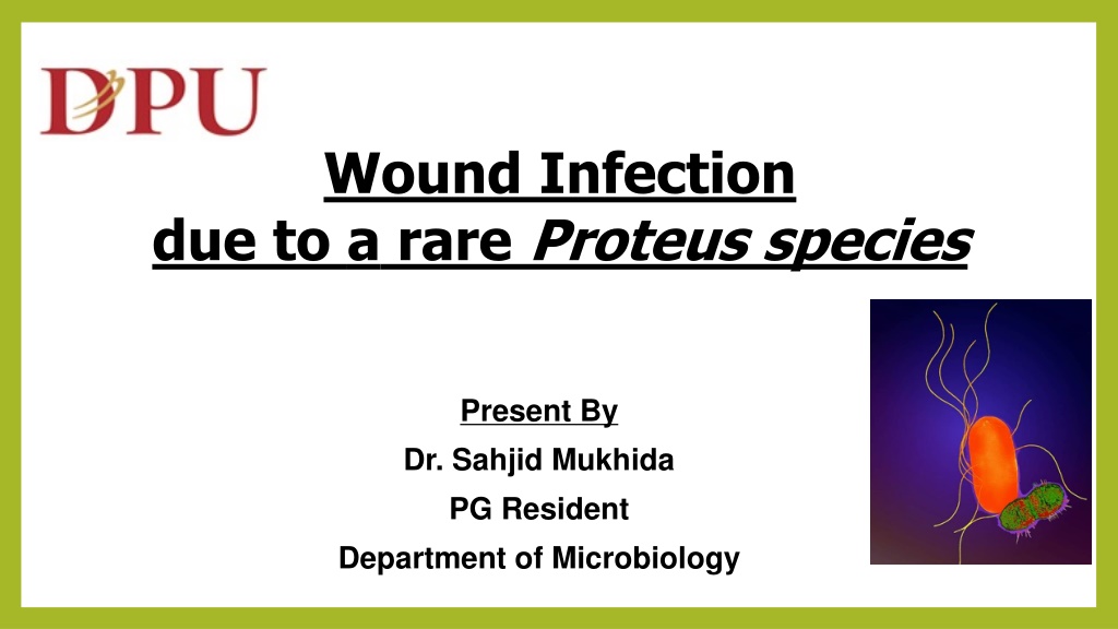
Wound Infection due to a rare Proteus species Present By Dr. Sahjid Mukhida PG Resident Department of Microbiology
CASE Details On 9th of October 2020, A 65 Years/Male presented with Punched out ulcer over left 2nd toe Size: 2x1 cm Discharge: Purulent, foul smelling, blood stained Appearance : Black discoloration with thickened skin around the ulcer site Ulcer base at meta tarsal bone
History K/C/O Type II diabetes 6 years (Tab: Metformin) CAD - angioplasty- 2 years back A bleb like swelling on left 2nd toe before 1 month, burst and formed an ulcer Initially small in size, progressed to the current size Previous discharge from wound was yellowish, foul smelly and non bloody Another ulcer developed below the left 2nd toe
Continue.. There was no Numbness Restriction of movements Local temperature rise, Fever, chill, rigors, cough, breathlessness, calf pain, edema Comorbidities not present Bronchial Asthma, Epilepsy Polyuria, polydypsia, blurred vision, DVT
On Admission Systemic evaluation CVS- S1S2 present, No additional heart sound CNS: No focal neurological deficit RS: B/L air entry- present, No added sounds present GIT: No organomegaly Lab investigation Hb: 10.4g/dLCK-Mb: 51 IU/L TLC: 10400/ ? BSL: 315 mg/dL2D ECHO: EF: 35%, Mild TR/ Mild MR HbA1C : 9 mmol/mol Troponin I: <10 Pg/dL ECG: T wave depression in V3-V6
Management 9th October : After admission, patient was started on empirical antibiotic therapy with Amoxycillin + Clavulanic acid 13th October : Tissue sample from infection site was sent for culture and sensitivity. 14th October: Due to progressive wound size, Left 2nd toe amputation was done Proteus vulgaris Pseudomonas aeruginosa Sensitive Resistant Sensitive Resistant Amikacin, Gentamycin, Ceftazidime, Ciprofloxacin, Piperacillin + Tazobactum Ceftazidime + Clavulanic acid, Norfloxacin, Ceftazidime + Tazobactum, Cefoxitin, Cefotaxime, Ceftrixone, Amoxycillin + Clavulanic acid Ampicillin Amikacin, Gentamycin, Ceftazidime, Carbenicillin, Ciprofloxacin, Piperacillin, Piperacillin + Tazobactum Ceftazidime + Clavulanic acid, Ceftazidime + Tazobactum, Cefoxitin, Cefotaxime Ceftrixone Amoxycillin+Clavulanic acid After this susceptibility report, antibiotic were changed from Amoxycillin+ Clavulanic Acid to Piperacillin+Tazobactum and Metronidazole.
Cont.. Pseudomonas aeruginosa ( 17th October 2020) Sensitive Resistant Amikacin, Gentamycin, Ciprofloxacin, Ceftazidime, Ceftazidime + Clavulanic acid, Ceftazidime + Tazobactum, Piperacillin, Piperacillin + Tazobactum Cefoxitin, Cefotaxime, Ceftrixone Amoxycillin + Clavulanic acid Regular wound cleaning, skin debridement and dressing were done but wound was not healing. Amputation was planned upto meta tarsal but due to progression of the necrosis, below knee amputation was performed on 28th October 2020. After below knee amputation, Piperacillin + Tazobactum was replaced by Cefoperazone + Sulbactam (1.5g)/BD.
4th November: Due to fever spike, Urine sample was sent for culture and sensitivity and it was reported as No growth . 7th November: As wound was not-healing, a repeated swab was sent for culture and sensitivity which also isolated Pseudomonas aeruginosa but different susceptibility testing. Pseudomonas aeruginosa Sensitive Resistant Amikacin, Gentamycin Colistin, Tigecycline, Ofloxacin, Tobramycin Carbenicillin, piperacillin, Ciprofloxacin, Cefoxitin, Cefotaxime, Ceftrixone, Ceftazidime, Ceftazidime + Clavulanic acid, Ceftazidime + Tazobactum, Piperacillin+Tazobactum, Meropenem, Imipenem, Amoxycillin + Clavulanic acid After above susceptibility report, antibiotic treatment was changed from Cefoperazone+ Sulbactam to Amikacin.
17th November: Even after prolonged antibiotics treatment, infection was not subsiding at the wound site and another swab was repeated. These are the 2 isolates: Acinetobacter species Proteus species Sensitive Resistant Sensitive Resistant Colistin, Tigecycline Ampicilin, Norfloxacin, Amikacin, Cotrimoxazole, Chloramphenicol, Cefoxitin, Cefotaxime, Ceftrixone, Ceftazidime, Ceftazidime + Clavulanic acid, Piperacillin+Tazobactum, Meropenem, Imipenem,, Amoxycillin + Clavulanic acid, Ofloxacin, Gentamycin Tigecycline Ampicilin, Norfloxacin, Amikacin, Cotrimoxazole, Chloramphenicol, Cefoxitin, Cefotaxime, Ceftrixone, Ceftazidime, Meropenem, Colistin Ceftazidime + Clavulanic acid, Ofloxacin, Ceftazidime + Tazobactum, Pipercillin+Tazobactum, Meropenem, Imipenem, Gentamycin, Amoxycillin+Clavulanic acid
Culture and sensitivity test process .. Pus (Swab) samples were processed for gram stain, Z-N stain, culture on blood agar and MacConkey agar. Gram stain showed gram negative bacilli in the smear. Blood Agar : Swarming was seen. Gram Stain MacConkey Agar: Smooth, Non Lactose Fermenting colonies with distinct odor Blood agar Swarming MacConkey Agar NLF colonies
Identification (Automation) Bio chemical reaction did not match with the common Proteus spp. Isolate was subjected to automation (Vitek 2c) for species identification and ABST. Proteus penneri was identified which was resistant to most antibiotic groups including Tigecycline (MIC - 2) and Colistin (MIC >16 )
Bio-Chemical Reaction 1 P. vulgaris P. mirabilis Test P. penneri Gram Negative Rods Gram Negative Rods Microscopic morphology Gram Negative Rods Beta Beta Hemolysis (sheep blood agar) Beta Positive Positive Urease Positive Positive Negative Indole production Negative Positive Negative Esculin hydrolysis Negative Positive Negative Acid from Maltose Positive Positive Negative Acid from Sucrose Positive Negative Variable Citrate Negative Negative Positive Ornithine decarboxylase Negative Positive Positive Hydrogen sulfide production Negative Positive Negative Salicin Negative
Bio Chemical reactions I MR C U TSI G S L M P. vulgaris P. mirabilis P. penneri
Further samples received for Culture & sensitivity test Date Type of sample Results Organism if any Sensitive drug Resistance drug 17/11/20 Pus Growth P. penneri Acinetobactor spp Tigecyclin All drugs including colistin 23/11/20 Pus Growth P. penneri P. aeruginosa All drugs are resistant including Colistin (MIC >16) and Tigecycline (MIC 2) 25/11/20 Pus Growth 01/12/20 P. penneri All drugs are resistant including Colistin (MIC >16) and Tigecycline (MIC 2) Pus Growth Pus 05/12/20 Cip, Pen-G, Va, Linezolid Erythromycin, Clindamycin, Cotrimoxazole S. pyogenes
Clinical course After Proteus penneri was reported with multi drug resistance (Tigecycline MIC-2), Tigecycline was prescribed but the drug could not be administered due to poor patient compliance Regular dressing and tissue debridement were performed twice a day for 10 days and it continued for once a day As the infection was not subsiding, decision for above knee amputation was taken but patient relatives did not gave consent. On 9th December 2020 mid night patient had ARDS- symptoms, was shifted to ICU for management, but unfortunately patient could not be resuscitated. Pulmonary embolism was established as the cause of death
Discussion Surgical site infection is one of the major cause of hospital associated infections2 P. penneri is an underreported pathogen. Proteus penneri named individually in 1982 from Proteus vulgaris bio-group3 Gram negative, facultative anaerobic, Non Lactose fermenting, non capsulated, pleomorphic, motile, rod-shaped bacterium1 It is an Invasive pathogen and can cause hospital associated infections like UTI or open wound infections4 In our case, it was isolated after 20 days of below knee amputation so it may be a possible hospital associated infection Number of reports of this organism are few, as it is often isolated along with other organisms like Pseudomonas aeruginosa, Acinetobacter species and other gram negative organism.
Cont.. Proteus species are known to be intrinsically Resistant to Nitrofurantoin, Tetracycline, Tigecycline and polymyxins. Proteus penneri is also known to be intrinsically Resistant to other -lactams and macrolides. Though it is known to be Sensitive to group of antibiotics like aminoglycosides, Carbapenems, aztreonam, quinolones and co-trimoxazole.6 Feglo et al.7 and Pandey et al8 reported MDR Proteus penneri from wound site infection. In our case P. penneri isolated was multi-drug resistant including Carbapenems and colistin4 Mortality in this case may be due to the presence of co-morbidities present
Take home message Rapid and correct identification of such rare and resistant strains are of utmost importance as they can be significant nosocomial threat. Automation guide to identify this type of rare isolates and drug sensitivity to prompt treatment Hospital associated bacterial infection like Proteus penneri which have proven pathogenicity and it need continuous emphasis on effective infection control practices to warrant appropriate therapeutic plan
References Koneman EW, Allen SD, Janda WM, et al; Color Atlas & Textbook of Diagnostic Microbiology, 7th edition,Philadelphia;Lippincott:2017 1. Kaistha N, Bansal N, Chander J. Proteus pennerilurking in the intensive care unit: An important often ignored nosocomial pathogen. Indian J anaesth. 2011 Jul-aug; 55(4):411-413 2. O Hara CM, Brenner FW, Miller JM. Classification, identification and clinical significance of Proteus, Providencia and Morganella.Clin Microbiol Rev.2000;13:534 46. 3. Krajden S, Fuksa M, Petrea C, Crisp LJ, Penner JL. Expanded clinical spectrum of infections caused by Proteus penneri.J Clin Microbiol. 1987;25:578 9 4. Stock I. Natural antibiotic susceptibility of Proteus spp., with special reference to P. mirabilis and P. penneri strains. J Chemother. 2003 Feb;15(1):12-26. doi: 10.1179/joc.2003.15.1.12. PMID: 12678409. 5. CLSI. Performance standards for Antimicrobial susceptibility testing, 30 edition CLSI supplement M100 Wayne, PA: Clinical Laboratory Standards Institute 2020 6. Feglo PK, Gbedema SY, Quay SN, Adu-Sarkodie Y, Opoku-Okrah C. Occurrence, species distribution and antibiotic resistance of Proteusisolates: A case study at a Komfo Anokye Teaching Hospital (KATH) in Ghana. Int J Pharm Sci Res 2010;1:347-52 7. Pandey A, Verma H, Asthana AK, Madan M. Extended spectrum beta lactamase producing Proteus penneri: A rare missed pathogen?. Indian J Pathol Microbiol 2014;57:489-91 8.
ACKNOWLEDGEMENT Surgery department Dr. D. Y. Patil Medical College, Hospital and Research Centre, Dr. D. Y. Patil Vidyapeeth, Pimpri, Pune, Maharashtra
Thank You Very Much !
