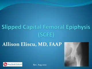
Slipped Capital Femoral Epiphysis: Adolescent Hip Disorder
Slipped capital femoral epiphysis (SCFE) is a condition affecting the proximal femoral physis, leading to slippage of the metaphysis relative to the epiphysis. It is most commonly seen in adolescent males, particularly those who are obese. Treatment usually involves percutaneous pin fixation. Risk factors include obesity, acetabular retroversion, and femoral retroversion. Associated conditions may include hypothyroidism, renal osteodystrophy, and growth hormone deficiency. The pathophysiology involves mechanical forces acting on a susceptible physis during adolescence, leading to slippage through the hypertrophic zone. Radiographs are essential for diagnosis, and prompt surgical intervention is often necessary to prevent complications.

Download Presentation

Please find below an Image/Link to download the presentation.
The content on the website is provided AS IS for your information and personal use only. It may not be sold, licensed, or shared on other websites without obtaining consent from the author. Download presentation by click this link. If you encounter any issues during the download, it is possible that the publisher has removed the file from their server.
E N D
Presentation Transcript
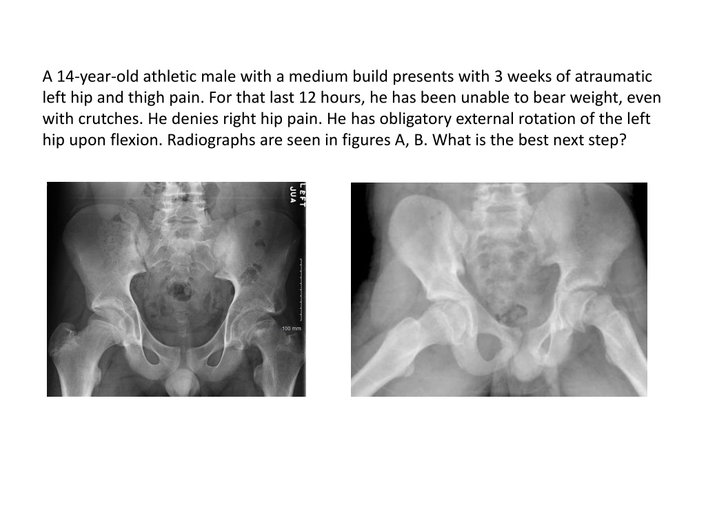
A 14-year-old athletic male with a medium build presents with 3 weeks of atraumatic left hip and thigh pain. For that last 12 hours, he has been unable to bear weight, even with crutches. He denies right hip pain. He has obligatory external rotation of the left hip upon flexion. Radiographs are seen in figures A, B. What is the best next step?
Overview Condition of the proximal femoral physis that leads to slippage of the metaphysis relative to the epiphysis Most commonly seen in adolescent obese males Treatment is usually percutaneous pin fixation
Epidemiology Most common disorder affecting adolescent hips 10 per 100,000(USA) More common in obese children males African Americans,Pacific islanders, Latinos
Epidemiology During period of rapid growth (10-16 years) 13 for boys 12 for girls Associated with puberty Left hip is more common Bilateral in (~25%)
Risk factors Obesity-single greatest risk factor Acetabular retroversion and femoral retroversion
Associated conditions Hypothyroidism -most common etiology of nonidiopathic SCFE Renal osteodystrophy Growth hormone deficiency Panhypopituitarism Indications for endocrine workup child is < 10 years old weight is < 50th percentile Down syndrome
Pathophysiology Adolescence-perichondrial ring thins and weakens Physis is still vertical in this age group Mechanical forces acting on a susceptible physis Slippage occurs though the hypertrophic zone of the physis
Pathoanatomy Angulation-metaphysis translates anterior and externally rotates Epiphysis remains in the acetabulum Periosteum remains intact (chronic SCFE) In acute SCFE, periosteum can be partially torn
Loder Classification Based on ability to bear weight Stable-Able to bear weight with or without crutches Minimal risk of osteonecrosis (<10%) Unstable-Unable to ambulate (not even with crutches) High risk of osteonecrosis (25-35%) Provides prognostic information
Southwick Slip Angle Classification Based on femoral epipyseal-diaphyseal angle difference Mild Moderate 30-50 Severe Based on the degree of difference between the affected and unaffected hip < 30 > 50
Grading System -- based on percentage of slippage Grade I 0-33% of slippage Grade II 34-50% of slippage Grade III >50% of slippage
Symptoms Groin and thigh pain -most common presentation knee pain can lead to missed diagnosis Limp antalgic gait externally rotated foot progression angle Patients prefer to sit in a chair with affected leg crossed over the other Duration symptoms for weeks to months before diagnosis is made Acute on chronic-severe pain,unable to bear weight
Physical exam Abnormal gait antalgic, waddling, externally rotated gait or Trendelenburg gait Decreased hip motion Loss of hip IR, abduction, and flexion Obligatory external rotation during passive flexion of hip (Drehmann sign) Weakness thigh atrophy
Radiographs AP & frog-leg lateral of right and left hip lateral radiograph is best way to identify a subtle slip
Radiographs Klein's line Line drawn along superior border femoral neck Will intersect less of the femoral head or not at all in SCFE
Radiographs Epiphysiolysis (growth plate widening or lucency) Blurring of proximal femoral metaphysis (metaphyseal blanch sign of Steel) seen on AP due to overlapping of the metaphysis and posteriorly displaced epiphysis MRI may help diagnose a preslip condition
Treatment-Stable slip Operative-percutaneous in situ fixation indications both stable and unstable slips one vs. two cannulated screws is controversial benefit of 2 screws needs to be considered in the face of greater screw related complications contralateral hip prophylactic fixation- controversial
Treatment Open epiphyseal reduction and fixation controversial unstable and severe slips technique capital realignment via the modified Dunn procedure
Complications Osteonecrosis of femoral head increased risk with unstable slips (~24-47%), most common predictor operative complication (4-6%) hardware placement in posteriosuperior femoral neck has the greatest risk of disrupting the vascular supply Chondrolysis (0-2%) Residual proximal femoral deformity & limb length discrepancy Degenerative arthritis