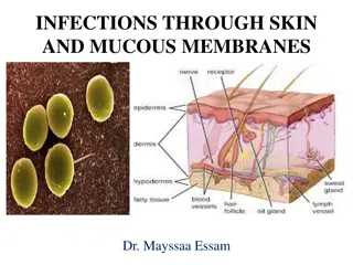
Understanding Skin and Soft Tissue Infections: A Comprehensive Overview
Skin and soft tissue infections are common and can vary in severity. Recognizing the types, causes, and treatment options is crucial. Learn about the anatomical structure, bacterial involvement, diagnostic procedures, and management approaches in this detailed guide.

Download Presentation

Please find below an Image/Link to download the presentation.
The content on the website is provided AS IS for your information and personal use only. It may not be sold, licensed, or shared on other websites without obtaining consent from the author. Download presentation by click this link. If you encounter any issues during the download, it is possible that the publisher has removed the file from their server.
E N D
Presentation Transcript
Microbiology - Skin and Soft-Tissue Infections Team 437 Please check our editing file frequently. Red : Important! Black: Doctors slides Grey : Extra Info Green: Notes Msk editing file
Objectives Describe the anatomical structure of skin and soft tissues. 1. Differentiate the various types of skin and soft tissue infections and their clinical presentation. 2. Name bacteria commonly involved in skin and soft tissue infections 3. Describe the pathogenesis of various types of skin and soft tissue infections 4. Recognize specimens that are acceptable and unacceptable for different types of skin and soft tissue 5. infections Describe the microscopic and colony morphology and the results of differentiating bacteria isolates in 6. addition to other non-microbiological investigation Discuss antimicrobial susceptibility testing of anaerobes including methods and antimicrobial agents to be 7. tested. Describe the major approaches to treat of skin and soft tissue infections 8. either medical or surgical.
Key to developing an adequate differential diagnosis requires: Diagnostic and therapeutic: Investigation: History: Surgical exploration or debridement diagnostic (very important to determine the infection and whether it is deep or superficial ) and therapeutic (if it's severe it might need amputating Antibiotics treatment CBCs, Chemistry Swab, biopsy or aspiration (Aspirate :For pus or exudate) Radiographic procedures (X-rays, CT, MRI) ( primary to determine if there is bone infection or if there is gas.) Level of infection and the presence of gas or abscess. Patient s immune status, the geographical locale, travel history, recent trauma or surgery, previous antimicrobial therapy, lifestyle, animal exposure or bites Physical examination: To determine the severity of infection
Introduction: Soft tissue definition: anything other than bones. Soft tissue infections : It is a Common disease. Can be mild to moderate or severe. Muscle or bone and lungs or heart valves infection. Most common Cause are: Staphylococcus aureus and streptococcus. Emerging antibiotic resistance among: -Staphylococcus aureus (methicillin resistance). -Methicillin drug is the laboratory name for cloxicillin drug -Streptococcus pyogenes (erythromycin resistance). You should differentiate between epidermis, dermis, subcutaneous infections and is it invading other tissues like muscles or bones to choose the right management. The more the organism has powerful toxins and enzyme the more it spread deeply, and he may devolve gas gangrene. For example : clostridium perfringase Important to know the: 1- Name of the syndrome. 2- Organisms causing it. 3- Clinical presentation. 4- Antibiotics.
Impetigo - (pyoderma) It s a common skin infection, in Children 2 5 years in tropical or subtropical regions. Very superficial in epidermis Always caused by -hemolytic streptococci ((Nonbullous) and, or staphylococcus aureus bollus ( rarely alone) Treatment: Characteristics: Cefazolin: covers Both Staphylococcus and streptococcus. Cloxacillin: covers Staphylococcus only Erythromycin: covers streptococcus only Mupirocin : It may lead to immune complication in some cases. Systemic symptoms are usually absent. Consists of discrete purulent lesions Blister if it rupture, it s produce fluid (honey cust Exposed areas of the body( face and extremities) Skin colonization- Inoculation by abrasions, minor trauma, or insect bites Poststreptococcal glomerulonephritis*. (anti DNAse B) :(antideoxyribonuclease B) ; if there is a lot of AntiDNAse in the test then it means he have impetigo . If patient has cellulitis, 1stdrug you think of = Cefazolin *Group A streptococci are sensitive to penicillin Pyoderma : skin disease that is pyogenic (has pus). Bullous: Involves the formation of bullae (blisters).
Cutaneous abscesses. Collections of pus within the dermis and deeper skin tissues. Typically polymicrobial (caused by several types of microorganisms )S. aureus alone in 25 % Characteristics: Treatment: Painful, tender, and fluctuant Multiple lesions: cutaneous gangrene. severely impaired host defenses. extensive surrounding cellulitis. high fever. Incision and evacuation of the pus.( ) Antibody: Cloxacillin (S.Aureus only) Diagnosis: Gram stain, culture and systemic antibiotics
MSSA :Methicillin sensitive Staphylococcus aureus MRSA : Methicillin resistant Staphylococcus aureus Furuncles and carbuncles Furuncles (or boils ) are infections of the hair follicle (folliculitis ), usually caused by S. aureus, in which suppuration extends through the dermis into the subcutaneous tissue. One or separated hair follicles. Carbuncle: extension to involve several adjacent follicles with coalescent inflammatory mass. Present in back of the neck, especially in diabetics. usually caused by S. aureus Treatment: Outbreaks of furunculosis caused by MSSA* and MRSA*. Characteristics: Larger furuncles and all carbuncles require incision and drainage. Mupirocin ointment: eradicate staphylococcal carriage and nasal colonization. Systemic antibiotics are usually unnecessary extends through the dermis into the subcutaneous tissue (so it is Deeper than Cutaneous abscesses.) Carbuncle present in the back of neck especially diabetes patients Families, prisons ,sports teams Inadequate personal hygiene Repeated attacks of furunculosis Presence of S. aureus in the anterior nares- 20-40% Nose swap Decolonization + topical antibiotics
Erysipelas and Cellulitis Diffuse spreading skin infections, excluding infections associated with underlying suppurative foci Most of the infections arise from streptococci, often group A, but also from other groups, such as B, C, or G. Erysipelas Affects the upper dermis (raised-clear line of demarcation) Red, tender, painful plaque Infants, young children- Well Demarcated Swelling Edema Redness Caused by: -hemolytic streptococci ( group A or S. pyogenes) Treated by: Penicillin-IV or oral. Erysipelas: in the whole epidermis and dermis and it is caused mainly by group A streptococci 8
Cellulitis Acute spreading infection involves the deeper dermis and subcutaneous tissues. Caused by: o -hemolytic streptococci, Group A streptococci, and group B streptococci-in diabetics o S. aureus : commonly causes cellulitis- penetrating trauma. o Haemophilus influenzae periorbital cellulitis in children Risk factors: Obesity, venous insufficiency, lymphatic obstruction (operations), preexisting skin infections- ulceration, or eczema, CA-MRSA (Community Associated MRSA): Carry Panton-Valentine leukocidin gene More sensitive to antibiotics Can lead to severe skin and soft tissue infection or septic shock
Cellulitis Clinical diagnosis Symptoms and Signs: o High WBCs, blood culture rarely needed o Aspiration and biopsy might be needed in diabetes mellitus, malignancy, animal bites, neutropenia (Pseudomonas aeruginosa ),immunodeficiency, obesity and renal failure o Observe for progression to severe infection(increased in size with systemic manifestation ie . fever, leukocytosis) Treatment: o cover streptococcus and staphylococcus o Penicillin, cloxacillin, cefazolin (cephalexin),clindamycin o Vancomycin or linazolid in case of MRSA o Clindamycin, TMP-SMZ for CaMRSA
Summary 11
Necrotizing fasciitis (Flesh-eating disease) It is a rare deep skin and subcutaneous tissues infection Necrotizing fasciitis Type 1 Type 2 Caused by multiple organisms caused by only Group A strept Polymicrobial monomicrobial Most common in the arms, legs, and abdominal wall and is fatal in 30%-40% of cases. Fournier's gangrene (testicular), Necrotizing cellulitis Mortality as high as 73 % if untreated
Causing microbes: Risk factors Pathophysiology Signs and symptoms Group A streptococcus -Immune-suppression Destruction of skin and muscle by releasing toxins Streptococcal pyogenic exotoxins Superantigen Non-specific activation of T-cells Overproduction of cytokines Severe systemic illness (Toxic shock syndrome) : Edema will press on the nerves (causing a severe pain) and blood vessels which will lead to necrosis and death of the supplied tissue Rapid progression of severe pain with fever , chills (typical) Swelling , redness, hotness, blister, gas formation, gangrene and necrosis Blisters with subsequent necrosis , necrotic eschars Diarrhea and vomiting (very ill) Mortality as high as 73% if left untreated Shock organ failure Hypotension It produces toxins that causes a septic shock Clostridium perfringens (gas in tissues) It s a normal flora of the GI -A wound can causes the Clostridium perfringens to spread in other tissues -It has a high virulent factor we call it {Phospholipase} which causes a necrosis of the fascia of the muscle ,it can lead to Death due to hypotension. Staphylococcus aureus or CA- MRSA (Streptococcus pyogenes) -Chronic diseases: ( diabetes, liver and kidney diseases, malignancy -Trauma:(laceration, cut, abrasion, contusion, burn, bite, subcutaneous injection, operative incision) -Recent viral infection rash (chickenpox) -Steroids -Alcoholism -Malnutrition Bacteroides fragilis Vibrio vulnificus (liver function) Gram-negative bacteria (synergy). E. coli, Klebsiella, Pseudomonas Fungi -Idiopathic
Diagnosis of Necrotizing fasciitis A delay in diagnosis is associated with a grave prognosis and increased mortality to check for renal failure Diagnosis Radiographic studies Surgery debridement amputation Clinical-high index of suspicion Microbiology Blood tests X-rays : subcutaneous gases Culture &Gram's stain ( blood, tissue, pus aspirate) WBC CT or MRI To evaluate the spread CBC Susceptibility tests ESR BUN (blood urea nitrogen) to check for renal failure.
Treatment of Necrotizing fasciitis If clinically suspected patient needs to be hospitalized OR require admission to ICU. 1. Antibiotics combinations: Penicillin-clindamycin-gentamicin. (Given together, and they re the most important) Ampicillin/sulbactam. Cefazolin plus metronidazole. Piperacillin/tazobactam. Clostridium perfringens - penicillin G. Start intravenous antibiotics immediately. Antibiotic selection based on bacteria suspected broad spectrum antibiotic combinations against: methicillin-resistant Staphylococcus aureus .(A) anaerobic bacteria. Gram-negative and gram-positive bacilli. 2. Hyperbaric oxygen therapy (HBO) treatment. Surgeon consultation: Extensive Debridement of necrotic tissue and collection of tissue samples. Can reduce morbidity and mortality. Why clindamycin? Because it s a ribosome inhibitor which will lead to inhibition of toxic formation Surgery Antibiotics Penicillin + Clindamycin Removing of fascia and dead tissue (don t close the wound) (: Perfusion of the tissue is better than penicillin Clindamycin is better used in the Lag phase of the infection
Pyomyositis: Definition: Acute bacterial infection of skeletal muscle, usually caused by Staph. Aureus. - No predisposing penetrating wound, vascular insufficiency or contiguous infection. - Most cases occur in the tropics ( - 60% of cases outside of tropics have predisposing risk factors (RF): Diabetes mellitus, ethyl alcohol, liver disease, steroid prescriptions, HIV, hematologic malignancy. ). History: - Blunt trauma or vigorous exercise (50%), then period of swelling without pain. - 10-21 days later : pain, tenderness, swelling and fever. - Pus can be aspirated from muscle. - 3rdstage: sepsis, later metastatic abscesses if untreated. Diagnosis: X-ray, US, MRI or CT. Treatment: Surgical drainage and antibiotics. The doctor barely explained this slide
Take home points Most commonly caused by Staphylococcus aureus and Streptococcus pyogenes Risk factors for developing SSTIs include breakdown of the epidermis, surgical procedures ,crowding, comorbidities, venous stasis, lymphedema Most of the infection are mild and can be managed on an outpatient basis In case Most SSTIs can be managed on an outpatient basis, although patients with evidence of rapidly progressive infection, high fevers, or other signs of systemic inflammatory response should be monitored in the hospital setting. Superficial SSTIs typically do not require systemic antibiotic treatment and can be managed with topical antibiotic agents, heat packs, or incision and drainage. Systemic antibiotic agents that provide coverage for both Staphylococcus aureus and Streptococcus pyogenes are most commonly used as empiric therapy for both uncomplicated and complicated deeper infections.
Quiz Please take a look at team 436 SAQ 1-Which of the following organism cause gas in tissue ? A- s.aureus B- c.perfringens C- group A streptococcus D- E.coli 2- Which of the flowing disease treat with penicillin? A-Erysipelas B-cellulitis C -Necrotizing fasciitis D-impetigo 3- Know as flesh eating disease? A-Erysipelas B-cellulitis C-Necrotizing fasciitis D-impetigo 4- Which of the following best treatment to necrotizing fasciitis? A-penicillin with clindamycin B-surgery C-ampicillin D-metronidazole 5- Which of the following disease localized in the epidermis ? A-Erysipelas B-cellulitis C-Necrotizing fasciitis D-impetigo 6- Which of the following disease cause infection to the skeletal muscle? A-Erysipelas B-pyomyositis C-Necrotizing fasciitis D-impetigo 1- B, 2- A, 3-C, 4-B, 5-A, 6-B
Some videos for understanding the lecture: Team members: 1- Impetigo https://www.youtube.com/watch?v=h Q9xv2cTAyw 2- abscesses https://www.youtube.com/watch?v=pL 6rP8C1e7w 3- MRSA https://www.youtube.com/watch?v=tI7a Xjgy_Vk Team leaders:


")