Understanding Wound Infections: A Comprehensive Overview

Explore the various stages of wound infections on a continuum from contamination to systemic infection. Learn how to identify and manage each stage, from presence of microbes to invasion of surrounding tissues. Discover the subtle signs of local and spreading infections, and the serious implications of systemic infection. Understand the concept of biofilm and its impact on wound healing.

Download Presentation

Please find below an Image/Link to download the presentation.
The content on the website is provided AS IS for your information and personal use only. It may not be sold, licensed, or shared on other websites without obtaining consent from the author. Download presentation by click this link. If you encounter any issues during the download, it is possible that the publisher has removed the file from their server.
E N D
Presentation Transcript
Wound Infections The Basics
Overview Wound Infection Continuum Biofilm Assessment: NERDS & STONEES Management 2
Wound Infection Continuum Contamination Colonization Local Infection Spreading Infection Systemic Infection 3
Contamination Presence of non-proliferating microbes within a wound at a level that does not evoke a host response All wounds are contaminated due to bacterial presence on the skin Host responses respond and destroy via phagocytosis 4
Colonization Presence of microbial organisms in the wound that undergo limited proliferation without evoking a host response Microbial growth to a non-critical level Wound healing not impeded or delayed 5
Local Infection Bacteria or microbes move deeper into the wound and proliferate at a rate that evokes a response Contained to one location, system, or structure Erythema, local warmth, swelling, purulent discharge, delayed healing, increasing or new pain, increasing malodour Subtle signs: hypergranulation, bleeding, epithelial bridging and pocketing, breakdown 6
Spreading Infection Invasion of the surrounding tissue by infective organisms that have spread from the wound. Signs and symptoms spread beyond the wound border Extending in duration +/- erythema, lymphangitis, crepitus, wound breakdown +/- satellite lesions, malaise, loss of appetite, inflammation, swelling of lymph glands 7
Systemic Infection Microbes spreading throughout he body via vascular or lymphatic system Systemic inflammatory response, sepsis, organ dysfunction 8
Biofilm Not typically visible Criteria indicative of biofilm: Failure of appropriate antibiotics, recalcitrance to appropriate antimicrobial, recurrence of delayed healing on cessation of antibiotics, delayed healing despite optimal wound management and health support, increased exudate/moisture, low-level chronic inflammation, low- level erythema, poor granulation/friable hypergranulation, secondary signs of infection 9
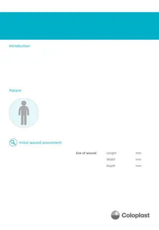
Assessment Healthy individuals with acute wounds present with OVERT or CLASSIC signs and symptoms of infection Immunocompromised and those with chronic wounds: SUBTLE or COVERT Friable, bright red granulation tissue Increasing malodour New or increasing pain or change in sensation Epithelial bridging and pocketing in granulation tissue Delayed wound healing beyond expectations Wound breakdown/enlargement or new ulcerations in periwound 10
NERDS (local infection) Non-Healing: wounds that are not 20-40% smaller in 4 weeks according to patient history or existing documentation Exudative Wound: Increase in wound exudate can be indicative of bacterial pro-inflammatory damage and leads to periwound maceration. More than 50% of the dressing stained with exudate Red and Bleeding Wound: wound bed tissue is bright red with exuberant granulation tissue. Tissue bleeds easily with gentle manipulation Debris: presence of discoloured granulation tissue, slough, and necrotic tissue Smell from the wound: unpleasant or sweet, sickening odour 11
STONEES (Spreading Infection) Size is bigger: wound increasing in size Temperature increased: increased periwound margin temperature by more than 3 F difference between two mirror image sites Os (probes to or exposed bone): wounds that have exposed bone or that probed to bone at he time of examination New areas of breakdown: new areas of breakdown or satellite lesions Eyrthema/Edema: reddened skin in periwound area, presence of swelling in periwound area Exudate: increased amount of drainage Smell: unpleasant or sweet, sickening odour 12
Validation Combining three signs Sensitivity for scant to light bacterial growth 73.3% Sensitivity for moderate to heavy bacterial growth 90% Wounds with elevated temperature were 8x more likely to have moderate to heavy bacterial growth 13
Management Optimize individual host response Reduce wound microbial load Promote environment and general measures Regular reassessment 14
Optimize Host Control and optimize co-morbidities Minimize risk factors that increase infection risk Optimize nutritional and hydration status Treat systemic symptoms (pain, fever) Promote psychosocial support Provide antibiotic therapy as appropriate Promote interdisciplinary team approach 15
Reduce Wound Microbial Load PPE and aseptic technique to reduce cross-contamination Facilitate wound drainage Periwound protection and hygiene Optimize wound bed: Debridement to remove necrotic tissue and disrupt biofilm Cleanse at each dressing change Appropriate dressing to manage exudate level If necessary topical antiseptic for short period 16
Dressings and Antimicrobials Select a dressing to match the appropriate wound and individual Healable wound and autolytic debridement: alginate, hydrogel, hydrocolloid, acrylics Local infection: Silver, Iodides, PHMB, Honey Persistent Inflammation: anti-inflammatory Moisture balance: foams, hydrofibers, alginates, highly absorbant Nonhealable, maintenance: chlorhexidine, providone-iodine 17
Promote environmental and general measures Wound care in clean environment Use proper aseptic technique Store equipment and supplies appropriately Provide education to individual and care givers Review local policies and procedures regarding infection control and prevention 18
Regular re-assessment Evaluate interventions: Has pain decreased? Has exudate decreased? Has malodour resolved? Has erythema and edema decreased? Is there a reduction in non-viable tissue? Is the wound reducing in size or depth? Monitor the periwound Consider referrals if limited to no improvement. 19
IDSA Guidelines 2012 - Table 8 Paper specific to the diabetic foot Table 8 outlines pathogen, antibiotic agent and dosing recommendations 21
Antibiotics 22
Antibiotics Continued (Dow et al 1999) Presentation Organisms Antibiotic Duration Wound <4 weeks old, mild cellulitis, no systemic infection or bone involvement S. Aureus Strep Cephalexin 500mg PO QID, or Clindamycin 300mg PO TID 14 days (outpatient) Wound <4 weeks old, extensive cellulitis, systemic response S. Aureus Strep Cloxacillin or Oxacillin 2g q6h IV (step down to oral) 14 days total (initially inpatient) Wound >4 weeks old, deep tissue infection, no systemic response S. Aureus Strep Coliforms Anaerobes Amoxi-Clav 500/125mg PO TID, or Cephalexin 500mg PO QID + Flagyl 500mg PO BID, or Cotrimoxazole 160/800mg PO BID + Flagyl or Clindamycin, or Clindamycin 300mg PO TID + Levofloxacin 500mg PO OD 2-12 weeks (outpatient) Wound >4 weeks old, deep infection with systemic response S. Aureus Strep Coliforms Anaerobes Pseudomona s Clindamycin 600mg IV q8h + Cefotaxime 1g IV q8h (or Ceftriaxame 1gm IV q24h), or Piperacillin 3g IV q6h + Gentamicin 5mg/kg IV q24 h, or Pip-Taz 4.5g IV q8h, or Clindamycin 600mg IV q8h + Levofloxacin 500mg IV q24h, or Imipenem 500mg IV q6h 14 days IV (prolonged oral therapy if bone or joint involvement, initially inpatient management) 23
Infection Vs. Inflammation Characteristic Inflammation Infection Edges or discoloration diffuse and indistinct. May be intense. Red stripes/streaking indicates infection Erythema Well-defined borders, not as intense Elevated temp Palpable increase at peri-wound Systemic fever Specific odors are related to some bacteria, i.e. sweet smell of pseudomonas or ammonia odor of Proteus Odor may be present due to necrotic tissue and/or type of dressing in use Exudate: Odor Exudate: Amount Usually minimal and gradually decreases over 3-5 days post injury Usually moderate- large. Exudate does not decrease, rather may increase Exudate: Character Serous Sang Serous Purulent Pain Variable may be tender post injury Pain is persistent, continues Edema/ Induration Slight swelling and firmness at peri- wound post injury is normal May indicate infection if edema and induration are localized and accompanied by warmth 24
Case Example 25
Case Example 56 year old female with history of varicose veins, HTN, Hyperlipidemia, Obesity. Presents to clinic with a wound to her right medial lower leg. Client reports that she was out gardening and a branch scratched her leg Wound is one week old Signs and Symptoms? Interventions? 26
Follow up 4 weeks later Referred to Community nursing clinic: put a foam dressing on the wound twice a week and advised patient not to shower At her follow up appointment you notice the following: Signs and Symptoms? What went wrong? 27
References Dow, G. et al. 1999. Infection in chronic wounds: controversies in diagnosis and treatment. Ostomy Wound Management 45, pp. 46-62. International Wound Infection Institute. 2016. Wound infection in clinical practice. Wounds International. Lipsky, B.A. et al. 2012. 2012 Infectious diseases society of America clinical practice guideline for the diagnosis and treatment of diabetic foot infections. Clinical Infectious Diseases 54(12), pp. 132-173. Sibbald, R.G. 2011. Special considerations in wound bed preparation 2011: an update. Advances in Skin Wound Care 24, pp 415-436. Woo, K.Y. and Sibbald, R.G. 2009. A cross-sectional validation study of using NERDS and STONEES to assess bacterial burden. Ostomy Wound Management 55(8), pp. 40-48. 29


")
")


")