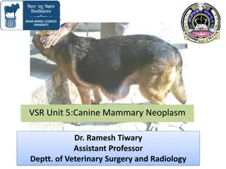
Canine Castration Techniques and Examination Guide
The physical examination of the scrotum, testicles, prepuce, and penis is vital in evaluating for abnormalities. Castration techniques such as open prescrotal castration are detailed, emphasizing the importance of proper procedure and care. Images and step-by-step instructions further illustrate the process.

Download Presentation

Please find below an Image/Link to download the presentation.
The content on the website is provided AS IS for your information and personal use only. It may not be sold, licensed, or shared on other websites without obtaining consent from the author. Download presentation by click this link. If you encounter any issues during the download, it is possible that the publisher has removed the file from their server.
E N D
Presentation Transcript
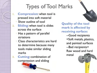
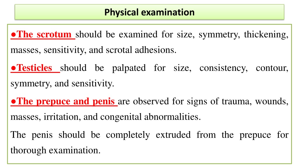
Physical examination The scrotum should be examined for size, symmetry, thickening, masses, sensitivity, and scrotal adhesions. Testicles should be palpated for size, consistency, contour, symmetry, and sensitivity. The prepuce and penis are observed for signs of trauma, wounds, masses, irritation, and congenital abnormalities. The penis should be completely extruded from the prepuce for thorough examination.
technique Castration technique 1-Prescrotal castration A-Open prescrotal Castration. B-closed prescrotal castration 2- Scrotal ablation
A-Open Prescrotal Castration. Position the patient in dorsal recumbency. Verify the presence of both testicles in the scrotum. Clip and aseptically prepare the caudal abdomen and medial thighs. Avoid irritating the scrotum with clippers or antiseptics. Drape the surgical area to exclude the scrotum from the field. Apply pressure on the scrotum to advance one testicle as far as possible into the prescrotal area. Incise skin and subcutaneous tissue along the median raphe over the displaced testicle. Continue the incision through spermatic fascia to exteriorize the testicle. Incise the parietal vaginal tunic over the testicle. Do not incise the tunica albuginea because this would expose the testicular parenchyma. Place a hemostat across the vaginal tunic where it attaches to the epididymis. Digitally separate the ligament of the tail of the epididymis from the tunic while applying traction with the hemostat on the tunic. Further exteriorize the testicle by applying caudal and outward traction. Identify the structures of the spermatic cord. Individually ligate the vascular cord and ductus deferens, then place an encircling ligature around both. Many surgeons ligate the ductus deferens and pampiniform plexus together. Use 2-0 or 3-0 absorbable suture (e.g., polyglactin 910 [Vicryl], polydioxanone [PDS], poliglecaprone 25 [Monocryl], polyglyconate [Maxon], or glycomer 631 [Biosyn]) for ligatures. Advance the second testicle into the incision, incise the fascial covering, and remove the testicle as described. Appose the incised dense fascia on either side of the penis with interrupted or continuous sutures. Close subcutaneous tissue with a continuous pattern. Appose skin with an intradermal, subcuticular, or simple interrupted suture pattern.
technique A, To perform an open canine castration, advance one testicle into the prescrotal area by applying pressure over the scrotum. Make an incision over the testicle. B, Incise the spermatic fascia and parietal vaginal tunic. C, Place a hemostat across the tunic where it attaches to the epididymis and digitally separate the ligament of the tail of the epididymis from the tunic. D, Ligate the ductus deferens individually, and then encircle circumferential ligature. Apply a Carmalt forceps distal to the ligatures and transect between the clamp and ligatures. and vascular with cord both a proximal