Understanding the Posterior Pituitary Gland and Its Hormones
The posterior pituitary gland, a key part of the endocrine system, plays a vital role in hormone secretion. It controls the release of oxytocin and vasopressin, influencing social bonding, reproduction, and childbirth. Learn about the anatomy, function, and disorders associated with this important gland.

Download Presentation

Please find below an Image/Link to download the presentation.
The content on the website is provided AS IS for your information and personal use only. It may not be sold, licensed, or shared on other websites without obtaining consent from the author. Download presentation by click this link. If you encounter any issues during the download, it is possible that the publisher has removed the file from their server.
E N D
Presentation Transcript
Group 2 MLS514 presentation OVERVIEW ON POSTERIOR PITUITARY GLAND
Presented By Group 2 ONOKPE OGHENEFEJIRO DANDUTSE HAJARA TIJJANI (Presenter) 15/MHS06/023 OGUNYEMI TOSIN ESHEGBE EZEKIEL 15/MHS06/051 15/MHS06/043 14/MHS06/022 2
OUTLINE Introduction Oxytocin Vasopressin Disease associated with posterior pituitary Diagnosis Treatment Reference 3
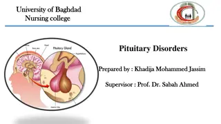
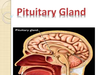
PITUITARY GLAND The pituitary gland is about the size of a pea and is situated in a bony hollow, behind the bridge of the nose. It is attached to the base of the brain by a thin stalk. The hypothalamus is situated immediately above the gland. 4
The pituitary gland is often called the master gland because it controls several other hormone glands in your body. It secretes hormones from both the front part (anterior) and the back part (posterior) of the gland. Hormones are chemicals that carry messages from one cell to another through the bloodstream (TPF, 2018). 5
Posterior Pituitary Gland It is the posterior lobe of the pituitary gland which is part of the endocrine system. The posterior pituitary is not glandular as is the anterior pituitary. it is largely a collection of axonal projections from the hypothalamus that terminate behind the anterior pituitary, and serve as a site for the secretion of oxytocin and vasopressin directly into the blood (Malenka et al., 2009). 7
Two hormones are classically considered as being related to the posterior pituitary: oxytocin and vasopressin. These hormones are created in the hypothalamus and released in the posterior pituitary. After creation, they are stored in neurosecretory vesicles regrouped into Herring bodies before being secreted in the posterior pituitary via the bloodstream. 8
OXYTOCIN Oxytocin (Oxt) is a peptide hormone and neuropeptide. It is normally produced in the hypothalamus and released by the posterior pituitary. It plays a role in social bonding, sexual reproduction, childbirth, and the period after childbirth (Yang et al., 2013). 9
Oxytocin is released into the bloodstream as a hormone in response to stretching of the cervix and uterus during labor and with stimulation of the nipples from breastfeeding (Chiras, 2012). This helps with birth, bonding with the baby, and milk production (HEV, 2010). 10
For men, oxytocin function is less important, but it does have a role to play in moving sperm. It also appears to affect the production of testosterone in the testes. Most hormones create negative feedback loops after they are released, but oxytocin is one of the few that exhibit positive feedback loops (Ellis et al.,2019). 11
This means that the release of oxytocin leads to actions that stimulate even more of a release of oxytocin. VASOPRESSIN Vasopressin, also called antidiuretic hormone (ADH) or arginine vasopressin (AVP) it is a hormone synthesized as a peptide prohormone in neurons in the hypothalamus, and is converted to AVP (Anderson, 2012). 12
It then travels down the axon of that cell, which terminates in the posterior pituitary, and is released from vesicles into the circulation in response to extracellular fluid hypertonicity retention of water (Marieb, 2014). AVP has two primary functions First, it increases the amount of solute-free water reabsorbed back into the circulation from the filtrate in the kidney tubules of the nephrons (Caldwell and Young, 2006). 13
Second, AVP constricts arterioles, which increases peripheral vascular resistance and raises arterial blood pressure (Babar, 2013). A third function is possible, some AVP may be released directly into the brain from the hypothalamus. It may play an important role in social behavior, sexual motivation and pair bonding, and maternal responses to stress (Insel, 2010). 14
An example of negative feedback in vasopressin is when the level of water in the blood falls, negative feedback ensures that the amount of ADH rises and also as the level of water in the blood rises negative feedback ensures that the amount of ADH falls. 15
Syndrome of inappropriate antidiuretic hormone secretion Syndrome of inappropriate antidiuretic hormone secretion (SIADH) is a condition in which the body makes too much antidiuretic hormone (ADH). This hormone helps the kidneys control the amount of water the body loses through the urine. SIADH causes the body to retain too much water. 17
Clinical Manifestations Excess ADH increases reabsorption of water into the circulation. ECF volume expands, plasma osmolality declines and GFR increases. Sodium levels decline (dilutional hyponatremia) This hyponatremia causes muscle cramps, weakness etc. Increased body weight without edema. 18
CAUSES Medicines, such as certain type 2 diabetes drugs, seizure drugs, antidepressants, heart and blood pressure drugs, cancer drugs, anesthesia Disorders of the brain, such as injury, infections, stroke Brain surgery in the region of the hypothalamus Lung disease, such as tuberculosis, cancer, chronic infections Rare diseases of the hypothalamus or pituitary Cancer of the lung, small intestine, pancreas, brain. 19
DIAGNOSIS Based on clinical and laboratory findings of low serum osmolality and low serum sodium. Urinalysis Decreased effective serum osmolality - <275 mOsm/kg of water Urinary sodium concentration high - over 40 mEq/L with adequate dietary salt intake (Gross, 2012). 20
TREATMENT Removing the underlying cause when possible. Mild and asymptomatic hyponatremia is treated with adequate solute intake (including salt and protein) and fluid restriction starting at 500 ml per day of water with adjustments based on serum sodium levels (Christianto et al., 2010). Central diabetes insipidus It is also called neurogenic diabetes insipidus, is a type of diabetes insipidus due to a lack of vasopressin (ADH) production in the brain (Chitturi et al., 2008). 21
Causes Genetic The most rare form of central DI is familial neurogenic diabetes insipidus. This form of DI is due to an inherited mutation of the arginine vasopressin-neurophysin II (AVP- NPII) gene, inherited in an autosomal dominant manner (DIF, 2006). Acquired The lack of vasopressin production usually results from some sort of damage to the pituitary gland. It may be due to damage to the brain. 22
DIAGNOSIS Common rule outs include: diabetes mellitus, chronic kidney disease, hypokalemia and hypercalcemia. Once these conditions have been ruled out a water deprivation test is employed to confirm the diagnosis of CDI. 23
TREATMENT The disorder is treated with vasopressin analogs such as desmopressin. Nonetheless, many times desmopressin alone is not enough to bring under control all the symptoms. 24
REFERENCES Anderson, D.A. (2012). Dorland's Illustrated Medical Dictionary (32nd ed.). Elsevier. Babar, S.M. (2013). SIADH associated with ciprofloxacin. The Annals of Pharmacotherapy. 47 (10): 1359 1363 Chiras, D.D. (2012). Human Biology (7th ed.). Sudbury, MA: Jones & Bartlett Learning. p. 262. Chitturi, S., Harris, M., Thomsett M.J. (2008). Utility of AVP gene testing in familial neurohypophyseal diabetes insipidus. Clin. Endocrinol. 69 (6): 926 930 Caldwell, H.K., Young, W.S. III (2006). Oxytocin and Vasopressin: Genetics and Behavioral Implications. In Lajtha A, Lim R (eds.). Handbook of Neurochemistry and Molecular Neurobiology: Neuroactive Proteins and Peptides (3rd ed.). Berlin: Springer. pp. 573 607. Diabetes Inspidus. Library of the National Medical Society. 2008. http://www.medical-library.org/journals4a/diabetes_insipidus.htm Diabetes Insipidus Foundation (2006). Familial Neurogenic Diabetes Insipidus: a disease caused by a traffic jam. http://diabetesinsipidus.org/4di_familial.htm Archived 2020-02-21 Ellis, J.A., Brown, C.M., Barger, B., Carlson, N.S. (2019). Influence of Maternal Obesity on Labor Induction: A Systematic Review and Meta-Analysis. J Midwifery WomensHealth. 64(1):55-67. 25
Gross, P. (2012). Clinical management of SIADH. Therapeutic Advances in Endocrinology and Metabolism. 3 (2): 61 73. Human Evolutionary Biology (2010). Cambridge University Press. p. 282. Insel, T.R. (2010). The challenge of translation in social neuroscience: A review of oxytocin, vasopressin, and affiliative behavior. Neuron. 65 (6): 768 779 Malenka, R.C., Nestler, E.J., Hyman, S.E (2009). "Chapter 10: Neural and Neuroendocrine Control of the Internal Milieu". Molecular Neuropharmacology: A Foundation for Clinical Neuroscience (2nd ed.). New York: McGraw-Hill Medical. pp. 246, 248 259. Marieb, E. (2014). Anatomy & physiology. Glenview, IL: Pearson Education, Inc. Sch rer, L., Wolf, S., Lumenta, C.B. (2010). Water and Electrolyte Regulation. In Lumenta, Christianto B., Di Rocco, Concezio, Haase, Jens. (eds.). Neurosurgery. European Manual of Medicine. pp. 611 615 Yang, H.P., Wang, L., Han, L., Wang, S.C. (2013). Nonsocial functions of hypothalamic oxytocin. 54:746-812 26


. Clinical management of SIADH.")