Understanding Phototransduction in Vision: Rods, Cones, and Light Adaptation
This lecture delves into the intricacies of photo transduction in vision, focusing on the functional properties of rods and cones in different lighting conditions, the convergence of visual pathways, phototransduction processes in light and dark, synaptic mediators in the retina, and more. Explore the differentiation between rods and cones, their structural characteristics, and the photochemistry of vision through rhodopsin regeneration. Gain insights into conditions like nyctalopia and the remarkable adaptability of our eyes to varying light environments.

Download Presentation

Please find below an Image/Link to download the presentation.
The content on the website is provided AS IS for your information and personal use only. It may not be sold, licensed, or shared on other websites without obtaining consent from the author. Download presentation by click this link. If you encounter any issues during the download, it is possible that the publisher has removed the file from their server.
E N D
Presentation Transcript
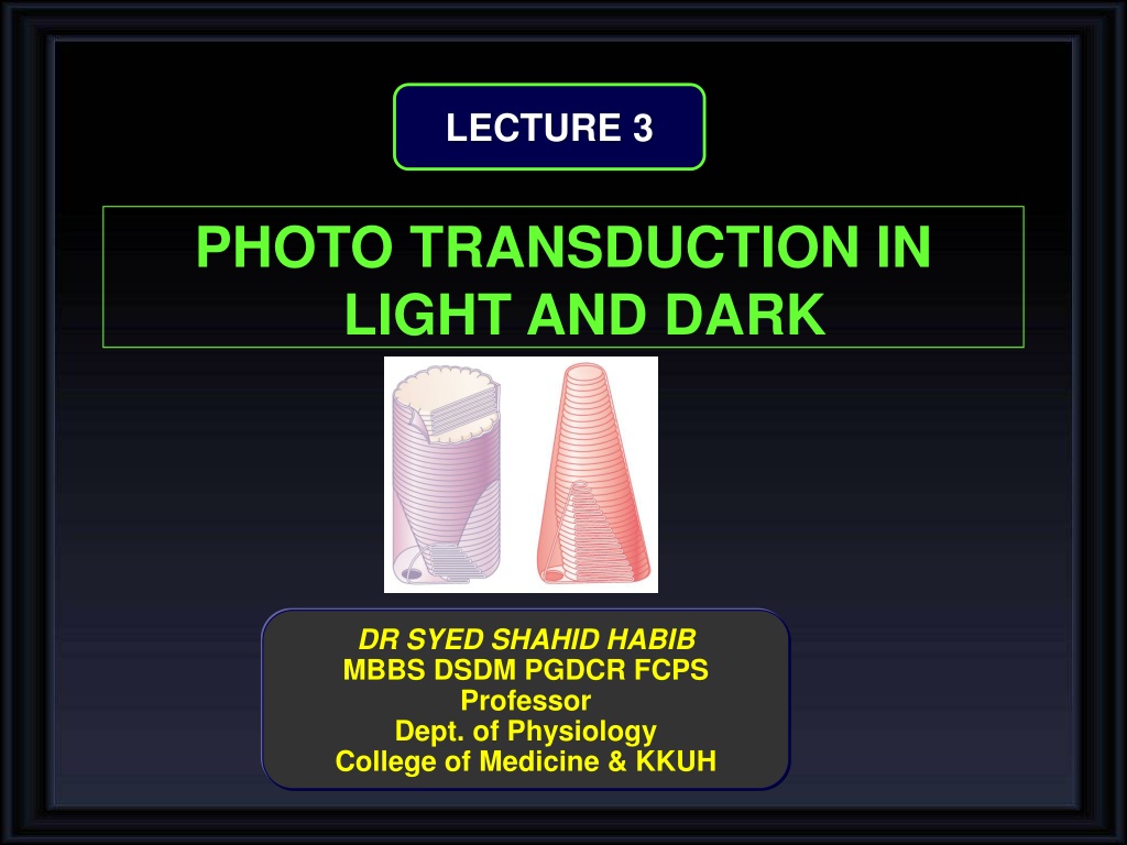
LECTURE 3 PHOTO TRANSDUCTION IN LIGHT AND DARK DR SYED SHAHID HABIB MBBS DSDM PGDCR FCPS Professor Dept. of Physiology College of Medicine & KKUH
OBJECTIVES At the end of this lecture you should be able to: Explain functional properties of rods and cones in scotopic and photopic vision Know the convergence and its value Describe phototransduction process for rods and cones in light and dark and the ionic basis of these responses Enumarate Synaptic mediators at retina Describe Rhodopsin regeneration Define nyctalopia, dark and light adaptation
Low convergenc in cones: cones synapse with one bipolar cell one ganglion cell It increases visual acuity & decreases sensitivity to light High convergence of rods: 300:1- It decreases visual acuity & increases sensitivity to light
Receptors of vision (Rods&cones) Outer segment (modified cilia) has disks full of photosensitive pigment (rhodopsin) react with light to initiate action potential -In cones is conical , small and contain 3 types of rhodopsin - in rods it is big, rode like and contain one type of rhodopsin -There are Na channels in the outer segment Inner segment full of mitochondria ( source of energy for Na-K pump), it is thick in cones There is Na-K pump in inner segment
Comparison of Rods and Cones Rods Cones Abundant in the periphery of the retina About 120 million Contain Scotopsin Best for low light conditions See black/white and shades of gray Abundant in fovea About 6 million Contain Photopsin (3Types) Best for bright light conditions See all colors
Photochemistry of Vision 1-In Rods: it is rhodopsin: [Scotopsin protein (opsin) + retinal (retinene 1 = Vit A)] Called visual purple (Rhodopsin of the rods most strongly absorbs green-blue light and, therefore, appears reddish-purple 2- In cones there are 3 types of Photopsins (I,II & III) : [Photopsin protein (opsin) + retinal (retinene 1 = Vit A)] - It is stored in disks of rods at outer segment - It forms (90% of its protein ) -In dark rhodopsin is in 11-cisretinal form (INACTIVE) It is light sensitive form which increase sensitivity of rods to light
RHODOPSIN NYCTALOPIA (night blindness) Deficiency of Vitamine A (main source of retinal of rhodopsin) cause rods pigments Also causes xerophthamia (Dry Eyes)
Sodium Current in Rods In Dark 2. Cancels Negativity (-40 mv) 1.Creates Negativity (-70 to -80 mv Sodium Current is flowing in Dark
PHOTOTRANSDUCTION In dark Na+ current leads to Depolarization flow to synaptic endings to steadily release glutamate Response in bipolar cells( depolarization) Stimulate ganglion cells AP in optic nerve Vision in dark called Dark vision (Scotopic Vision) Steady State Release of Glutamate Release of Glutamate
Rhodopsin Retinal Visual Cycle
Photochemistry of Color Vision by the Cones Photopsins Retinal Visual Cycle The cones are about 30 to 300 times less sensitive than rods Photopsins
SEQUENCE OF EVENTS IN PHOTOTRANSDUCTION OF RODS AND CONES
SEQUENCE OF EVENTS IN RODS AND CONES ACTIVATED BY LIGHT 1. The Photon transforms 11-cis retinal of rhodopsin to metarhodopsin II, [active form of rhodopsin in 11- trans retinal form] 2. The activated rhodopsin Protein] 3. Activated transducin activates phosphodiesterase. 4. Activated phosphodiesterase 5-GMP Decrease in cGMP 5. Leads to Hyperpolarization 6. Rhodopsin kinase inactivates metarhodopsin II activates transducin [G converts cGMP to closes Na Channels Glutamate
Glutamate binds to specific receptor sites on the bipolar cells, associated with specific types of ion channels. Depending on the kinds of ion channels present, the bipolar cell may become depolarized (On Center) or it may become Hyperpolarized (Off Center)
Note: This may be due to lateral inhibition by Horizontal cells to sharpen the edges of a stimulus and improve discrimination
Rodes &cones potentials are graded, local potential (generator potential) propagated as AP in ganglion cells. Only Ganglion cell action potential (All or none AP) are transmitted to optic nerve.
DARK ADAPTATION If a person moves from brightly lighted surroundings to a dim lighted area the retinas slowly become more sensitive to light as the individual becomes accustomed to the dark [20 min] for dark vision (only gross features but no details or colors Rapid (5 minutes) due to adaptation of cones in fovea (sensitivity of cones to light increases) More light sensitive pigments In Visual threshold In Visual sensitivity Na channels remain open Na current continues Less rapid (20 min) due to adaptation of rodes in the peripheral retina (sensitivity of rodes to light increases)
LIGHT ADAPTATION If a person moves from dim to enlightened area light seems intensely and even uncomfortably bright until the eyes adapt to the increased illumination and the visual threshold rises in 5 minutes. Less light sensitive pigments In Visual threshold In Visual sensitivity Na channels remain close Na current decreases Why radiologists & aircraft pilots wear red goggles in bright light? Red light mainly stimulates the cones & rods to some extent, so red goggles for rods act as dimlight, so with it rods are adapted to darkness & form large amounts of rhodopsin while the person is in bright light & when person enters a dark place he can see well & do not have to wait for 20 minutes for dark adaptation.


")