Understanding Episcleritis and Scleritis in Ophthalmology
 undefined


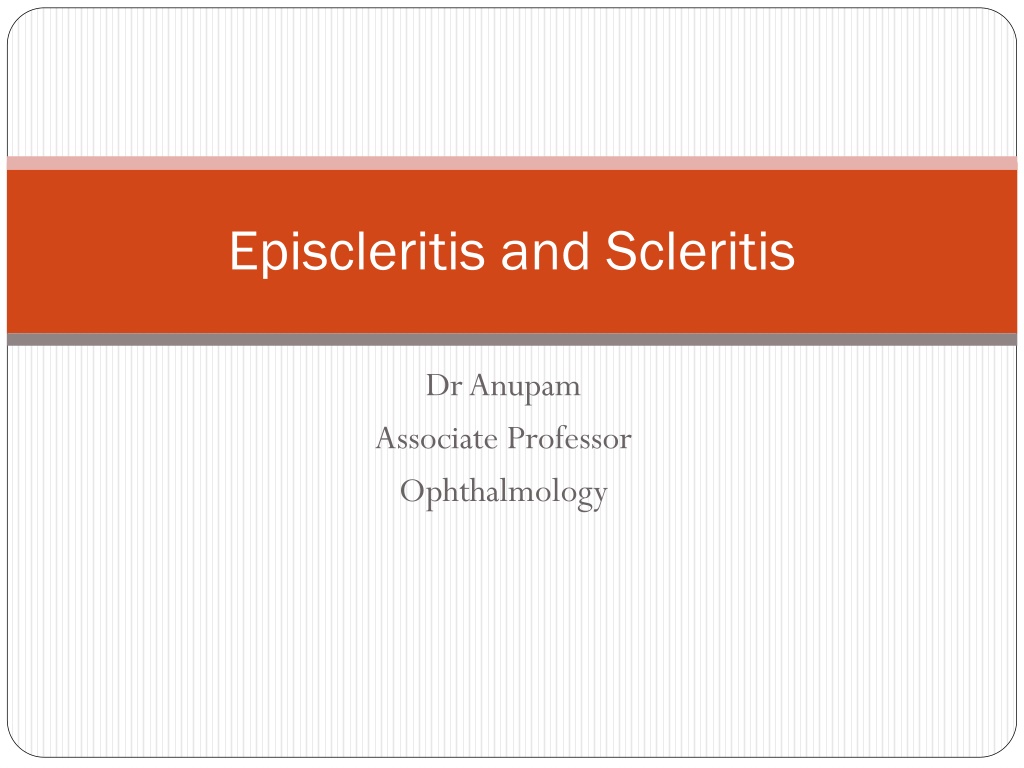
Dr Anupam
Associate Professor
Ophthalmology
Episcleritis and Scleritis


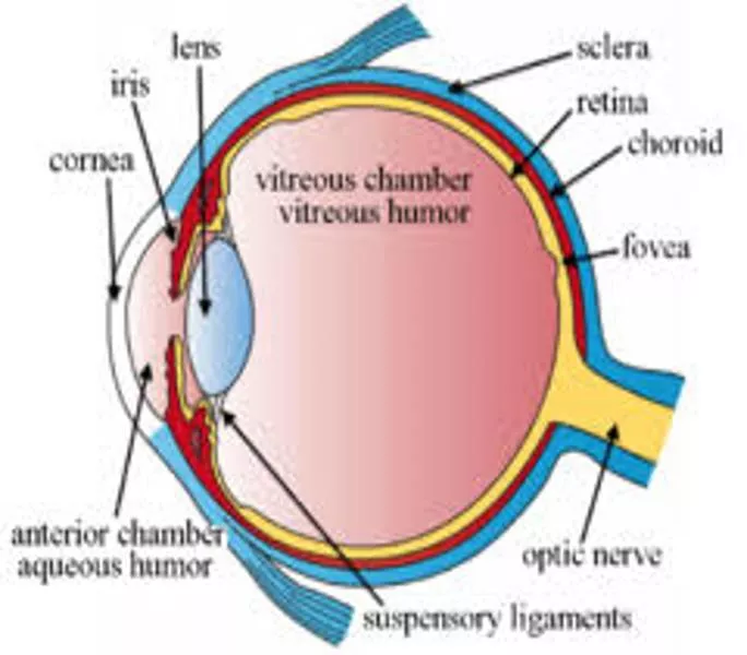
Applied Anatomy
Sclera forms the posterior five-
sixth opaque part of the of the
eyeball.
Its whole outer surface is
covered by Tenon's capsule and
bulbar conjunctiva.
Its inner surface lies in contact
with choroid with a potential
suprachoroidal space in between.
It is generally thinner in children & females.
Sclera is thickest posteriorly (1mm) and is thinnest at the
insertion of extraocular muscles (0.3 mm).
Lamina cribrosa is a sieve-like sclera from which fibres of
optic nerve pass.
Microscopic structure
1. Episcleral tissue.
2. Sclera proper.
3. Lamina fusca
.
Vasculature:
1. Conjunctival vessels
2. Episcleral vessels
3. Deep vascular plexus
EPISCLERITIS
Benign recurrent inflammation of the episclera,
Common
Benign
Self-limiting
Recurrent
Never progresses to scleritis
Rarely associated with systemic disease
Etiology
Exact etiology is not known.
It is found in association with gout, rosacea and psoriasis.
It has also been considered a hypersensitivity reaction to
endogenous tubercular or streptococcal toxins.
Pathology
Histologically, there occurs localised lymphocytic infiltration
of episcleral tissue associated with oedema and congestion of
overlying Tenon's capsule and conjunctiva.
Clinical picture
Symptoms.
Redness, mild ocular discomfort, burning sensation or
foreign body sensation.
Rarely, mild photophobia and lacrimation may occur.
Signs
Simple or diffuse episcleritis
– Sectoral redness
– Diffuse redness
– Resolves in 1-2 weeks
Nodular episcleritis
– Focal, raised, nodular
– Sclera uninvolved
– Longer to resolve
Vessels remain radial and mobile
Palpation of the globe often elicits marked tenderness in
scleritis, but generally not in episcleritis.
Phenylephrine diagnostic test
:
Hyperemia usually blanches with topical phenylephrine
(2.5%) in episcleritis but not in scleritis.
Management
Mild cases
– Usually no specific Rx
– If discomfort
• Lubricant
• Topical NSAID eg acular (keterolac trimethamine)
• Mild topical corticosteroid
Or systemic Ibuproven/aspirin
Investigate in recurrent cases.
SCLERITIS
Scleritis refers to a chronic inflammation of the sclera proper. It is
a comparatively serious disease which may cause visual impairment
and even loss of the eye if treated inadequately.
Relatively rarer than episcleritis
Usually bilateral
More common in females
Associated with connective tissue disorders in upto 50% of cases.
Granulomatous inflammation
Mild to blinding spectrum
Clinical classification of Scleritis
It can be classified as follows:
I.
Anterior scleritis (98%)
1. Non-necrotizing scleritis (85%)
(a) Diffuse
(b) Nodular
2. Necrotizing scleritis (13%)
(a) with inflammation
(b) without inflammation (scleromalacia perforans)
II.
Posterior scleritis (2%)
Associated systemic conditions
Rheumatoid Arthritis
1:200 develop scleritis
Connective Tissue Disease
Wegener granulomatosis
Systemic lupus erythematosus
Polyarteritis nodosa
Ankylosing spondilytis
Associated systemic conditions
Herpes Zoster Ophthalmicus
Metabolic disorders like gout and thyrotoxicosis
Granulomatous diseases like
•
Tuberculosis
,
•
Syphilis,
•
Sarcoidosis,
•
Leprosy
Miscellaneous
Surgically induced
Infectious
Idiopathic
Clinical features
Symptoms
Pain
Redness
Photophobia
Lacrimation
Diminution of vision
Clinical features
Signs
1. Non-necrotizing anterior diffuse
scleritis.
Commonest variety,
Widespread inflammation involving a
quadrant or more of the anterior sclera.
The involved area is raised and salmon
pink to purple in colour
Clinical features
Signs
2. Necrotizing anterior nodular
scleritis.
characterised by one or two hard,
purplish elevated scleral nodules, usually
situated near the limbus
Sometimes, the nodules are arranged in a
ring around the limbus
(
annular
scleritis).
Clinical features
Signs
3. Necrotizing scleritis with inflammation.
The affected necrosed area is thinned out and sclera becomes
transparent and ectatic with uveal tissue shining through it.
It is usually associated with anterior uveitis.
4.
Anterior necrotizing scleritis without inflammation
(scleromalacia perforans).
Usually associated with seropositive RA
Painless scleral thinning due to ischaemia.
Posterior Scleritis
Defined as primarily arising posterior to the equator
Painful or painless diminution of vision
Proptosis
Restricted ocular movements
Disc or macular edema
Choroidal folds or detachment
Uveal effusion syndrome
Retinal detachment
Scleral thickening seen on CT or USG B scan.
Investigation
1. TLC, DLC and ESR
2. Serum levels of complement (C3), immune complexes,
rheumatoid factor, antinuclear antibodies and L.E cells for an
immunological survey.
3. FTA - ABS, VDRL for syphilis.
4. Serum uric acid for gout.
5. Urine analysis.
6. Mantoux test.
7. X-rays of chest, paranasal sinuses, sacroiliac joint and orbit
to rule out foreign body especially in patients with nodular
scleritis.
Management
(A) Non-necrotising scleritis.
Steroid eye drops and systemic indomethacin 100 mg
daily for a day and then 75 mg daily.
B) Necrotising scleritis.
It is treated by topical
steroids and heavy doses of oral steroids tapered
slowly.
Immuno-suppressive agents like methotrexate or cyclophos-
phamide.
Subconjunctival steroids are contraindicated because they
may lead to scleral thinning and perforation
STAPHYLOMAS
Ectasia or bulging of the outer coats (cornea, sclera or both)
of the eye with incarceration of the uveal tissue.
Due to weakening of the eye wall resulting from any
degenerative or inflammatory condition of the same.
Types:
A.
Anterior (involves cornea)
B.
Intercalary (with in 2mm of limbus)
C.
Ciliary (2-8mm behind the limbus
D.
Equatorial (14mm behind the limbus)
E.
Posterior (posterior to equator)
A: Intercalary staphyloma
B. Ciliary staphyloma
Equatorial staphyloma
Posterior staphyloma
Management
Treat the underlying cause like, scleritis, RA, vit A def or
cornal ulcer.
Local excision and patch graft of cornea or sclera
Enucleation with implant.
Thank You
Episcleritis and scleritis are inflammatory conditions affecting the episclera and sclera of the eye, respectively. Episcleritis is benign, self-limiting, and rarely progresses to scleritis, presenting with redness and mild discomfort. Scleritis, on the other hand, is a more serious condition that can lead to vision loss and systemic diseases. Understanding the etiology, pathology, clinical presentation, and management of these conditions is crucial for eye health.

Download Presentation

Please find below an Image/Link to download the presentation.
The content on the website is provided AS IS for your information and personal use only. It may not be sold, licensed, or shared on other websites without obtaining consent from the author. Download presentation by click this link. If you encounter any issues during the download, it is possible that the publisher has removed the file from their server.
E N D
Presentation Transcript
Episcleritis and Scleritis Dr Anupam Associate Professor Ophthalmology
Applied Anatomy Applied Anatomy Sclera forms the posterior five- sixth opaque part of the of the eyeball. Its whole outer surface is covered by Tenon's capsule and bulbar conjunctiva. Its inner surface lies in contact with choroid with a potential suprachoroidal space in between.
It is generally thinner in children & females. Sclera is thickest posteriorly (1mm) and is thinnest at the insertion of extraocular muscles (0.3 mm). Lamina cribrosa is a sieve-like sclera from which fibres of optic nerve pass.
Microscopic structure Microscopic structure 1. Episcleral tissue. 2. Sclera proper. 3. Lamina fusca. Vasculature: 1. Conjunctival vessels 2. Episcleral vessels 3. Deep vascular plexus
EPISCLERITIS Benign recurrent inflammation of the episclera, Common Benign Self-limiting Recurrent Never progresses to scleritis Rarely associated with systemic disease
Etiology Exact etiology is not known. It is found in association with gout, rosacea and psoriasis. It has also been considered a hypersensitivity reaction to endogenous tubercular or streptococcal toxins. Pathology Histologically, there occurs localised lymphocytic infiltration of episcleral tissue associated with oedema and congestion of overlying Tenon's capsule and conjunctiva.
Clinical picture Clinical picture Symptoms. Redness, mild ocular discomfort, burning sensation or foreign body sensation. Rarely, mild photophobia and lacrimation may occur.
Signs Simple or diffuse episcleritis Sectoral redness Diffuse redness Resolves in 1-2 weeks Nodular episcleritis Focal, raised, nodular Sclera uninvolved Longer to resolve
Vessels remain radial and mobile Palpation of the globe often elicits marked tenderness in scleritis, but generally not in episcleritis. Phenylephrine diagnostic test: Hyperemia usually blanches with topical phenylephrine (2.5%) in episcleritis but not in scleritis.
Management Management Mild cases Usually no specific Rx If discomfort Lubricant Topical NSAID eg acular (keterolac trimethamine) Mild topical corticosteroid Or systemic Ibuproven/aspirin Investigate in recurrent cases.
SCLERITIS SCLERITIS Scleritis refers to a chronic inflammation of the sclera proper. It is a comparatively serious disease which may cause visual impairment and even loss of the eye if treated inadequately. Relatively rarer than episcleritis Usually bilateral More common in females Associated with connective tissue disorders in upto 50% of cases. Granulomatous inflammation Mild to blinding spectrum
Clinical classification of Scleritis It can be classified as follows: I. Anterior scleritis (98%) 1. Non-necrotizing scleritis (85%) (a) Diffuse (b) Nodular 2. Necrotizing scleritis (13%) (a) with inflammation (b) without inflammation (scleromalacia perforans) II. Posterior scleritis (2%)
Associated systemic conditions Associated systemic conditions Rheumatoid Arthritis 1:200 develop scleritis Connective Tissue Disease Wegener granulomatosis Systemic lupus erythematosus Polyarteritis nodosa Ankylosing spondilytis
Associated systemic conditions Associated systemic conditions Herpes Zoster Ophthalmicus Metabolic disorders like gout and thyrotoxicosis Granulomatous diseases like Tuberculosis, Syphilis, Sarcoidosis, Leprosy Miscellaneous Surgically induced Infectious Idiopathic
Clinical features Clinical features Symptoms Pain Redness Photophobia Lacrimation Diminution of vision
Clinical features Clinical features Signs 1. Non-necrotizing anterior diffuse scleritis. Commonest variety, Widespread inflammation involving a quadrant or more of the anterior sclera. The involved area is raised and salmon pink to purple in colour
Clinical features Clinical features Signs 2. Necrotizing anterior nodular scleritis. characterised by one or two hard, purplish elevated scleral nodules, usually situated near the limbus Sometimes, the nodules are arranged in a ring around the limbus (annular scleritis).
Clinical features Clinical features Signs 3. Necrotizing scleritis with inflammation. The affected necrosed area is thinned out and sclera becomes transparent and ectatic with uveal tissue shining through it. It is usually associated with anterior uveitis. 4. Anterior necrotizing scleritis without inflammation (scleromalacia perforans). Usually associated with seropositive RA Painless scleral thinning due to ischaemia.
Posterior Scleritis Defined as primarily arising posterior to the equator Painful or painless diminution of vision Proptosis Restricted ocular movements Disc or macular edema Choroidal folds or detachment Uveal effusion syndrome Retinal detachment Scleral thickening seen on CT or USG B scan.
Investigation Investigation 1. TLC, DLC and ESR 2. Serum levels of complement (C3), immune complexes, rheumatoid factor, antinuclear antibodies and L.E cells for an immunological survey. 3. FTA -ABS, VDRL for syphilis. 4. Serum uric acid for gout. 5. Urine analysis. 6. Mantoux test. 7. X-rays of chest, paranasal sinuses, sacroiliac joint and orbit to rule out foreign body especially in patients with nodular scleritis.
Management Management (A) Non-necrotising scleritis. Steroid eye drops and systemic indomethacin 100 mg daily for a day and then 75 mg daily. B) Necrotising scleritis. It is treated by topical steroids and heavy doses of oral steroids tapered slowly. Immuno-suppressive agents like methotrexate or cyclophos- phamide. Subconjunctival steroids are contraindicated because they may lead to scleral thinning and perforation
STAPHYLOMAS STAPHYLOMAS Ectasia or bulging of the outer coats (cornea, sclera or both) of the eye with incarceration of the uveal tissue. Due to weakening of the eye wall resulting from any degenerative or inflammatory condition of the same. Types: A. Anterior (involves cornea) B. Intercalary (with in 2mm of limbus) C. Ciliary (2-8mm behind the limbus D. Equatorial (14mm behind the limbus) E. Posterior (posterior to equator)
A: Intercalary staphyloma B. Ciliary staphyloma Equatorial staphyloma Posterior staphyloma
Management Treat the underlying cause like, scleritis, RA, vitA def or cornal ulcer. Local excision and patch graft of cornea or sclera Enucleation with implant.