Single-Cell Transcriptomics of TAZ-Deficient Murine Corneal Endothelium
This study explores the single-cell transcriptomics of TAZ-deficient murine corneal endothelium in the context of Fuchs Endothelial Corneal Dystrophy (FECD), a polygenic disease affecting millions globally. The research utilizes a TAZ-deficient mouse model to mimic late-onset FECD, revealing reduced corneal endothelial density, altered morphology, and changes in gene expression associated with disease progression and potential therapeutics. The findings shed light on the molecular mechanisms underlying FECD pathology and the need for alternative non-surgical treatments in the absence of a cure beyond corneal transplants.
Download Presentation
Please find below an Image/Link to download the presentation.
The content on the website is provided AS IS for your information and personal use only. It may not be sold, licensed, or shared on other websites without obtaining consent from the author. Download presentation by click this link. If you encounter any issues during the download, it is possible that the publisher has removed the file from their server.

Presentation Transcript
Single Cell Transcriptomics of the TAZ-deficient Murine Corneal Endothelium Raneesh Ramarapu Mentor: Dr. Sara Thomasy Comparative Ophthalmology and Vision Sciences Laboratory (COVSL)
Corneal Endothelium Hexagonal single cell layer Non-proliferative in most species Active cells that constantly pump out water maintain corneal transparency Ref [1]
The Pathology of Interest: Fuchs Endothelial Corneal Dystrophy (FECD) Ref [2]
The Pathology of Interest: Fuchs Endothelial Corneal Dystrophy (FECD) Polygenic Disease >300 million human patients globally under the age of 30 [3] Ref [2]
The Pathology of Interest: Fuchs Endothelial Corneal Dystrophy (FECD) Polygenic Disease >300 million human patients globally under the age of 30 [3] Characterized by [4,5] Accelerated Loss of CEn Altered Descemet Membrane (DM) stiffness Aberrant ECM accumulation Guttae Ref [3]
The Pathology of Interest: Fuchs Endothelial Corneal Dystrophy (FECD) Only treatment is corneal transplant Low accessibility Need for alternative non- surgical therapeutics! Ref [3]
Our Model of FECD: TAZ-deficient mice COVSL modelled late-onset FECD using Wwtr1 deficient mice [6]
Our Model of FECD: TAZ-deficient mice COVSL modelled late-onset FECD using Wwtr1 deficient mice [6] Reduced CEn density abnormal CEn morphology Softer DM Thinner corneas Altered expression and localization of Na/K-ATPase and ZO-1
Wwtr1 (TAZ) Encodes transcriptional co- activator with PDZ-binding motif (TAZ) Multifunctional Gene [7-13] Mechanotransducer of DM stiffness Ref [7]
Wwtr1 (TAZ) Encodes transcriptional co- activator with PDZ-binding motif (TAZ) Multifunctional Gene [7-13] Mechanotransducer of DM stiffness Central orchestrator of multiple pathways Hippo Wnt TGF-Beta NF-kB
Goal of the project Define the transcriptomic landscape of TAZ deficiency
Goal of the project Define the transcriptomic landscape of TAZ deficiency We have single cell transcriptomic data of 3 genotypes of 3 age groups: Wildtype (WT): 2-, 6-, 11-month-old Wwtr1 Heterozygousknockout (TAZ HET): 2-, 6-, 11-month-old Wwtr1 Homozygous knockout (TAZ KO): 2-, 6-, 11-month-old
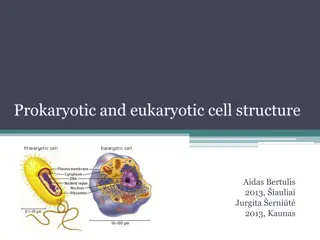
Single Cell RNA-sequencing High dimensionality data analysis Sequence all the RNA from individual cells Allows us to assess heterogenous tissues such as the cornea
ScRNA-seq Analysis in a nutshell Allows us to engage with and interpret multidimensional complex data by
ScRNA-seq Analysis in a nut shell Allows us to engage with and interpret multidimensional complex data by 1. Dimensionality reduction Ref [14]
ScRNA-seq Analysis in a nut shell Allows us to engage with and interpret multidimensional complex data by 1. Dimensionality reduction But how do we define cell types? Ref [14]
ScRNA-seq Analysis in a nut shell Allows us to engage with and interpret multidimensional complex data by 1. Dimensionality reduction But how do we define cell types? 2. K-means Clustering Ref [14] Ref [15]
ScRNA-seq Analysis in a nut shell Allows us to engage with and interpret multidimensional complex data by 1. Dimensionality reduction But how do we define cell types? 2. K-means Clustering We now have statistically defined cell types that can be further analyzed
WT, TAZ HET and TAZ KO CEn cells clustered separately from the CEp and Limbus These were validated with previously described markers
WT 2MO versus KO 2MO CEn KO WT
Gene Ontology Compiles the 1000s of upregulated genes between WT and KO Compares it to a database of genes categorized and labelled by function and networks
Gene Ontology Based on comparisons to multiple databases and Electron microscopy work, we focused on 1. Endoplasmic reticulum stress 2. Mitochondrial function
WT2MO v KO2MO KO downregulated functional markers Slc4a11 anion exchanger Slc4a4 Na/bicarbonate transporter Aqp1/5 Aquaporins
WT2MO v KO2MO KO downregulated functional markers Interestingly it downregulates type I collagens responsible for DM stiffness
WT2MO v KO2MO KO downregulated functional markers Interestingly it downregulates type I collagens responsible for DM stiffness Although fibroblastic marker fibronectin is upregulated
WT2MO v KO2MO KO downregulated functional markers KO upregulates ER Stress transcripts Hspa5 is integral to the ER stress response [16]
WT2MO v KO2MO KO downregulated functional markers KO upregulates ER Stress transcripts Calnexin regulates mitochondrial function with ER stress, calcium movement and ER stress associated cell death [17,18]
WT2MO v KO2MO KO downregulated functional markers KO upregulates ER Stress transcripts KO upregulates calcium binding genes These genes are upregulated with ER stress and calcium binding [19, 20]
WT2MO v KO2MO KO downregulated functional markers KO upregulates ER Stress transcripts KO upregulates calcium binding gene KO upregulates genes associated with stress metabolism
WT2MO v KO2MO KO downregulated functional markers KO upregulates ER Stress transcripts KO upregulates calcium binding gene KO upregulates genes associated with stress metabolism Slc2a1 glucose transporter Pkm Phosphofructokinase Fgfr1 associated with metabolic changes in stress [21] Ldha lactate dehydrogenase
WT2MO v KO2MO KO downregulated functional markers KO upregulates ER Stress transcripts KO upregulates calcium binding gene KO upregulates genes associated with stress metabolism KO upregulates mitochondrial genes
Putting it together Improper TAZ signaling negatively impacts the CEn healing response due to cytoplasmic retention This coupled with TAZ s key role in orchestrating multiple pathways likely causes the trifecta of ER stress, change in metabolism and mitochondrial upregulation
WT11MO vs KO11MO KO WT
WT11MO vs KO11MO KO11MO has only downregulated genes except RIK gene (unknown function, maybe associated with mitophagy/mitochondrial function) GO indicates KO11MO are likely low functioning WT11MO cells (downregulation of protein synthesis and regulation of apoptosis)
Conclusions We have a greater understanding of the mechanisms driving CEn dysfunction in FECD in the TAZ model With age, TAZ dysfunction causes a reduction in cell function (at least at the transcriptomic level)
Future direction Validate these transcriptomic differences through qPCR and localization through fluorescent in situ hybridization Functionally validate the pathways of interest using immunohistochemistry
Acknowledgements I d like to thank Drs. Sara Thomasy and Brian Leonard for their incredible support both in and outside the lab I would like to thank the members of the wet lab portion (Sangwan Park, Nayeli Echeverria, Michelle Ferneding, Sophie Le and Monica Ardon) for procuring this data I would also like to thank Dr. Vijay Raghunathan for his critical comments that improved these projects Financial support was provided by the Students Training in Advanced Research (STAR) Program through the NIH T35 OD010956-22 grant
References 1. Meir, Y.-J. J., Chen, H.-C., Chen, C.-C. & Ma, H.-K. D. Revisiting Existing Evidence of Corneal Endothelial Progenitors and Their Potential Therapeutic Applications in Corneal Endothelial Dysfunction. Adv. Ther.37, 1034 1048 (2020) Aiello F, Gallo Afflitto G, Ceccarelli F, Cesareo M, Nucci C. Global Prevalence of Fuchs Endothelial Corneal Dystrophy (FECD) in Adult Population: A Systematic Review and Meta-Analysis. J Ophthalmol. 2022;2022: 3091695. Xia D, Zhang S, Nielsen E, et al. The ultrastructures and mechanical properties of the Descement's membrane in Fuchs endothelial corneal dystrophy. Sci Rep. 2016; 6: 23096. Matthaei M, Hribek A, Clahsen T, Bachmann B, Cursiefen C, Jun AS. Fuchs Endothelial Corneal Dystrophy: Clinical, Genetic, Pathophysiologic, and Therapeutic Aspects. Annu Rev Vis Sci. 2019;5: 151 175. Cui, Z. et al. Pathological molecular mechanism of symptomatic late-onset Fuchs endothelial corneal dystrophy by bioinformatic analysis. PLoS One13, e0197750 (2018) Park S, Leonard B, Kim S, Mccorkell ME, Murphy CJ, Raghunathan V, et al. Evaluation of function-related corneal endothelial markers in TAZ deficient mice over time. Invest Ophthalmol Vis Sci. 2021;62: 840 840. Varelas X, Sakuma R, Samavarchi-Tehrani P, Peerani R, Rao BM, Dembowy J, et al. TAZ controls Smad nucleocytoplasmic shuttling and regulates human embryonic stem-cell self-renewal. Nat Cell Biol. 2008;10: 837 848. Dupont S, Morsut L, Aragona M, Enzo E, Giulitti S, Cordenonsi M, et al. Role of YAP/TAZ in mechanotransduction. Nature. 2011;474: 179 183. Okumura N, Nakamura T, Kay EP, Nakahara M, Kinoshita S, Koizumi N. R-spondin1 regulates cell proliferation of corneal endothelial cells via the Wnt3a/ -catenin pathway. Invest Ophthalmol Vis Sci. 2014;55: 6861 6869. Morgan JT, Murphy CJ, Russell P. What do mechanotransduction, Hippo, Wnt, and TGF have in common? YAP and TAZ as key orchestrating molecules in ocular health and disease. Exp Eye Res. 2013;115: 1 12. Okumura N, Hashimoto K, Kitahara M, Okuda H, Ueda E, Watanabe K, et al. Activation of TGF- signaling induces cell death via the unfolded protein response in Fuchs endothelial corneal dystrophy. Sci Rep. 2017;7: 6801. Okumura N, Minamiyama R, Ho LT, Kay EP, Kawasaki S, Tourtas T, et al. Involvement of ZEB1 and Snail1 in excessive production of extracellular matrix in Fuchs endothelial corneal dystrophy. Lab Invest. 2015;95: 1291 1304. Losick VP, Jun AS, Spradling AC. Wound-Induced Polyploidization: Regulation by Hippo and JNK Signaling and Conservation in Mammals. PLoS One. 2016;11: e0151251 https://www.jkobject.com/blog/umap-explanation/ https://towardsdatascience.com/k-means-a-complete-introduction-1702af9cd8c Cai, Yu, Jyothi Arikkath, Lu Yang, Ming-Lei Guo, Palsamy Periyasamy, and Shilpa Buch. 2016. Interplay of Endoplasmic Reticulum Stress and Autophagy in Neurodegenerative Disorders. Autophagy 12 (2): 225 44. Gu rin, R., Arseneault, G., Dumont, S. & Rokeach, L. A. Calnexin is involved in apoptosis induced by endoplasmic reticulum stress in the fission yeast. Mol. Biol. Cell19, 4404 4420 (2008) Guti rrez, T. et al. The ER chaperone calnexin controls mitochondrial positioning and respiration. Sci. Signal.13, (2020) Ren, Bingyu, Yanmei Huang, Chen Zou, Yingying Wu, Yuru Huang, Jiazuan Ni, and Jing Tian. 2019. Transcriptional Regulation of Selenoprotein F by Heat Shock Factor 1 during Selenium Supplementation and Stress Response. Cells 8 (5). https://doi.org/10.3390/cells8050479. Shin, Young-Hee, Hana Cho, Bo Young Choi, Jonghoon Kim, Jaeyoung Ha, Sang Won Suh, and Seung Bum Park. 2021. Phenotypic Discovery of Neuroprotective Agents by Regulation of Tau Proteostasis via Stress- Responsive Activation of PERK Signaling. Angewandte Chemie 60 (4): 1831 38. Nies VJ, Sancar G, Liu W, van Zutphen T, Struik D, Yu RT, Atkins AR, Evans RM, Jonker JW, Downes MR. Fibroblast Growth Factor Signaling in Metabolic Regulation. Front Endocrinol (Lausanne). 2016 Jan 19;6:193. doi: 10.3389/fendo.2015.00193. PMID: 26834701; PMCID: PMC4718082. 2. 3. 4. 5. 6. 7. 8. 9. 10. 11. 12. 13. 14. 15. 16. 17. 18. 19. 20. 21.


")
")