Bowel Anastomotic Leaks: Salient Findings on CT - Pictorial Review at IRIA 2023, Amritsar
Anastomotic leaks post gastrointestinal surgery can have serious consequences if undiagnosed. This pictorial review presented at the 75th Annual Conference of the Indian Radiological and Imaging Association in Amritsar focuses on the importance of timely CT imaging interpretation for detecting and diagnosing bowel anastomotic leaks. The review compiles salient imaging findings from 49 CT studies, categorizing leaks as free/generalized or contained. The study aims to enhance understanding and recognition of these critical post-surgical complications.

Uploaded on Sep 25, 2024 | 0 Views
Download Presentation

Please find below an Image/Link to download the presentation.
The content on the website is provided AS IS for your information and personal use only. It may not be sold, licensed, or shared on other websites without obtaining consent from the author. Download presentation by click this link. If you encounter any issues during the download, it is possible that the publisher has removed the file from their server.
E N D
Presentation Transcript
75 75th Indian Radiological and Imaging Association Indian Radiological and Imaging Association IRIA 2023, Amritsar IRIA 2023, Amritsar thAnnual Conference of the Annual Conference of the review of of Title Title of of the the poster: poster: Bowel salient salient findings findings on Bowel anastomotic anastomotic leaks: on Computed Computed Tomography leaks: A A Pictorial Tomography (CT) Pictorial review (CT) (1) Author Author : : Dr Dr Shipra Shipra Kumari Kumari (1) Email: Email: Cancerian2147@gmail.com Cancerian2147@gmail.com Dr . . Arjun Arjun Kalyanpur(4) Kalyanpur(4) Co Co- -Authors: Authors: Dr Dr . . Pallavi Pallavi Rao Rao (2), (2), Dr Dr . . Anjali Anjali Agrawal Agrawal (3), (3), Dr (2), Bangalore Bangalore Institution: Institution: Teleradiology Teleradiology Solutions Solutions (1,3,4)and (1,3,4)and Image Image Core Core Lab Lab (2),
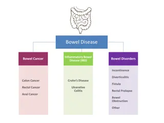
the 75 75th thAnnual Annual Conference Indian Indian Radiological Radiological and Conference of of the and Imaging IRIA IRIA 2023, Imaging Association Association 2023, Amritsar Amritsar INTRODUCTION INTRODUCTION Anastomotic leaks can occur in early and late post-operative phase when enteric anastomosis fails (1). Anastomotic leak can be classified as: a. free/generalized leak, b. contained leak (2). Undiagnosed anastomotic leak carries a poor outcome (3). Knowledge of accurate interpretation of CT imaging characteristics is vital for a timely and accurate diagnosis of anastomotic leak. LEARNING LEARNING OBJECTIVES OBJECTIVES To assess the salient imaging findings of bowel anastomotic leak on Computed Tomography (CT) 1 To compile a pictorial review depicting features of anastomotic leak after gastrointestinal tract surgery 2 Image Source: my.clevelandclinic.org/health/diseases/22324-anastomotic-leak
the 75 75th thAnnual Annual Conference Indian Indian Radiological Radiological and Conference of of the and Imaging IRIA IRIA 2023, Imaging Association Association 2023, Amritsar Amritsar and Methods Methods Materials Materials and Keyword search on post-surgical anastomotic leak from emergency teleradiology reports Retrospective evaluation of 49 CT abdomen and pelvis studies 49 cases cases Total Total contrast contrast and Non Non contrast contrast CT Contrast Contrast CT and non CT 17 CT 32 32 cases non- -contrast contrast CT: 17 cases cases cases CT: 49 CT: : Contrast Contrast CT IV IV contrast contrast 17 Oral Oral contrast contrast 4 4 cases Oral Oral and and IV IV contrast Rectal Rectal contrast contrast 1 1 case 17 cases cases cases contrast 10 Images were assessed for salient imaging findings of bowel anastomotic leak and the results were analyzed and complied in a pictorial review 10 cases cases case
75 75th Indian Radiological and Imaging Association Indian Radiological and Imaging Association IRIA 2023, Amritsar IRIA 2023, Amritsar thAnnual Conference of the Annual Conference of the Other imaging findings in Anastomotic Other imaging findings in Anastomotic Leak Leak 5.39 5.39 4.9 4.9 3.43 3.43 2.94 2.94 1.47 1.47 0.98 0.98 1 fluid Wall Thickening Thickening obstruction Intra Intra- -abdominal abdominal Free Freefluid Bowel Bowel Wall Leakage Leakage of of intraluminal intraluminal Contrast Contrast Agent Agent Bowel Bowel obstruction Fistula Fistula contained contained anastomotic anastomoticleakage leakage The graph represents the percentages of imaging findings. It demonstrates that extra-luminal air was the most common imaging finding seen in 14.21% of patients. Other common findings include focal collection or abscess (13.7%), peritonitis (12.3%), bowel wall thickening (5.4%) and intra-abdominal free fluid (2.9%). Few other imaging findings included fistula in 2% of the cases and bowel obstruction in one case. Out of 14 examinations performed after administration of enteric contrast, 10 cases were positive for extravasation of intra-luminal contrast (70 %, and 4.9% of total number of studies) CASE EXAMPLES TO FOLLOW CASE EXAMPLES TO FOLLOW
75 75th Indian Radiological and Imaging Association Indian Radiological and Imaging Association IRIA 2023, Amritsar IRIA 2023, Amritsar thAnnual Conference of the Annual Conference of the CASE:2 CASE:2 CASE:1 CASE:1 CASE:2 An 84-year-old female patient presents with acute non localized abdominal pain. CT scan of abdomen and pelvis with IV and oral contrast shows anastomotic sutures in the pelvis, a multi-loculated rim enhancing fluid collection in the rectouterine pouch adjacent to the sigmoid anastomosis, suggestive of anastomotic leak with an abscess (green). CASE:2 A 55-year-old female patient presents with c/o abdominal pain. CT scan of abdomen and pelvis without contrast shows diffuse wall thickening of the stomach/ proximal jejunum with defect at the site of anastomosis (red) and extra-luminal air loculi and fluid in the left upper abdomen (yellow), suggestive of anastomotic leak.
75 75th Indian Radiological and Imaging Association Indian Radiological and Imaging Association IRIA 2023, Amritsar IRIA 2023, Amritsar thAnnual Conference of the Annual Conference of the CASE:4 CASE:4 CASE:3 CASE:3 A 71-year-old female with complaints of nausea and vomiting. CT scan of abdomen and pelvis with oral contrast shows anastomotic sutures in the sigmoid colon, extra-luminal air loculi (yellow) around sigmoid anastomosis with minimal extra luminal oral contrast (blue), consistent with anastomotic leak. A 43-year-old female, post laparoscopic sigmoid colectomy presents with abdominal pain and signs of sepsis. CT abdomen and pelvis with rectal contrast demonstrates extra luminal air loculi (yellow) adjacent to sigmoid anastomosis, suggestive of anastomotic leak with mesenteric fat stranding (red) in the pelvis.
CASE:5 CASE:5 nnual Conference of the nnual Conference of the n Radiologi n Radiologi I I CASE:6 CASE:6 ation ation CASE:2 A 57-year-old male patient presents abdominal pain. abdomen and pelvis with oral and IV contrast shows extra-luminal extravasation of oral contrast in right lower quadrant suggestive of anastomotic Extra-luminal air fluid level is also seen in the pelvis (yellow). with lower scan of CT A 59-year-oldfemale, post right colectomy presents with acute pain abdomen. CT scan of abdomen and pelvis with rectal contrast shows extra-luminal contrast (yellow) and air (pink) in the peritoneal cavity, suggestive of anastomotic leak. Small bowel ileus (blue) with mesenteric fat stranding (red) is also noted. (pink), leak.
75 75th Indian Radiologic Indian Radiologic CASE:7 CASE:7 thAnnual Conference of the Annual Conference of the CASE:8 CASE:8 n n IRIA IRIA A 53-year-old female presents with signs of sepsis. CT scan of abdomen and pelvis with oral and IV contrast demonstrates a large extra luminal contrast and air in the anterior aspect of the abdomen, consistent anastomotic leak. fistulous communication extravasated contrast with the abdominal wall wound (green). There mesenteric fat stranding (red). past A 42-year-old history of gunshot injury to the abdomen, post laparoscopy presenting with acute upper abdominal pain, distension, nausea, vomiting and diarrhea. CT scan of abdomen and pelvis with IV contrast shows high grade small bowel obstruction transition at the level anastomosis (blue). Fluid collection adjacent to anastomosis (green), suggestive of anastomotic leak. There is adjacent bowel wall thickening suggestive of inflammation. male with exploratory with is of There with (red) (blue) is also
75 75th Indian Radiological and Imaging Association Indian Radiological and Imaging Association IRIA 2023, Amritsar IRIA 2023, Amritsar thAnnual Conference of the Annual Conference of the CASE:9 CASE:9 A 72-year-old female, post colectomy and gastric bypass presents with wound infection. CT scan of abdomen and pelvis with oral contrast shows extravasation of contrast at the anastomotic site extending to anterior abdominal anastomotic leak (yellow) and an entero-cutaneous fistula (blue) forming localized collections in epigastric and umbilical regions. Peritonitis (red) and distal ileal wall thickening are also noted. with wall, consistent
75 75th Indian Radiological and Imaging Association Indian Radiological and Imaging Association IRIA 2023, Amritsar IRIA 2023, Amritsar thAnnual Conference of the Annual Conference of the CONCLUSION CONCLUSION Abdominal CT is an accurate non-invasive test in the detection of bowel anastomotic leak using specific findings of leakage of intraluminal contrast agent, extra-luminal air and focal fluid collection or abscess adjacent to the anastomosis. Other supportive findings include intra-abdominal free air and adjacent fat stranding. The diagnosis of bowel anastomotic leak may also be suspected on unenhanced CT based on the presence of extra-luminal air and focal fluid collection adjacent to the anastomosis. REFERENCES REFERENCES Samji K B, Kielar A Z et.all, Anastomotic Leaks After Small- and Large-Bowel Surgery: Diagnostic Performance of CT and the Importance of Intraluminal Contrast Administration. American Journal of Roentgenology: 1259-1265. 10.2214/AJR.17.18642 https://radiopaedia.org/articles/colonic-anastomotic-leak?lang=us Lynn ET, Chen J, Wilck EJ, El-Sabrout K, Lo CC, Divino CM. Radiographic findings of anastomotic leaks. Am Surg. 2013 Feb;79(2):194-7. Khoury W, Ben-Yehuda A, Ben-Haim M, Klausner J M, Szold O. Abdominal computed tomography for diagnosing postoperative lower gastrointestinal tract leaks. J Gastro intest Surg2009; 13:1454 1458 Golub R, Golub RW, Cantu R, et al. A multivariate analysis of factors contributing to leakage of intestinal anastomosis. J Am Coll Surg. 1997;184:364 372. Kornmann VN, Treskes N, Hoonhout LH, Bollen T L, van Ramshorst B, Boerma D. Systematic review on the value of CT scanning in the diagnosis of anastomotic leakage after colorectal surgery. Int J Colorectal Dis 2013; 28:437 44 1. 2. 3. 4. 5. 6.