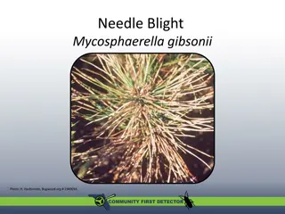
Understanding Akabane Disease: Causes, Symptoms, and Prevention


Akabane disease
(AKA)
Enzootic bovine arthrogryposis
and hydranencephaly
By
Dr/ Marawan Elfky
Definition
Arthropod-transmitted viral disease.
Ch. by congenital abnormalities in
calves, lambs
and kids in a form:
Arthrogryposis
(joint immobility or ankylosed)
Hydranencephaly
(absence of cerebral
hemisphere)
In adult,
abortion and mummification, still
birth or premature birth.
Etiology
Akabane virus (ssRNA)
Family (Bunyaviridae)
Genus (Orthobunyavirus).
Simbu serogroup.
Can be isolated in suckling mice (I/C) and cell
cultures
Sensitive to heat and UV light.
Epidemiology
1.
Distribution
:
Tropical and subtropical areas and
recognized in Australia, Israel, Japan, and Korea
(middle east showed antibodies only)
.
2.
Host rang:
Wide range of domesticated ruminants and wildlife.
Cattle, sheep and goat are frequently infected.
Asymptomatic infection (horses, buffalo, deer, and
pigs).
3.
Seasonal incidence:
Summer and early
autumn.
4.
Transmission:
a.
Source:
Infected blood contain virus.
b.
Mode:
1.
Mainly horizontal transmission
:
through biting
midges (Culicoides)
2.
Vertical transmission is also important (less).
1)
Deaths in young animals.
2)
Abortion, mummification and still birth in adults.
5. Economic and zoonotic importance
Not Zoonotic
Pathogenesis
•
After insect biting viremia occurs for 3-5 d then the
virus penetrate placenta & multiply causing abortion or
still birth or birth of calves or lambs with
congenital
anomalies.
•
The most susceptible stages of gestation in small
ruminants range from
1-2 months
and in cattle from
3
-6 months.
•
In the latter stages of gestation the incidence of
abnormalities declines to a very low level.
Clinical signs
I.P is short (1-6 d.) with low morbidity and mortality.
A.
Adult;
usually asymptomatic except some signs
including fever, abortion, still birth or premature birth
and dystocia.
B.
Calves, lambs and kids;
Congenital anomalies as
arthrogryposis and hydranencephaly.
1. Arthrogryposis (3-4 m. of pregnancy):
Calves are born
underweight
or small in size and
usually die at birth.
Initially,
only one or two joints
may be affected on a
single limb.
Later,
severe fixation
of multiple joints on all limbs and
muscular atrophy so the calves are unable to raise or
stand or walk,
Sever deformities of vertebral column
(incomplete
closure of spinal canal or lateral deviation of the spine).
2. Hydranencephaly (5-6 m. of pregnancy):
Calves have varying degrees of
cavitation of cerebral
hemispheres,
ranging from porencephaly to severe
hydranencephaly.
Lack of intelligence, blindness, absence of dam seeking
reflex, torticollis and may survive for months if they
are hand reared but will not grow.
Some calves with micrencephaly show unable to stand,
incoordination, ataxia, paralysis, blindness, deafness,
nystagmus, dullness, slow suckling and dysphagia.
P/M lesions (Muscles and CNS)
Arthogryposis;
affected joints cannot be straightened
Subcutaneous haemorrhages
in fetus.
Hypoplasia
of the skeletal muscle
Opthalmia and cataracts
Hydranencephaly and or microencephaly
Agenesis of the brain and agenesis or hypoplasia of
the spinal cord
Diagnosis
1- Field diagnosis;
depends on case history, clinical signs
and P/M lesions.
2. Lab. Diagnosis;
A.
Sample:
Fetus, fetal membranes and fluids
Brain, CSF, caught culicoides
Blood and paired serum (preclostral sera)
Diagnosis
B. Laboratory procedures:
Serological investigation using ELISAs, AGIDT.
Virus isolation on in cell lines including hamster lung
(HmLu-1) and baby hamster kidney (BHK-21) cells.
Detection of the viral nucleic acid using RT-PCR.
Laboratory animal’s inoculation, intracranial injection
of mice or hamster cause non-suppurative encephalitis
Differential Diagnosis
The disease should be differentiated from all causes
of congenital anomalies as:
(Manganese deficiency,
inherited arthrogryposis, Border disease, BT, BVD
and poisoning as in datura etromaitum, tobacco and
lupines)
Treatment
No treatment
Control & vaccination
Vector control,
such as covering breeding sites
and using insect repellents and insecticide
treatments.
Disinfection
using hypochlorite, glutaraldehyde,
70% alcohol, hydrogen peroxide.
Introduction of stock from non-endemic areas.
Vaccination using live attenuated or inactivated
vaccine is available in endemic localities
.
Akabane disease, also known as Enzootic Bovine Arthrogryposis and Hydranencephaly, is a viral illness transmitted by arthropods that primarily affects calves, lambs, and kids. The disease can lead to congenital abnormalities such as joint immobility and absence of the cerebral hemisphere. It is caused by the Akabane virus, a single-stranded RNA virus belonging to the Bunyaviridae family. The virus is prevalent in tropical and subtropical regions and has been identified in various countries. Understanding the epidemiology, transmission, and clinical signs of Akabane disease is crucial for its prevention and management.

Download Presentation

Please find below an Image/Link to download the presentation.
The content on the website is provided AS IS for your information and personal use only. It may not be sold, licensed, or shared on other websites without obtaining consent from the author. Download presentation by click this link. If you encounter any issues during the download, it is possible that the publisher has removed the file from their server.
E N D
Presentation Transcript
Akabane disease (AKA) Enzootic bovine arthrogryposis and hydranencephaly By Dr/ Marawan Elfky
Definition Arthropod-transmitted viral disease. Ch. by congenital abnormalities in calves, lambs and kids in a form: Arthrogryposis (joint immobility or ankylosed) Hydranencephaly (absence of cerebral hemisphere) In adult, abortion and mummification, still birth or premature birth.
Etiology Akabane virus (ssRNA) Family (Bunyaviridae) Genus (Orthobunyavirus). Simbu serogroup. Can be isolated in suckling mice (I/C) and cell cultures Sensitive to heat and UV light.
Epidemiology 1. Distribution: Tropical and subtropical areas and recognized in Australia, Israel, Japan, and Korea (middle east showed antibodies only). 2. Host rang: Wide range of domesticated ruminants and wildlife. Cattle, sheep and goat are frequently infected. Asymptomatic infection (horses, buffalo, deer, and pigs).
3. Seasonal incidence: Summer and early autumn. 4. Transmission: a. Source: Infected blood contain virus. b. Mode: 1. Mainly horizontal transmission: through biting midges (Culicoides) 2. Vertical transmission is also important (less).
5. Economic and zoonotic importance 1) Deaths in young animals. 2) Abortion, mummification and still birth in adults. Not Zoonotic
Pathogenesis After insect biting viremia occurs for 3-5 d then the virus penetrate placenta & multiply causing abortion or still birth or birth of calves or lambs with congenital anomalies. The most susceptible stages of gestation in small ruminants range from 1-2 months and in cattle from 3 -6 months. In the latter stages of gestation the incidence of abnormalities declines to a very low level.
Clinical signs I.P is short (1-6 d.) with low morbidity and mortality. A. Adult; usually asymptomatic except some signs including fever, abortion, still birth or premature birth and dystocia. B. Calves, lambs and kids; Congenital anomalies as arthrogryposis and hydranencephaly.
1. Arthrogryposis (3-4 m. of pregnancy): Calves are born underweight or small in size and usually die at birth. Initially, only one or two joints may be affected on a single limb. Later, severe fixation of multiple joints on all limbs and muscular atrophy so the calves are unable to raise or stand or walk, Sever deformities of vertebral column (incomplete closure of spinal canal or lateral deviation of the spine).
2. Hydranencephaly (5-6 m. of pregnancy): Calves have varying degrees of cavitation of cerebral hemispheres, ranging from porencephaly to severe hydranencephaly. Lack of intelligence, blindness, absence of dam seeking reflex, torticollis and may survive for months if they are hand reared but will not grow. Some calves with micrencephaly show unable to stand, incoordination, ataxia, paralysis, blindness, deafness, nystagmus, dullness, slow suckling and dysphagia.
P/M lesions (Muscles and CNS) Arthogryposis; affected joints cannot be straightened Subcutaneous haemorrhages in fetus. Hypoplasia of the skeletal muscle Opthalmia and cataracts Hydranencephaly and or microencephaly Agenesis of the brain and agenesis or hypoplasia of the spinal cord
Diagnosis 1- Field diagnosis; depends on case history, clinical signs and P/M lesions. 2. Lab. Diagnosis; A. Sample: Fetus, fetal membranes and fluids Brain, CSF, caught culicoides Blood and paired serum (preclostral sera)
Diagnosis B. Laboratory procedures: Serological investigation using ELISAs, AGIDT. Virus isolation on in cell lines including hamster lung (HmLu-1) and baby hamster kidney (BHK-21) cells. Detection of the viral nucleic acid using RT-PCR. Laboratory animal s inoculation, intracranial injection of mice or hamster cause non-suppurative encephalitis
Differential Diagnosis The disease should be differentiated from all causes of congenital anomalies as: (Manganese deficiency, inherited arthrogryposis, Border disease, BT, BVD and poisoning as in datura etromaitum, tobacco and lupines) Treatment No treatment
Control & vaccination Vector control, such as covering breeding sites and using insect repellents and insecticide treatments. Disinfection using hypochlorite, glutaraldehyde, 70% alcohol, hydrogen peroxide. Introduction of stock from non-endemic areas. Vaccination using live attenuated or inactivated vaccine is available in endemic localities.
")


:")
:")
:")
:")
")