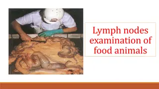
Understanding the Lymphatic System and Immune Response
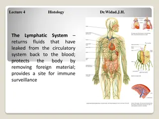
The lymphatic system, a vital component of the immune system, consists of lymphocytes, plasma cells, and other cells within a framework of reticular cells and fibers. It includes lymphoid organs like the bone marrow, thymus, lymph nodes, and spleen. Diffuse lymphatic tissue and lymphatic nodules are crucial in immune response, while structures like Peyer's patches and tonsils play specific roles in mucosal immunity. Understanding these components is essential for comprehending the body's defense mechanisms.

Download Presentation

Please find below an Image/Link to download the presentation.
The content on the website is provided AS IS for your information and personal use only. It may not be sold, licensed, or shared on other websites without obtaining consent from the author. Download presentation by click this link. If you encounter any issues during the download, it is possible that the publisher has removed the file from their server.
E N D
Presentation Transcript
Histology Immune System: Lymphatic System
Immune System Lymphatic tissue consists predominantly of lymphocytes. These and a variable number of plasma cells, macrophages, and other cells occur among a framework of reticular cells and fibers. In H&E preparations lymphatic tissue appears purple presence of numerous small lymphocytes, each with a basophilic nucleus and little cytoplasm. because of the
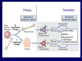
The components are: Lymphocytes T cells B cells Plasma cells Bone marrow Thymus Lymph Node Mucosa-associated lymphoid tissue (MALT) Spleen
NOTE: The bone marrow and thymus are considered as primary immune/lymphoid components of the functioning immune systems. because they contain the stem cells that will develop into T cells, B cells and natural killer cells and lymphatic
Diffuse lymphatic tissue is characterized by a moderate concentration of scattered Iymphocytes. A round, aggregation of mostly small, densely packed lymphocytes is called a lymphatic nodule. A nodule may contain a central pale area, the germinal center. Because the majority of cells of the germinal center are larger lymphocytes with more cytoplasm and lightly staining nuclei, this region appears pale in contrast to the dense corona (marginal zone, peripheral zone) of small lymphocytes. Diffuse lymphatic tissue and lymphatic nodules are components of most lymphatic organs. They also appear in the connective tissue of the digestive, respiratory, urinary, and reproductive organs, among other locations. oval, or irregularly circumscribed
Peyer's patches Aggregations of lymphatic nodules form Peyer's patches in the lamina propria and submucosa of the small intestine, ileum. particularly the
Tonsils with crypt Tonsils are collections of lymphatic nodules and diffuse lymphatic tissue. They occur in the connective tissue below the epithelium in specific regions of the tongue, pharynx, and larynx. Follicular tonsils are characterized by deep invaginations of the surface epithelium called crypts. A crypt together with its associated lymphatic tissue is a tonsillar follicle. Collectively, several follicles form the tonsil. Examples of tonsils with crypts include the following: lingual tonsils of the horse, pig, and cow; paraepiglottic tonsils of the pig, sheep, and goat; palatine tonsils of the horse, pig, and ruminant.
Tonsils without crypts Tonsils without crypts have a smooth, somewhat bulging surface, but lack deep invaginations of the epithelium. Examples of these are the tubal tonsils or ruminants, paraepiglottic tonsil of the cat, and the palatine tonsils of carnivores. folded, or the
lymph node A lymph node is organized into a cortex and medulla. The cortex consists of lymphatic nodules surrounded by diffuse lymphatic tissue. Extensions of the latter tissue into the medulla are called medullary cords. Lymphocytes, other leukocytes, macrophages, and plasma cells can be found in the medullary cord. A capsule of connective tissue, with some smooth muscle and elastic fibers, covers the lymph node. Parts of the capsule extend inward as trabeculae. Afferent lymphatic vessels penetrate subcapsular sinus. Cortical sinuses connect the subcapsular sinus to medullary sinuses. The latter lead to efferent lymphatic vessels at the hilus. The various sinuses are less cellular than the parenchyma and appear pale by comparison. They are lined by a discontinuous endothelium and are spanned by a network of cytoplasmic processes of reticular cells. They contain some free cells such as lymphocytes and macrophages. the capsule to join the
Blood vessels enter and leave the node mostly from the region of the hilus. Unique blood postcapillary venules are found in the deep cortex. They are lined by elongated cells that appear cuboidal when cut in cross section. Lymphocytes migrate cells. The amount or cortical and medullary tissue can vary from that of the "typical" lymph node. The lymph node of the pig, for example, is characteristically atypical location of the cortical and medullary tissue, as well as the flow of lymph, being reversed vessels called between these arrangement of with the
Hemal nodes occur Hemal blood vessels of ruminants. They are characterized blood-filled sinuses cellular cords. tissue and muscle form the capsule and trabeculae (which are sparse). Hemal nodes lack lymphatic vessels. Hemolymph nodes, in contrast to hemal possess lymphatic Their sinuses receive a mixture of blood and lymph. nodes along by between Connective some smooth nodes, vessels.
Thymus Located posterior to the sternum in the anterior part of the mediastinum, the thymus is a bi-lobed nodular organ that is very large in the yong, reaching maximum size at puberty then becoming smaller in a process called involution. Immature T lymphocytes move from the bone marrow into the thymus where they become immunocompetent T cells. These T cells then leave the thymus, go into the circulation and eventually find their way to lymph nodes, mucosa-associated lymphoid tissue or the spleen.
Mucosa-associated lymphoid tissue (MALT) MALT is really connective tissue located beneath mucous membranes in which the lymphocyte is the predominant cell type. Examples occur in the respiratory, gastrointestinal, urinary and reproductive tracts. The exact extent of these aggregations of lymphocytes is not easily discernible because they have no distinct capsule like that of lymph nodes. However, they are like lymph nodes in that they often have a pale-staining germinal center lymphocytes like the germinal centers in lymph nodes. The larger aggregations contain B and T cell zones and antigen processing cells; the smaller, more scattered MALT components such as those in the intestines and respiratory tract are mostly T lymphocytes. Some B cells and plasma cells are also present. containing actively dividing
Distribution of MALT In the digestive system: in the wall of the pharynx - tonsils (palatine, lingual, pharyngeal) in the wall of the small intestine - aggregate lymphoid nodules in the wall of the colon-aggregate lymphoid nodules in the walls of the appendix In the reproductive system: in the wall of the vagina
Spleen The spleen has a capsule that is rich in smooth muscle and elastic fibers. In horses and cows two or three layers of muscle are oriented perpendicular to each other, while in carnivores, pigs, sheep, and goats the muscle fibers are interwoven. The capsule is thickest in the horse and cow and thinnest in carnivores. Trabeculae project into the interior of the spleen from the capsule. They tend to be especially large in cows and sheep.
Spleen The parenchyma of the spleen is divisible into the white and red pulp. Dense accumulations of lymphocytes, arranged around central arteries, form the periarterial lymphatic sheaths (PALS). These, along with lymphatic nodules, comprise the white pulp. White pulp appears purple in H&E preparations because of the high concentration of numerous small lymphocytes. Red pulp, because of the large numbers of erythrocytes it contains in its reticular meshwork and blood vessels, is stained red in H&E preparatIons.
Lymphatic tissues in bird Lymph nodes do not occur in the chicken . However, diffuse lymphatic tissue and lymphatic nodules are widespread. The bursa of Fabricius is a sac like dorsal diverticulum of the proctodeum that is unique to birds. It is characterized by tall, thick mucosal folds (plicae) filled with numerous polyhedral follicles. Each follicle, composed of lymphatic tissue, is divided into a cortex and medulla. A layer of undifferentiated epithelial cells occupies the periphery of the medulla, which is separated from the cortex by a capillary layer. The bursa is lined by a pseudostratified columnar epitheliwn, except at the apex of each follicle, which is covered by an epithelial tuft of simple columnar cells.


")