Radiation Monitoring and Personnel Dosimetry: A Comprehensive Guide
This chapter delves into the importance of personnel dosimetry in radiation protection, covering topics such as dosimeter placement, types of dosimeters, radiation survey instruments, and calibration tools. It highlights the necessity for monitoring radiation exposure regularly to ensure safety within allowable limits. Various dosimeters, including film badges, OSL dosimeters, pocket ionization chambers, and TLDs, are explained in detail, along with their specific uses. The chapter also discusses the role of radiation survey instruments and their calibration in radiographic and fluoroscopic procedures.

Download Presentation

Please find below an Image/Link to download the presentation.
The content on the website is provided AS IS for your information and personal use only. It may not be sold, licensed, or shared on other websites without obtaining consent from the author. Download presentation by click this link. If you encounter any issues during the download, it is possible that the publisher has removed the file from their server.
E N D
Presentation Transcript
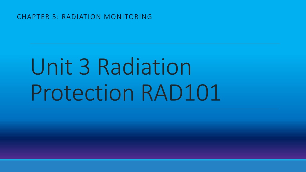
CHAPTER 5: RADIATION MONITORING Unit 3 Radiation Protection RAD101
Objectives- Chapter 5 1) State the reason why a radiation worker should wear a personnel dosimeter, and explain the function and characteristics of such devices. 2) Identify the appropriate location on the body where the personnel dosimeter(s) should be worn during the following procedures or conditions: (1) routine computed radiography, digital radiography, or conventional radiographic procedures, (2) fluoroscopic procedures, (3) special radiographic procedures, (4) pregnancy. 3) Describe the various components of the film badge, optically stimulated luminescence (OSL) dosimeter, pocket ionization chamber, and thermoluminescent dosimeter (TLD), and explain the use of each of these devices as personnel monitors. 4) Explain the function of radiation survey instruments. 5) List three gas-filled radiation survey instruments. 6) Explain the requirements for radiation survey instruments. 7) Explain the purpose of the following instruments: (1) ionization chamber-type survey meter (cutie pie), (2) proportional counter, (3) Geiger-M ller (GM) detector. 8) Identify the radiation survey instrument that can be used to calibrate radiographic and fluoroscopic x-ray equipment.
Personnel Monitoring Can t see, hear or feel radiation so we have instruments designed to detect ionizing radiation Devices can measure effect on film( density) or air( ionization ) Monitor on a regular basis Required to ensure radiation exposure levels are kept within allowable limits ( Efd) Personnel dosimetry- any occupational worker exposed to radiation Personnel dosimeter- provides the detecting over a period of time, but does not protect REQUIRED when risk is > 10% of annual EfD 50mSv / 5rem ( 5 mSv or .5 rem ) Most provide when dose could reach 1% per month of annual limit ( 0.04mSv or 4mrem)
Placement of Dosimeter Routine Placement collar level anterior-to provide maximum does measurements of thyroid, neck and head Should be consistent placement Don t wear during personal testing With Apron Outside the apron at collar level thyroid/eye dose Head, neck and eyes receive 10 -20 X higher than trunk of body 2ndMonitor w/ apron Used for techs with high readings . Ie: fluoroscopy, CAC, IR Worn at waist level UNDER apron Fetal Badge Records dose to abdomen for estimate of embryo-fetal dose Worn under apron during fluoro exams Extremity Extremity dosimeter or thermoluminescent dosimeter (TLD) ring Worn by techs doing hands-on fluoro
Personnel Monitoring Reports Must be maintained to meet state and federal regulations List Deep, eye and shallow doses for each person monitored Report is based on length of time worn Monthly, bimonthly, quarterly, yearly Can provide TEDE- total effective dose equivalent at end of year Control badge reading Legal document- require signature before exposure records can be released to other individuals, new employers Must be maintained in perm record- length may vary on policies and exposure area
Report contents Contain Personal data- name, SSN , gender DOB Badge type and location Radiation type, x, beta or neutron Monthly dose received Quarterly accumulated dose Cumulative occupational exposure history Doses received: Deep dose equivalent ( DDE) Dose at depth of 1 cm in soft tissue( whole body dose) Shallow dose equivalent ( SDE) Dose at depth of .007 cm deep in soft tissue( skin dose) Eye dose equivalent ( LDE ) (EDE) External exposure to lens of the eye- depth of 0.3 cm M minimal or no exposure
Characteristics of Dosimeters 1. Portability- lightweight / easy to carry 2. Ruggedness- durable 3. Sensitivity- detecting radiation 4. Reliability- consistent 5. Low cost
Types 1. OSL- optically stimulated luminescence dosimeters 2. Film badges 3. TLDs - Thermoluminescent dosimeters 4. Pocket Ionization chambers
1. Optically Stimulated Luminescence - OSL Most common- has replaced the film badge Contains aluminum oxide detector Al2O3 Contain filters aluminum, tin, and copper Identifies static, contaminant and dynamic exposure to low energy x-rays - beta particles too- Deep , eye and shallow doses Read by laser light- luminesces in proportion to amount of radiation exposure 1- 3 month wear time Can be read in-house or mailed out Use control monitor- provides base reading
Optically Stimulated Luminescence -OSL ADVANTAGES: Preloaded/self-contained packet Color coded Tamperproof from heat, moisture and pressure Can be reanalyzed Increased accuracy- as low as 1mrem DISADVANTAGES: Only accurate for the area of body worn Does not work if not worn
2. Film Badge Still used although OSL has taken their place 3 Parts Plastic film holder Low atomic number for passage of radiation Metal filters Aluminum or copper Helps determine energy of radiation and from what direction radiation was received from ( posterior or anterior) Helps determine if mostly scatter or a single exposure from primary beam Film packet- radiation-dosimetry film Sensitive from .1 mSv to 5000 mSv less .1 mSv are reported as minimal (M) Darkens just like regular film if exposed to radiation Density is measured
Film Badge Causes of Inaccurate reading Leaving badge on apron Washing Left in car- cosmic radiation and heat from sun Not correctly inserting film into plastic holder ADVANTAGES: Inexpensive Easy to handle Easy to process
3. Thermoluminescent Dosimeter Resemble film badge Contain crystalline form of lithium fluoride ( LiF) functional material Radiation interacts with LiF to change physical properties (emit light- luminescence) Light intensity is proportional to amount of radiation that interacts with crystals TLD analyzer use to measure radiation
Thermoluminescent Dosimeter ADVANTAGES LiF reacts similarly to human tissue- more accurate 5 mR Humidity, pressure and temp changes do not affect Can be worn for 3 months Can be re-used after crystals read DISADVANTAGES: Twice the cost of film badge Can be read only once- info destroyed after read Batch/group readings
4. Pocket Ionization Chamber(pocket dosimeter) Most sensitive Use is uncommon in diagnostic imaging Fountain pen shape- cylindrical- clip on end to hook on pocket 2 types Self reading- contains built electormeter most common of two Non-self reading Components: 2 electrodes one + , one Quartz fiber functions as indicator When electrodes are exposed to gamma or x-rays + end becomes ionized and discharges -
Pocket Ionization Chamber ADVANTAGES: Can be as sensitive as 0-200mR Immediate exposure Used in nuclear power plant tours, people assisting in dept DISADVANTAGES: Cost $150 for initial setup / unit Require special charge stations for accuracy Charge can leak or escape- false readings Trauma to device can cause false readings No permanent legal readings Must be read daily
AREA Monitoring Categorized as Radiation survey instruments 3 Gas filled types: 1. Ionization chamber based instruments 2. Proportional Counter 3. Geiger-Muller (GM) tube May or may not have calibrated readouts Not all equally sensitive Very basic to complex No cumulative exposure readings detect presence of radiation
Requirements 1. Portability- one person operation 2. Durable 3. Reliable 4. Need to be as reactive as human tissue is ionizing radiation 5. Sensitive to all types of ionizing radiations- increases usefulness 6. Direction and energy of radiation should not affect response 7. Should be cost effective 8. Need annual calibration
Ionization Chamber-Type Survey Meter AKA: Cutie Pie- nicknamed because of small size 2 functions: rate meter device for exposure rate for area surveys integrating or cumulative exposure instrument Specifics: Measures x and gamma radiation Some measure beta Sensitivity Ranges: Can measure intensity ranging from 10- several 1000microGy ( 1mRoentgen/hr-several 1000mR / hr Can sum exposures from 1mR several R
Ionization Chamber-Type Survey Meter Uses: Monitoring x-ray machine installs long exposures Fluoroscopic scatter radiation exposure rates Exposure rates from patients receiving therapeutic doses Assess radioisotope storage facilities Quantifying the cumulative exposures outside of protective barriers
Ionization Chamber-Type Survey Meter Advantages: Can measure a wide range of radiation exposures in a few seconds Sensitive to broad range of radiation energies Disadvantages: Sensor is delicate Can be inaccurate if not warmed up properly Not useful for short exposure times
Proportional Counter No useful purpose in diagnostic imaging Lab setting for alpha and beta Small amounts of low level radioactive contaminants Advantages: Can discriminate between alpha and beta Disadvantage: Operator must stand close to source ( alpha can t travel far)
Geiger-Muller Detector Uses: Primarily used in portable radiation survey uses in Nuclear Medicine Detection of lost sources Low level radioactive contamination Can be used for barriers for defects Sensitivity: Can detect individual particles- individual electrons Can detect any area contaminated by radioactive material x, gamma, beta and sometimes alpha
Geiger-Muller Detector Components: Audible sound system to alert user- can use headphones or has speaker Metal encloses gas filled tube or probe- units functional sensor- Metal shield can be open or closed to discriminate between energies low energy radiations when closed Readings are in Milliroentgens/hr Check source on outside to verify consistency
Geiger-Muller Detector Disadvantages: Not dependent on energy- can t discriminate- causes GM to respond differently Can give false readings when in high intensity radiation area
Calibration and Regulation Must be calibrated to meet state and federal requirements for patient dose evaluation American Association of Physicists in Medicine Lists calibration labs available Used by medical physicists to perform standard measurements required by state, federal and health care accreditation organizations
During radiographic procedures when an apron is NOT worn , where should the dosimeter be worn A. Collar level B. Chest level C. Waist level D. Do not need to wear Response Counter
Where should the dosimeter be worn during procedures when an apron IS worn A. Collar level inside apron B. Collar level outside apron C. Waist level outside apron D. Waist level inside apron Response Counter
Which of the following is used to calibrate radiographic and fluoro equipment A. Proportional counter B. Geiger-Mueller counter C. Ionization chamber D. Pocket ionization chamber Response Counter
Which of the following is used to find lost radiation sources? A. Geiger-Mueller Counter B. Proportional counter C. Ionization Chamber- cutie pie D. TLD analyzer Response Counter
Which should be used in x-ray install to assess fluoro scatter? A. Geiger-Mueller B. Ionization chamber C. Proportional counter D. TLD Response Counter
Chapter 10 DOSE LIMITS FOR EXPOSURE TO IONIZING RADIATION
Objectives 1. List the four major organizations that share the responsibility for evaluating the relationship between radiation dose equivalents and induced biologic effects. 2. Explain the purpose of the Radiation Control for Health and Safety Act of 1968 and the consumer Patient Health and Safety Act of 1981. 3. Recognize the United States regulatory agencies responsible for enforcing established radiation dose equivalent limiting standards. 4. Define the "effective dose equivalent" limits. 5 . Explain the ALARA concept. 6. State the formula for determining the maximum permissible dose equivalent. 7. Solve problems utilizing the cumulative effective dose for the whole body. 8. Explain current radiation protection philosophy. 9. State in terms of traditional units and System International units the occupational radiation absorbed dose limits for combined whole- body exposure and for selective body area exposures. 10. State in terms of System International units the various absorbed dose limits which apply to the general population.
Exposure Levels Limits Must have exposure limits to the general public, patients and radiation workers Limits the risk of harmful biologic effects Nuclear Regulatory Rules and Regulations Categorized as: EfD- effective dose limits for occupational and non-occupational EqD- equivalent dose limits for tissues and organs EfD limiting system- calculations of risks
RISK General terms- the probability of injury, ailment or death resulting from an injury Medical Imaging- the possibility of inducing a radiogenic cancer or genetic defect after irradiation Radiation induced malignancy- the basis of the effective dose limiting system Genetic defect The benefit obtained from any diagnostic radiologic procedure must always be weighed against the risk National Council on Radiation Protection Report No 116 and International Commission on Radiological Protection Publication No 60- provide resources for recommendation on radiation dose limits and radiation protection- most up-to-date
Radiation Protection Standards Organizations Four major organizations responsible for evaluating radiation EqD and biologic effects Formulate risk estimates of somatic and genetic effects IRCP- International Commission on Radiological Protection General recommendations for occupational and public dose does not enforce each nation responsible for enforcing specific regulations NRCP- National Council on Radiation Protection and Measurement Recommends standards for US based on IRCP Publish NRCP Reports UNSCREAR-United Nations Scientific Committee on the Effects of Atomic Radiation Evaluates exposures on humans / radioactive material, machines and accidents Report and Research reports to IRCP NAS/NRC-BEIR- National Academy of Sciences/ National Research Council Committee on Biological Effects of Ionizing Radiation Reports and Research on groups radiation workers, Hiroshima, Nagasaki , Chernobyl victims Reports to IRCP
U.S. Regulatory Agencies NRC-Nuclear Regulatory Commission Oversees and Enforces standards of dose limits, inspection and testing on sites- nuclear energy industry Agreement States- Regulate Hospitals, radioactive material (nuc med) Agreement States- regulations give to health departments from NRC for self regulation Non-Agreement States- state and NRC enforce policies EPA-Environmental Protection Agency - facilitates the development and enforcement of regulation of radiation in the environment FDA- U.S. Food and Drug Administration Conducts an ongoing product radiation control program, regulating the design and manufacture of electronic products, including x-ray equipment OSHA- Occupation Safety and Health Administration- Functions as a monitoring agency in places of employment- mostly industry
Radiation Safety Program Facilities must have radiation safety program to ensure patient and worker safety Radiation Safety Committee- provides guidance Radiation Safety Officer- daily operations and formal annual review Medical physicist, health physicist, radiologist Approved by the NRC Organizations must provide Budgeting Oversee the development of policies and procedures Provide equipment for starting and continuing the program
RSO Responsibilities RSO must be properly trained and experienced to perform job Developing program to coincide with international radiation protection guidelines Ensure all persons are protected from unnecessary exposure to radiation Must be able to implement and enforce policies Identify problems, corrective actions, stop operations, verify implementation of corrective actions Reviews and maintains radiation monitoring records for all personnel Provide counseling to those receiving high doses
Radiation Control for Health and Safety Act of 1968 Protected public from hazards of unnecessary exposures such as x-ray Act also allowed establishment of Bureau of Radiologic Health under the FDA (later known as Center for Devices & Radiological Health - CDRH) sets up standards for manufacturing, installation & operation for machines * 1974 - Code of Standards for X-ray equipment- see page 213 - Box 10-4
ALARA Concept 1954 - Nat l Committee on Radiation Protection (NCRP) now known as Nat l Council on Radiation Protection & Measurements came up with concept that radiation exposures should be kept As low as reasonably achievable Radiologist and YOUR responsibility as a tech to do this optimization Use good safety rules: collimate, technique charts, shielding, no repeats * although an occupational exp person can receive up to 5 rem/yr, this doesn t mean you can receive 5 rem every year Dose response curve- as dose increase- effect increases- no threshold Effect dose
FDA White Paper Published Feb 2010 Launch of initiative to reduced unnecessary radiation exposure from medical imaging. Each patient should get the right imaging exam, at the right time, and with the right radiation dose 3 main objectives: 1. Promote safe use of medical imaging devices 2. Support informed clinical decision 3. Increase patient awareness
Consumer-Patient Radiation Health and Safety Act of 1981 standards for accreditation of educational programs & for people taking x-rays and certifying them No legal penalty exists for noncompliance Result: many states have not responded with appropriate legislation
CARE Bill Consumer Assurance of Radiologic Excellence Act of 2003 Purpose is to amend the Public Health Service Act to ensure the safety and accuracy of medical imaging examinations and radiation therapy treatments 6 States do not require licensure of any kind for person who perform or plan medical imaging exams and radiation therapy treatments
Goals for Radiation Protection NRCP Report No 116 ( replaces 91) Limitation of Exposure to Ionizing Radiation Provides the most recent guidance for radiation protection Sets the limits of radiation dose limits for members of the public including: Background Medical imaging Security Screening
Radiation Induced Responses 2 Categories for radiation responses: 1. Deterministic effects ( old term Non-Stochastic) 2. Stochastic ( probabilistic) effects
1. Deterministic Effects Biologic somatic effects of ionizing radiation ( to the body) verses genetic ( sperm/eggs) Directly related to dose received Exhibit a threshold- below threshold no or not normally a occurrence above threshold severity of damage increases as dose increases Occur only after a large dose of radiation Can occur as a result of long-term individual low doses of radiation over several years Usually do not occur with typical diagnostic x-ray Example- no radiodermatitis/erythema below a certain dose ( threshold) - above threshold dose the damage will be worse as the dose increases- ( Early Deterministic Somatic effect) Cataracts- the more the radiation/absorbed dose to the lens of the eye, the worse the cataract will become ( Late Deterministic Somatic effect)
Early and Late Deterministic Effects Early Deterministic Appear within minutes, hours, days, weeks Substantial dose is required to produce changes soon after irradiation Severity is dose related Examples: nausea , fatigue, erythema, epilation , depressed sperm count, temp or perm sterility in male and female. Acute radiation syndrome. Late Deterministic Slowly developing changes to the body from radiation exposure Months or years after continued exposure to high levels of radiation Examples : cataracts, fibrosis , organ atrophy, reduced fertility, sterility
2. Stochastic Effects Mutational, nonthreshold, random, all-or-none Biological somatic changes Mutations that effect our own individual cells when they divide ( non-genetic ) Chance of occurrence increases with radiation exposure NO MINIMUM safe dose exists ( non-threshold) could happen if one cell is damaged Examples : CANCER , genetic alteration ( hereditary)
Current Philosophy 1. Radiation dose received and biological effects have a linear, nonthreshold relationship The more dose the more chance of damage 2. No known level where you have a zero probability of having biological damage Small amount can cause damage 3. Can be beneficial in the best interests of the patient. Benefit must outweigh risk BEFORE exam is done Up to the Physician to determine this