Primary Malignant Melanoma of the Lacrimal Sac: A Rare Case Report


[
P
r
i
m
a
r
y
M
a
l
i
g
n
a
n
t
M
e
l
a
n
o
m
a
o
f
t
h
e
L
a
c
r
i
m
a
l
S
a
c
:
A
c
a
s
e
r
e
p
o
r
t
]
[
D
r
.
V
I
K
A
S
S
H
A
N
K
A
R
R
A
O
]
M
a
n
o
j
M
a
t
h
u
r
E
-
P
o
s
t
e
r
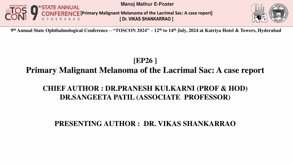
[EP26 ]
Primary Malignant Melanoma of the Lacrimal Sac: A case report
CHIEF AUTHOR : DR.PRANESH KULKARNI (PROF & HOD)
DR.SANGEETA PATIL (ASSOCIATE PROFESSOR)
PRESENTING AUTHOR : DR. VIKAS SHANKARRAO

INTRUDUCTION
•
Tumors of the lacrimal sac are rare
•
Most of them are benign and of epithelial origin.
•
Malignant melanoma of the lacrimal sac is even rarer accounting for 5% of lacrimal sac
tumors and 0.7% of ocular melanoma.
•
The disease has an insidious onset and decepts as a chronic dacryocystitis.
•
As a result, it is difficult to identify at an early stage.
•
It is one of the
HIDDEN THREAT
:
CANCER’S QUIET DECEPTION
•
An early diagnosis is essential, as it is potentially lethal progression if therapy is delayed
or inadequate.
Material and Methods
•
A 78-year-old woman came to our Ophthalmology OPD with a palpable mass at left
medial canthal region since 4 months.
•
H/O watering of left eye since then
•
Initially swelling was 10x10mm when she presented to OPD & increased to 50x50mm in
1month of duration .
•
One episode of bloody discharge from the left eye as well as from nose noted.
•
Swelling was not associated with pain or significant visual disturbance.
•
No previous h/o regional surgery or trauma or excessive sun exposure. There was no
history of a cutaneous primary melanoma.
•
CT-PARA NASAL SINUSES (Plain & Contrast) reported as
possible dacryocystocele
with secondary infection
•
Incision and Drainage was done and sample sent for HPE.
•
MRI – Brain and Orbit was done post I&D.
Left Eye Lacrimal Sac
Swelling of 5x5cm(pre-
op)
Highly pleomorphic tumor cells-round,oval vescicular nuclei.inta & extracellular
brown pigment s/o melanin.Focal areas of necrosis with Inflammation,mitosis0-1,
s/o Malignant Melanoma
Discussion
•
Malignant melanoma of Lacrimal Sac has insidious onset
and limited early visibility that generally result in delayed
diagnosis
•
Initially treated as Dacryocystocele as per CT-PNS Report.
•
HPE report confirmed Malignant melanoma post I&D.
•
Repeat MRI Brain and Orbit reported as well defined
altered signal intensity lesion, seen to abut the medial
margin of globe,mild scalloping of anteromedial margin of
left orbit.
•
Wide en-excision with transpositional frontal flap covering
the wound.
•
One episode of Raditherapy is given after 1 month of Post
Operative status.
Wide excision of Lacrimal sac
Closing wound with transposition
Frontal Flap
CONCLUSION
45 days of post operative
procedure. With LE Visual
Acuity 6/18.
•
Malignant melanoma of the head and neck represents approximately
8% of all melanomas that develop in the head and neck.
•
Generally behaves far more aggressively than cutaneous melanoma.
•
The resection margins were not free of tumor, therefore postoperative
radiotherapy was added to the treatment regimen
•
Early diagnosis is critical as tumor often presents with symptoms
similar to dacryocystitis and mimics as chronic-dacryocystitis.
•
Importance of vigilant clinical assessment and prompt biopsy is key
for early diagnosis.
Malignant melanoma of the lacrimal sac is a rare condition that poses challenges in early diagnosis due to its insidious onset. This case study discusses the presentation, diagnosis, and treatment of a 78-year-old woman with a palpable mass in the lacrimal sac, highlighting the importance of early identification and management of this potentially lethal disease.

Download Presentation

Please find below an Image/Link to download the presentation.
The content on the website is provided AS IS for your information and personal use only. It may not be sold, licensed, or shared on other websites without obtaining consent from the author. Download presentation by click this link. If you encounter any issues during the download, it is possible that the publisher has removed the file from their server.
E N D
Presentation Transcript
Manoj Mathur E-Poster [Primary Malignant Melanoma of the Lacrimal Sac: A case report] [ Dr. VIKAS SHANKARRAO ] 9thAnnual State Ophthalmological Conference TOSCON 2024 12thto 14thJuly, 2024 at Katriya Hotel & Towers, Hyderabad [EP26 ] Primary Malignant Melanoma of the Lacrimal Sac: A case report CHIEF AUTHOR : DR.PRANESH KULKARNI (PROF & HOD) DR.SANGEETA PATIL (ASSOCIATE PROFESSOR) PRESENTING AUTHOR : DR. VIKAS SHANKARRAO
INTRUDUCTION Tumors of the lacrimal sac are rare Most of them are benign and of epithelial origin. Malignant melanoma of the lacrimal sac is even rarer accounting for 5% of lacrimal sac tumors and 0.7% of ocular melanoma. The disease has an insidious onset and decepts as a chronic dacryocystitis. As a result, it is difficult to identify at an early stage. It is one of the HIDDEN THREAT : CANCER S QUIET DECEPTION An early diagnosis is essential, as it is potentially lethal progression if therapy is delayed or inadequate.
Material and Methods A 78-year-old woman came to our Ophthalmology OPD with a palpable mass at left medial canthal region since 4 months. H/O watering of left eye since then Initially swelling was 10x10mm when she presented to OPD & increased to 50x50mm in 1month of duration . One episode of bloody discharge from the left eye as well as from nose noted. Swelling was not associated with pain or significant visual disturbance. No previous h/o regional surgery or trauma or excessive sun exposure. There was no history of a cutaneous primary melanoma. CT-PARA NASAL SINUSES (Plain & Contrast) reported as possible dacryocystocele with secondary infection Incision and Drainage was done and sample sent for HPE. MRI Brain and Orbit was done post I&D.
Left Eye Lacrimal Sac Swelling of 5x5cm(pre- op) Highly pleomorphic tumor cells-round,oval vescicular nuclei.inta & extracellular brown pigment s/o melanin.Focal areas of necrosis with Inflammation,mitosis0-1, s/o Malignant Melanoma
Discussion Malignant melanoma of Lacrimal Sac has insidious onset and limited early visibility that generally result in delayed diagnosis Initially treated as Dacryocystocele as per CT-PNS Report. HPE report confirmed Malignant melanoma post I&D. Repeat MRI Brain and Orbit reported as well defined altered signal intensity lesion, seen to abut the medial margin of globe,mild scalloping of anteromedial margin of left orbit. Wide en-excision with transpositional frontal flap covering the wound. One episode of Raditherapy is given after 1 month of Post Operative status. Wide excision of Lacrimal sac Closing wound with transposition Frontal Flap
CONCLUSION Malignant melanoma of the head and neck represents approximately 8% of all melanomas that develop in the head and neck. Generally behaves far more aggressively than cutaneous melanoma. The resection margins were not free of tumor, therefore postoperative radiotherapy was added to the treatment regimen Early diagnosis is critical as tumor often presents with symptoms similar to dacryocystitis and mimics as chronic-dacryocystitis. Importance of vigilant clinical assessment and prompt biopsy is key for early diagnosis. 45 days of post operative procedure. With LE Visual Acuity 6/18.