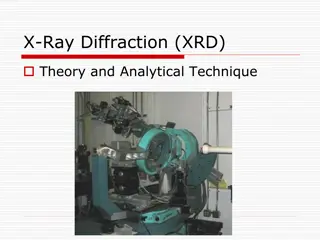
X-ray Diffraction Data Processing Tutorials with DAWN Software
Learn how to process X-ray diffraction data collected on I12-JEEP beamline using DAWN software through a series of tutorials. Discover features applicable to similar instruments and facilities, presented in 10-15 minute videos covering topics like data viewing, processing, calibration, and peak fitting. Enhance your understanding of metadata files, extraction, visualization, data processing pipelines, and more.

Download Presentation

Please find below an Image/Link to download the presentation.
The content on the website is provided AS IS for your information and personal use only. It may not be sold, licensed, or shared on other websites without obtaining consent from the author.If you encounter any issues during the download, it is possible that the publisher has removed the file from their server.
You are allowed to download the files provided on this website for personal or commercial use, subject to the condition that they are used lawfully. All files are the property of their respective owners.
The content on the website is provided AS IS for your information and personal use only. It may not be sold, licensed, or shared on other websites without obtaining consent from the author.
E N D
Presentation Transcript
Overview of (I12 Overview of (I12- -JEEP) DAWN tutorial videos JEEP) DAWN tutorial videos (Stefan Michalik, I12 (Stefan Michalik, I12- -JEEP BEAMLINE) JEEP BEAMLINE) The aim of tutorials is to show and remind (users) how to process X-ray diffraction data collected on I12-JEEP beamline using DAWN software (the Data Analysis WorkbeNch). Although, tutorials deal with data collected on I12 (reflected in using metafiles), presented features of DAWN are applicable for data collected at other similar instruments and facilities. For more information, see DAWN User Guide (https://diamondlightsource.atlassian.net/wiki/spaces/DT/pages/1378477/DAWN+User+Gui de ) The next few slides present the overview of the content of individual videos to help with tutorial browsing. The duration of videos is about 10-15 minutes. Finally, information how DAWN, I12-JEEP Beamline and Diamond Light Source can be acknowledged is reminded.
List of Tutorials List of Tutorials 1. DataVis Perspective: 2D PILATUS raw data viewing 2. Processing Perspective: 2D PILATUS raw data processing 3. Powder Calibration Perspective: Energy and Instrument calibration 4. Peak Fitting: Peak Fitting Tool 5. Peak Fitting: Function Fitting Tool 6. Processing Perspective: 2D Pilatus raw data processing Cake Remapping
Video 1: Video 1: DataVis Perspective 2D PILATUS raw data viewing What we will learn: Load and view a nxs file; e.g. File Open File or Recent Folders or Recent Files or just Drag and Drop a file into Data Files window (video at 01:28 min:sec) The nxs file is a metadata file containing a link to raw data images (tif files, cbf files, ) and information about the beamline itself before a scan (slit size, stage positions, scanning command ) and during a scan (sample temperature, time stamp, scan identification number, ). View metadata saved inside a nxs file (video at03:15), e.g.: Information about a scan command employed to collect this nxs file (video at 03:38) Finding information about the slit size during data collection in EH1 on I12 (video at 04:35) Usage of basic tools; e.g. metadata preview, zooming, automatic axis rescale, dimension checking (video at 05:05) DATA examples of scans collected on I12-JEEP Beamline at DLS 1. single shot scan of CeO2 (video at 06:06). 2. line scan through a amorphous sample ingot (video at06:15) 3. time scan during the sample heating (video at 07:56) Load a cbf/tif file to find differences in respect to a nxs file (video at 10:21)
Video 2: Video 2: Processing Perspective Processing Perspective 2D PILATUS raw data processing What we will learn: Use Processing Perspective (video at 00:35 min:sec). Define a basic pipeline of operations necessary for data processing: (video) Import Detector Calibration or Enter Diffraction Calibration (video at 03:10) Threshold mask (see video at 03:50) Powder Diffraction Intensity Corrections (video at 04:56) Azimuthal Integration to get an intensity curve v Q, 2theda or d (video at 05:32) Export to Text File to get an ASCII data type format file 2 columns (video at 08:40) Process data (video at 09:30) Save a defined pipeline of operations and upload it for a repeated usage (video at 11:34). Check processed data using DataVis (video at 12:56).
Video 3: Video 3: Powder Calibration Perspective Powder Calibration Perspective Energy and instrument calibration What we will learn: Use Powder Calibration Perspective. Perform X-ray energy and instrument calibration using multiple 2D diffraction patterns of a well-known NIST sample standard (CeO2, LaB6, Si and so on ) collected at various sample-to-detector distances with a well-defined offset Perform instrument calibration with a known X-ray energy (fixed energy mode) using a single 2D diffraction pattern of a well-known NIST sample standard (see video at09:23 min:sec). Given examples include: DAWN automatic and manual mode explanation (see video at 12:56 min:sec for manual mode). Using nxs files and files when a metadata file is not available (see vide at 06:27 min:sec). Export of a calibration file to be later used in Processing Perspective (at 10:59 min:sec).
Video 4: Peak Fitting Video 4: Peak Fitting Peak Fitting Tool What we will learn: Use Peak Fitting tools (pre-defined profile functions) Applying fitting on the whole dataset Given example includes: In situ measurement of the Au powder sample using a Linkam stage from room temperature up to 800 C. Data are integrated into Q-space A processed nxs file is used for a single peak fitting (video at 02:15 min:sec) and Peak fitting tool is shown ( at 02:50) Peak fitting tool applied on the whole dataset and fitting results viewed in DataVis Perspective (at 04:52) Simultaneous two peak fitting (video at 06:07) Export fitting results into an ASCII file (video at 08:03)
Video Video 5: Function Fitting Tool 5: Peak Fitting Peak Fitting What we will learn: Use Function Fitting tools (more functions available) Profile fitting function included into a pipeline (at 01:49 min:sec) Viewing fitting results in DataVis (at 03:50) Simultaneous two peak fitting (at 06:10) Given examples include: In situ measurement of the Au powder sample using a Linkam stage on I12 from room temperature up to 800 C. Data are integrated into Q-space and then selected peak/peaks are fitted.
Video 6: Video 6: Processing Perspective Processing Perspective Cake Remapping 2D (Pilatus) raw data processing Cake Remapping What we will learn: Use Cake Remapping Operation Export cake remapping results into a text file Use Fit Peak Image Rows Operation Given examples include: Data from loaded and unloaded an Al-based alloy to demonstrate the peak position variation as a function of an azimuthal angle.
DAWN DAWN references https://dawnsci.org/downloads/ https://dawnsci.org/downloads/ https://.../wiki/ https://.../wiki/DAWN+User+Guide DAWN+User+Guide If the analysis features available in DAWN are used significantly in your work, please cite: Data Analysis WorkbeNch (DAWN). Basham M. et al. (2015). J. Synchrotron Rad. 22, https://doi.org/10.1107/S1600577515002283
DAWN DAWN references references https://dawnsci.org/downloads/ https://dawnsci.org/downloads/ https://.../wiki/ https://.../wiki/DAWN+User+Guide DAWN+User+Guide If the 2D powder diffraction and processing tools are used, please cite: Processing two-dimensional X-ray diffraction and small-angle scattering data in DAWN 2. Filik J. et al. (2017). J. Appl. Cryst. 50, https://doi.org/10.1107/S1600576717004708.
Problem: a X Problem: a X- -ray energy and sample ray energy and sample- -to to- -detector distance are unknown distance are unknown detector It is not possible correctly to determine both the X-ray energy and sample-to-detector distance just from a single 2D diffraction image of a standard sample. One of these two parameters has to be fixed!!! Solution: collection of 2D diffraction images at multiple sample-to-detector distances with a well-defined offset. For more details see https://doi.org/10.1107/S0021889813022437. The presented algorithm is directly implemented in DAWN. Stefan Michalik stefan.michalik@diamond.ac.uk
I12 I12- -JEEP BEAMLINE reference JEEP BEAMLINE reference https://www.diamond.ac.uk/Instruments/Imaging-and-Microscopy/I12.html https://doi.org/10.1107/S1600577515003513
DLS acknowledgment DLS acknowledgment https://www.diamond.ac.uk/Users/Experiment https://www.diamond.ac.uk/Users/Experiment- -at at- -Diamond/After Diamond/After- -you you- -Leave.html Leave.html