Veterinary Physiology and the Functions of Blood
Veterinary physiology involves studying the functions of intact organisms, organs, tissues, and systems. Blood, consisting of plasma and formed elements, plays vital roles in nutrient transport, gas exchange, waste removal, hormone distribution, temperature regulation, water balance, pH maintenance, clotting, and immune defense. Important blood cells like erythrocytes, with their unique characteristics and hemoglobin composition, are crucial for oxygen transportation in the body.

Download Presentation

Please find below an Image/Link to download the presentation.
The content on the website is provided AS IS for your information and personal use only. It may not be sold, licensed, or shared on other websites without obtaining consent from the author.If you encounter any issues during the download, it is possible that the publisher has removed the file from their server.
You are allowed to download the files provided on this website for personal or commercial use, subject to the condition that they are used lawfully. All files are the property of their respective owners.
The content on the website is provided AS IS for your information and personal use only. It may not be sold, licensed, or shared on other websites without obtaining consent from the author.
E N D
Presentation Transcript
Veterinary Veterinary Physiology concerned with the intact organ or the whole organism and may be defined as the study of functions of the functions of all its parts like, systems, organs, tissues, components including the biophysical and biochemical process involved. Physiology- - It is the body integrated and the cells and cell
Blood It consist of a pale yellow fluid called plasma in which the formed elements, the red cells, the white cells & the platelets are suspended. FUNCTIONS:- It carries nutrients made available by the digestive tract to body tissues. It plays an important role in exchange of gases from blood to tissue and vice-versa. The waste products from various tissues are carried out to the kidney for excretion. Hormones are carried via blood from endocrine glands to other organs of the body. It play an important role in temperature control by transporting heat from deeper structures to the surface of the body Water balance is maintained partly by the blood.
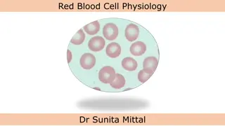
Buffer such as bicarbonate in the blood help to maintain a constant pH of tissues & body fluids. The clotting ability of blood prevents access loss of blood from injuries. It contains important factors for defense of the body against disease. BLOOD CELLS Erythocytes: Biconcave disks Averaging 7.5 m in diameter Thick 1.5 m circular margin & a thin centre Doesn`t have nucleus Shape can be remarkably changed into almost any shape Average volume is 87 6 cubic microns Average count is 7 11 million/cu mm
Hemoglobin: It is a complex organic compound composed of 4 red porphyrin pigments (hemes) each of which contains an atom of iron + globin consists of 4 amino acid chains. Its presence within the erythrocyte is responsible for its ability to transport O2& for the red color of the erythrocytes. Combines with O2to form oxyhemoglobin in lungs which readily give up its O2to tissue cells. It is not a true oxidation O2from lungs forms a loose combination with each iron atom of Hb & the product is oxy Hb (HbO2) When the blood reaches tissue deficient in O2, the loosely held O2of the Hb O2is given up readily.
Methemoglobin is the true oxidation present in ferric state (fe3+) Carboxyhemoglobin (CO + Hb) is a more stable compound Affinity of Hb for CO is 210 times greater than affinity for O2 but unable to carry the O2
Formation and fate of Hemoglobin: Iron is absorbed from the diet by epithelial cells of the duodenal mucosa, in mucosal blood capillaries, -globulin transferrin combines & carries the iron. Iron reserves in the bone marrow A small amount is used in myoglobin in the muscles Temporarily, it stores in liver & spleen as ferritin It is lost via urine, feces & sweat It is used other than in case of mensuration & developing fetus during pregnancy. In fetus, the nucleated RBC is produced by liver, spleen & lymph nodes In adults, it is formed in bone marrow & non nucleated In birds, the red cells have nucleus throughout the life
Contd Destruction occurs between 3 to 4 months Globin protein fraction of Hb is degraded to amino acids Fe is picked up by the globin transferrin and stored in the liver as ferritin further to form myoglobin or stored in tissue cells as hemosiderin Biliverdin & bilirubin, green pigment remains after the breakdown of Hb from liver to gall bladder is used for emulsification of fats and excreted from urine urobilinogen Accumulation in excess of bilirubin in vascular system is called Icterus or Jaundice This may be caused due to liver damage, anaplasmosis (parasitic infection) or some pathologic condition Seen as yellow in the visible membrane as
Hemolysis: It is a breakdown of red cells as Hb escapes into the plasma caused by the toxins, snake venoms, blood parasites, hypotonic solution & many chemical substances. The resulting Hb in the plasma gives it a reddish color & the condition called hemoglobinemia & if excreted through urine called hemoglobinuria (red water). Hemagglutination: It is a clumping of RBC results when blood injects in another species of animal. Clumping may occur within same species, if blood of the wrong type is used. So, blood matching is desirable before attempting transfusion Hematocrit value or PCV: It is the % by volume as of whole blood that is constituted by RBC. It is determined by filling a hematocrit tube with treated blood so that it will not clot & then centrifuge until the cells are packed in the lower end.
Erythrocyte Sedimentation Rate (ESR): When blood to which an anticoagulant has been added stands in a narrow tube the red cells from aggregates (rouleaux) which gradually sediment leaving a clear zone of plasma above. Can be measured after 1 hr of sedimentation Depends on concentration of fibrinogen, 2& globulins. Useful in detecting presence & activity of disease Also varies according to red cell count; lower the red cell greater the sedimentation rate Mean Corpuscular Volume (MCV): From the hematocrit & the red cell count, it is possible to count the MCV The range averages from 72 to 98 m3 Polycythemia: At high altitude, blood forming organs automatically produce large quantity of RBC`s.
Erythropoiesis: The process of formation of erythrocyte Regulation:- It is governed in bone marrow by the reduced level of O2in tissues & cells The glycoprotein (erythropoietin) of plasma & the enzyme of kidney (renal erythropoietic factor) combindely act to do so Concentration of RBC is controlled by ve feedback for the mechanism of erythropoiesis Other humoral factors governing erythropoietin are- Cobalamin & folic acid Copper pyridoxine
Anemia: It is a condition when number of red cells or the quantity of Hb is much decreased below normal. May be due to deficient blood formation Dietary deficiency of Fe, Cu, vitamins or amino acids May be due to hemorrhage occurs from wound or parasites (stomach worms or lice) Also by deficient secretion of intrinsic factor from stomach Genetically Viscosity of blood is reduced Reduced concentration of RBC`s It is measured in grams/100 ml of blood. Types of Anemia: Hypochromic- Depletion of Fe takes place & the Hb concentration in the red cell falls & appears hypochromic.
Contd Sickle Cell- Sickle shaped of the erythrocytes due to the presence of an abnormal Hb; HbS. It may be due to exposure to low O2tension. Sideroblastic- Massive accumulation of ferritin around the nucleus of erythroblast k/a ring sideroblast. It may be due to Pyridoxine. Thalassemia (Mediterrenian quantitative failure of synthesis of either or globulin chains of the Hb molecule. Pernicious Erythroblastosis fetalis etc. anemia)- It is due to
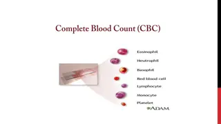
Leucocytes: They are nucleated and are capable of independent movement Classified in 2 types based on the presence of granules and nucleus Classification: They have granules within the cytoplasm and having life span of few hrs 1. Granulocytes N cells act as 1stline of defense & degraded in the form of Pus, E cells shows red staining under Eosin (allergic) & B cells shows blue stain under methylene blue (stasis of blood) Neutrophils Eosinophils Basophils Phagocytic in nature, largest WBC`s, variable in size & largest nucleated 2. Agrnulocytes M cells are phagocytic, acts in acute infections & also k/a Macrophage. L cells are variable in size, large nucleus, acts in cellular immunity, unit is cu mm, wrights stain. DLC % of WBC. Monocytes Lymphocytes
Plasma: Sample of untreated blood, permitted to slant position & cells settled down leaving a straw color fluid Water- 92%, Based on MW 7% are proteins & 1% consists of glucose, lipids, hormones, electrolytes, mineral salts, metabolic waste products etc. Functions of Plasma Proteins- Carrier- solubilized & carry Fe, thyroxine, cortisol etc. Also acts as storage pool, when needed leaves to act at targets Immunity- globulin is associated with immunity & resistance to disease (antibodies react with antigen to neutralize or to break it) Buffering- prevent to change in pH of blood because of ionized carboxyl & amides Maintenance of Osmotic pressure- It is 290mOsm/l at RT, mainly done by low MW colloids (proteins)
Serum: Plasma - Fibrinogen Contains antibodies, useful in prevention & treatment Immune/Hyperimmune serum- By inoculating repeatedly of a specific antigen in an animal as passive protection Blood pH: It refers to H+ion concentration & determines relative acidity or alkalinity of the solution. NaHCO3as buffer maintains blood within narrow limits. pH 7.35 7.45 pH 7 (neutral), pH >7 (acidic) & pH < 7 (alkaline) Blood clotting: When coagulation occurs a jelly mass results & shrinks to produce a firm clot & clear fluid, while the actual clot consists of fibrin. Blood vessel damaged or cut Reflex will be twitching to walls due to myogenic contract Spasm/sympathetic nerve reflex stimulate adrenergic fibre Constriction & narrow the vessel for reduce in blood flow
Platelet plug- Blood platelets exposed to endothelium of a cut vessel, they adhere to collagen & elastic fibres causes metamorphosis (swelling of BP) which becomes sticky & secrete ADP so as to trigger metamorphosis to make platelet aggregation Clotting/Coagulation- Begins between 15 sec to 2 min after injury & completes in 5 min followed by clot where fibroblasts change into fibrous tissue by 7 to 10 days with the help of different substances k/a factors. Fibrin- forms loose clot & present as precursor, fibrinogen. Dicoumarins- Antagonistic to vit. K, reduces the amount of prothrombin. Excess of this causes sweet clover disease. Warfarin- Rodent poison, animal dies due to internal bleeding. Heparin- It is a mucopolysaccharide, isolated from hepatic cells & stored in metachromatic granules of mast cells in the walls of blood vessels.
Coagulation time- Length of time from drawing a fresh blood sample until coagulation occurs. Specific gravity of blood- Index/ratio of weights of a substance compared to the wt. of an equal volume of water & measured by hydrometer Blood volume- It refers the total amount of blood in an animal body. Lymph: It is a clear, colorless fluid like blood plasma from which it derived. Consists numerous lymphocytes & few red cells Also inorganic salts Specific gravity is 1.015 Plasma picked up by lymphatics (capillary system) which are not absorbed in the capillary walls of the tissue spaces. Chyle, results from the absorption of lipids are derived from the lymph of the intestine Filtered by nodular structures called lymph nodes
Cerebrospinal fluid: Formed by choroid plexus in the ventricles of the brain. It circulates through sub-arachnoid space between the pia mater & arachnoids membrane. Resembles blood plasma from they are derived Less numbers of lipids, proteins, glucose & K+ Serves as cushioning agent Provides nutrition & lubrication to the brain & spinal cord Synovial fluid: It is a vascular CT consists of formed elements also k/a matrix. thick, tenacious liquid which is colorless to deep yellow varies with species & types of joints. Contains fibroblasts, macrophages, mast cells, fat cells & nerve fibres Provides lubrication (muco-polysaccharides & hyaluronic acid) Nourish the articular cartilages & reduce friction in joints
Serous fluid: It is found in body cavities include peritoneal, pericardial & pleural fluids. Reduces friction between apposed surfaces Nourish the sacs Prevents from the inflammation or infection by the production of serous fluids like pleuritis, peritonitis & pericarditis.
Blood clotting factors: I Fibrinogen II Prothrombin III Thromboplastin IV Calcium V Proaccelerin (labile factor, accelerator globulin) VII Proconvertin (stable factor, anti-prothrombin 1) VIII Anti-hemophilic factor (AHF-A, platelet co-factor) IX Christmas factor (AHF-B, component) X Stuart factor (anti-prothrombin III, stuart prower factor) XI Plasma thromboplastin anticedent (AHF-C) XII Hagemen factor (contact factor) XIII Fibrin stabilizing factor (plasma transglutaminase) plasma prothromboplastin


: When blood to")