Blood Components and Microhaematocrit Procedure in Physiology Lab
Blood is a complex fluid composed of various components like platelets, erythrocytes, and leukocytes suspended in plasma. Microhaematocrit is a technique used to measure hematocrit levels in blood. The process involves collecting blood in a capillary tube, centrifuging it to separate the layers, and analyzing the ratios of red blood cells, plasma, platelets, and leukocytes. Proper hygiene and technique are essential for accurate results.

Uploaded on Oct 10, 2024 | 0 Views
Download Presentation

Please find below an Image/Link to download the presentation.
The content on the website is provided AS IS for your information and personal use only. It may not be sold, licensed, or shared on other websites without obtaining consent from the author.If you encounter any issues during the download, it is possible that the publisher has removed the file from their server.
You are allowed to download the files provided on this website for personal or commercial use, subject to the condition that they are used lawfully. All files are the property of their respective owners.
The content on the website is provided AS IS for your information and personal use only. It may not be sold, licensed, or shared on other websites without obtaining consent from the author.
E N D
Presentation Transcript
Physiology lab Second stage Assisstant Physiology lab 2 2 Second stage Assisstant lect lect: : Zainab Zainab Mahdi Mahdi
Blood platelets, suspended in a liquid which are called plasma. Blood is a viscous, red liquid, consisting of : erythrocytes, leukocytes, When blood is collected in an anticoagulant and centrifuged, erythrocytes cells form a thin film on the surface of red blood cells and yellow plasma Hematocrit is the ratio of red blood cells to the whole blood volume. erythrocytes are submitted in the lower part of the vessel, white blood white blood yellow plasma occupy the top. Hematocrit It is usually expressed either as a percentage or as a decimal fraction (e.g. 41% or 0.41). The term comes from Greek roots hemat- (blood) and krites (edge).
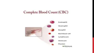
Microhaematocrit Electronic cell counting Microhaematocrit Electronic cell counting
Microhaematocrit tube (capillary tube) 75mm in length and 1mm in diameter which contains heparin and show a red ring at the end of the tube. Microhaematocrit centrifuge device. Plastic seal to seal one end of Microhaematocrit capillary tube. Microhaematocrit reader. lancet Cotton, alcohol.
Clean your finger with 70% alcohol and let dry. Prick finger with lancet, near the tip but not too close to the nail. Prick so that blood flows freely. Try squeezing up from your wrist if blood does not flow after pricking finger. Place the tip of a capillary tube onto a drop of blood on your finger. 4. Call your instructor to seal the tube. 5. The instructor will spin the tubes in a centrifuge ( 5 minutes at 10000 rpm). 6. Using a special reading device(since the 1. 2. 3.
Note that the blood has been separated into 3 layers: 1. An upper layer of clear plasma amber slightly yellow colored. It should not be pink or red which would indicate hemolysis of red cells in the sample or within the body in hemolytic diseases. 2. A greyish-white, thin layer (about 1 mm thick) called buffy layer , consisting of platelets (thrombocytes) above and leukocytes below it. 3. A bottom layer of red cells.
The normal values of PCV vary according to the age and sex of the individuals. The normal ranges are: Males: 40 % 54 % Females: 36 % 47 % Newborns: 55-68 %.
The percentage of the volume of blood occupied by the red cells constitutes hematocrit or packed cell volume. Hct % = {Height of RBCs (mm) / Height of RBCs and plasma (mm)} Hct plasma (mm)} 100 % = {Height of RBCs (mm) / Height of RBCs and 100 For example, if the height of packed red cells is 45 mm, then = 45/ 100 100 = 45 percent. It also means that out of 100 volumes of blood 45 volumes are red cells and 55 volumes are plasma. Thus, out of 1 liter of blood, 450 ml are red cells and 550 ml are plasma.
PCV is affected by the shape, & the number of the RBCs & the plasma volume. High number of circulating RBCs or decrease in plasma volume seen in cholera due to loss of water in the stool. Low PCV RBC or increase in plasma volume. High PCV PCV associated with either increase in Low PCV associated with either decrease in
An insufficient supply of healthy red blood cells (anemia A large number of white blood cells long-term illness, infection, leukemia, lymphoma or other disorders of white blood cells. Acute kidney disease production lead to less RBCs production by the bone marrow). Pregnancy fluid in blood. This could potentially lead to a small drop in hematocrit levels. 1. (anemia). A large number of white blood cells due to 2. Acute kidney disease (lower Erythropoietin 3. Pregnancy may lead to women having additional 4. 4.
Abnormal increase in red blood cells ( (erythrocytosis erythrocytosis) ) 1. A disorder, such as polycythemia too many red blood cells (in polycythemia it may rise to as high as 70 %). polycythemia vera vera that causes your body to produce 2. At higher altitudes hematocrit levels may increase over time. At higher altitudes, there is a lower oxygen supply in the air and thus 3. Low blood oxygen levels (hypoxia) (hypoxia) 4. Lung or heart disease more red blood cells in an effort to increase the amount of oxygen in the blood Lung or heart disease if the body senses low oxygen levels, it will make 5. 5. Dehydration Dehydration. 6. Burn Burn ( due to loss of plasma). 7. 7.
THANK YOU ! THANK YOU !


 75mm")
 75mm")